



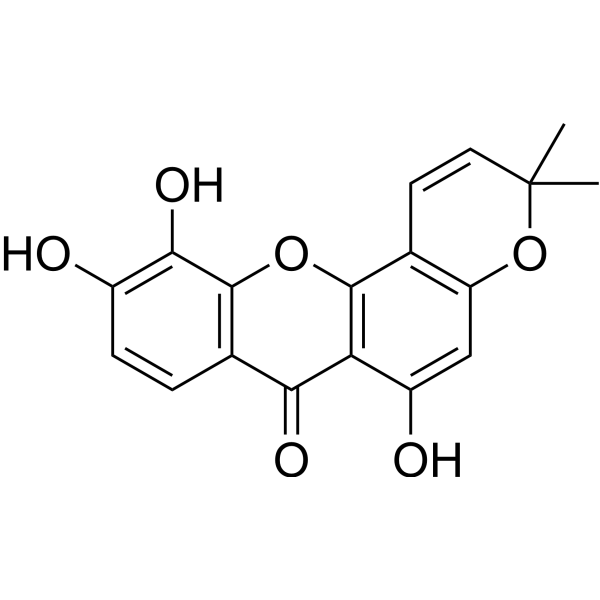
Isojacareubin

Based on 1 Customer Validation
Isojacareubin can be isolated from Hypericum japonicum. Isojacareubin covalently inhibits SARS-CoV-2 3CLpro. Isojacareubin also has anti-helicobacter activity. Isojacareubin inhibits PKC, and suppresses hepatocellular carcinoma metastasis and induces apoptosis.
For research use only. We do not sell to patients.
- Purity : 98.0%
- CAS No.: 50597-93-8
- Formula: C18H14O6
- Molecular Weight:326.30
-
Storage:
-20°C, sealed storage, away from moisture and light
* In solvent : -80°C, 6 months; -20°C, 1 month (sealed storage, away from moisture and light)

To place orders, for customer services and technical support, please contact: MedChemExpress USA
Tel: 609-228-6898 E-mail: [email protected] [email protected]


-
 Biological Activity
-
Chemical Information
- Protocols
- Purity & Documentation
- References
-
 Help & FAQs




Biological Activity
Description
Cellular Effect
|
Cell Line

|
Type | Value | Description | References |
|---|---|---|---|---|
| A549 | IC50 |
3.9 μM
Compound: 8
|
Cytotoxicity against human A549 cells assessed as reduction in cell viability incubated for 48 hrs by MTS assay
Cytotoxicity against human A549 cells assessed as reduction in cell viability incubated for 48 hrs by MTS assay
|
[PMID: 37650785] |
| BEAS-2B | IC50 |
21.1 μM
Compound: 8
|
Cytotoxicity against human BEAS-2B cells assessed as reduction in cell viability incubated for 48 hrs by MTS assay
Cytotoxicity against human BEAS-2B cells assessed as reduction in cell viability incubated for 48 hrs by MTS assay
|
[PMID: 37650785] |
| MDA-MB-231 | IC50 |
8.9 μM
Compound: 8
|
Cytotoxicity against human MDA-MB-231 cells assessed as reduction in cell viability incubated for 48 hrs by MTS assay
Cytotoxicity against human MDA-MB-231 cells assessed as reduction in cell viability incubated for 48 hrs by MTS assay
|
[PMID: 37650785] |
| SMMC-7721 | IC50 |
2.2 μM
Compound: 8
|
Cytotoxicity against human SMMC-7721 cells assessed as reduction in cell viability incubated for 48 hrs by MTS assay
Cytotoxicity against human SMMC-7721 cells assessed as reduction in cell viability incubated for 48 hrs by MTS assay
|
[PMID: 37650785] |
| SW480 | IC50 |
4 μM
Compound: 8
|
Cytotoxicity against human SW480 cells assessed as reduction in cell viability incubated for 48 hrs by MTS assay
Cytotoxicity against human SW480 cells assessed as reduction in cell viability incubated for 48 hrs by MTS assay
|
[PMID: 37650785] |


Chemical Information
-
CAS No. 50597-93-8
-
Appearance Solid
-
Molecular Weight 326.30
-
Formula C18H14O6
-
Color Light yellow to yellow
-
SMILES
O=C1C2=C(OC3=C1C=CC(O)=C3O)C(C=CC(C)(C)O4)=C4C=C2O
-
Structure Classification
-
Initial Source
-
Shipping
Room temperature in continental US; may vary elsewhere.
-
Storage
-20°C, sealed storage, away from moisture and light
* In solvent : -80°C, 6 months; -20°C, 1 month (sealed storage, away from moisture and light)


Purity & Documentation
-
Data Sheet (269 KB)
-
SDS (393 KB)
- English - EN (393 KB)
- Français - FR (393 KB)
- Deutsch - DE (393 KB)
- Norwegian - NO (393 KB)
- Español - ES (393 KB)
- Swedish - SV (393 KB)
- Italian - IT (393 KB)
- Korean - KR (393 KB)
- Portuguese - PT (393 KB)
-
Handling Instructions (2659 KB)
References
[2]. Khan A, et al. Discovery of Isojacareubin as a covalent inhibitor of SARS-CoV-2 main protease using structural and experimental approaches. J Med Virol. 2023 Feb;95(2):e28542. [Content Brief]
[3]. Yuan X, et al. Inhibition of protein kinase C by isojacareubin suppresses hepatocellular carcinoma metastasis and induces apoptosis in vitro and in vivo. Sci Rep. 2015 Aug 6;5:12889. [Content Brief]

Calculators
Concentration (start) × Volume (start) = Concentration (final) × Volume (final)


Isojacareubin Related Classifications
HY-N8306 Related Classifications
 Powered by Bioz