



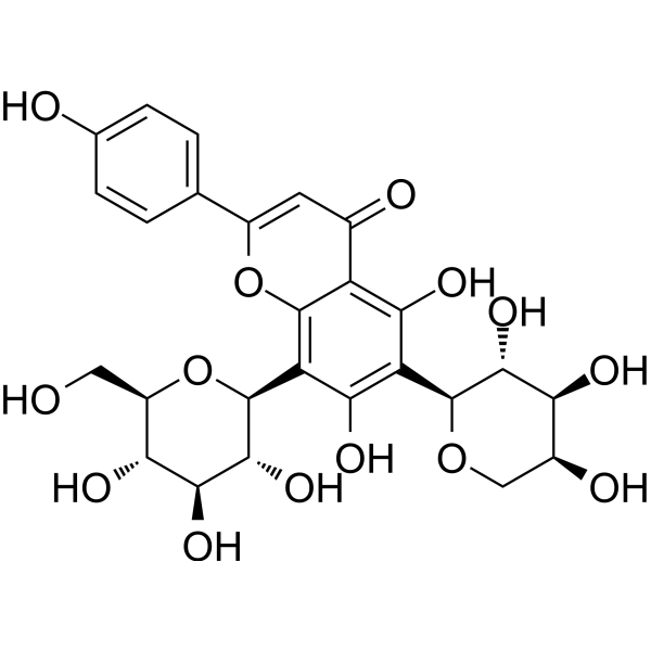
Isoschaftoside
Based on 1 publication(s) in Google Scholar
Isoschaftoside, a C-glycosylflavonoid from Desmodium uncinatum root exudate, can inhibit the growth of germinated S. hermonthica radicles. Isoschaftoside reduces reactive oxygen species (ROS) and induces proliferation in senescent cells. Isoschaftoside activates autophagy. Isoschaftoside can be used for anti-tumor, anti-inflammatory, antioxidant, antihypertensive, hepatoprotective and nematicidal study.
Nos produits utilisent uniquement pour la recherche. Nous ne vendons pas aux patients.
- Pureté : 99.95%
- CAS No.: 52012-29-0
- Formule: C26H28O14
- Masse moléculaire:564.49
-
Stockage:
4°C, protect from light
* In solvent : -80°C, 6 months; -20°C, 1 month (protect from light)

To place orders, for customer services and technical support, please contact: MedChemExpress USA
Tel: 609-228-6898 E-mail: [email protected] [email protected]


-
 Activité biologique
-
Chemical Information
-
Solvant et solubilité
- Protocole
- Pureté et documentation
- Références
-
 Help & FAQs



Publications Citing Use of MedChemExpress (MCE) Isoschaftoside
MoreVoir tous les produits spécifiques à Isoform Parasite
More

Activité biologique
Description
In Vitro
Isoschaftoside (4 μM, 12 days) suppresses ROS by reducing the expression of RAC2 and LINC00294 in senescent fibroblasts[1].
Isoschaftoside (0.25-4 μM, 12 days) significantly increases cell proliferation in senescent fibroblasts[1].
Isoschaftoside (1 μM, 12 days) significantly increases mitochondrial membrane potential (MMP), induces metabolic changes that restore mitochondrial function and reduces dependence on glycolysis in senescent fibroblasts[1].
Isoschaftoside (1 μM, 12 days) reduces DNA damage and fragmentation, activates the autophagy system to clear senescent fibroblasts, reduces the expression of inflammatory factors in senescent fibroblasts[1].
Isoschaftoside (20-600 μg/mL) possesses strong nematicidal activity against M. incognita (LC50 = 323.09 μg/mL)[2].
Isoschaftoside (1-500 μM, 24 h) reduces lipid deposition via activating autophagy flux, and suppresses the expression of light-chain 3-II (LC3-II) and SQSTM1/p62 in Palmitic acid (PA) (HY-N0830)-induced autophagy inhibition in HepG2 cells[3].
Isoschaftoside (0.05-2 mg/mL) considerably decreases renal Na+, K+-ATPase activation in basolateral membrane of the proximal tubule from pig kidney[4].
Isoschaftoside (0-1000 μM, 9-24 h) can suppress inflammatory responses in Lipopolysaccharides (HY-D1056) (LPS)-activated microglia[5].
MedChemExpress (MCE) has not independently confirmed the accuracy of these methods. They are for reference only.
-
Cell Line:human dermal fibroblasts
-
Concentration:0.25, 0.5, 1, 2, 4 μM
-
Incubation Time:12 days
-
Result:The increase in cell proliferation rate was most significant at a concentration of 1 μM.
-
Cell Line:human dermal fibroblasts
-
Concentration:1 μM
-
Incubation Time:12 days
-
Result:LC3B was noticeably activated.
Perinuclear mitochondria were reduced.
Showed significantly enhanced levels of autophagy.
Significant reduction in lipofuscin was observed.
-
Cell Line:HepG2 cells
-
Concentration:1, 10, 20, 50, 100, 200, 300, 400, 500 μM
-
Incubation Time:24 h
-
Result:When the concentration was higher than 200 μM, observed significant reductions in LC3-II expression.
Decreased LC3-II and p62 expression in Palmitic acid (PA) (HY-N0830)-induced autophagy.
-
Cell Line:BV2 microglial cells
-
Concentration:200 μM
-
Incubation Time:9 h or 24 h
-
Result:Significantly inhibited Lipopolysaccharides-induced iNOS, TNF-α, IL-1β, and COX2 protein expression.
Significantly reduced the Lipopolysaccharides-induced HIF-1α, HK2, and PFKFB3 expression.
Attenuated Lipopolysaccharides-induced phosphorylation of ERK1/2 and mTOR.
In Vivo
MedChemExpress (MCE) has not independently confirmed the accuracy of these methods. They are for reference only.
-
Animal Model:C57 mice fed with a high-fat diet (HFD) (4 weeks; male; C57BL/6JGpt)[3]
-
Dosage:20 mg/kg
-
Administration:Intraperitoneal injection (i.p.); daily for 4 weeks
-
Result:Significantly reduced the body weight and body size of the high-fat diet mice compared with the model group.
Reduced the visceral fat weight of high-fat diet mice.
Significantly attenuated hepatic lipid accumulation induced by a high-fat diet and decreased the number of intracellular lipid droplets and hepatocyte balloons.
Reversed the damage to liver function and the elevation of blood glucose caused by a high-fat diet.
The expression level of p62 and LC3-II was significantly lower than that in the HFD group.


Chemical Information
-
CAS No. 52012-29-0
-
Appearance Solid
-
Masse moléculaire 564.49
-
Formule C26H28O14
-
Color Light yellow to yellow
-
SMILES
O=C1C=C(C2=CC=C(O)C=C2)OC3=C([C@H]4[C@@H]([C@H]([C@@H]([C@@H](CO)O4)O)O)O)C(O)=C([C@H]5[C@@H]([C@H]([C@H](CO5)O)O)O)C(O)=C13
-
Structure Classification
-
Livraison
Room temperature in continental US; may vary elsewhere.
-
Stockage
4°C, protect from light
* In solvent : -80°C, 6 months; -20°C, 1 month (protect from light)


Publications (1)
-
Journal Impact Factor
-
Most Recent


Solvant et solubilité
In Vitro:
DMSO : 100 mg/mL (177.15 mM; Need ultrasonic; Hygroscopic DMSO has a significant impact on the solubility of product, please use newly opened DMSO)
Please refer to the solubility information to select the appropriate solvent. Once prepared, please aliquot and store the solution to prevent product inactivation from repeated freeze-thaw cycles.
Storage method and period of stock solution: -80°C, 6 months; -20°C, 1 month (protect from light). When stored at -80°C, please use it within 6 months. When stored at -20°C, please use it within 1 month.
Please refer to the solubility information to select the appropriate solvent. Once prepared, please aliquot and store the solution to prevent product inactivation from repeated freeze-thaw cycles.
Storage method and period of stock solution: -80°C, 6 months; -20°C, 1 month (protect from light). When stored at -80°C, please use it within 6 months. When stored at -20°C, please use it within 1 month.
Concentration (start) × Volume (start) = Concentration (final) × Volume (final)
In Vivo:
Select the appropriate dissolution method based on your experimental animal and administration route.
- For the following dissolution methods, please ensure to first prepare a clear stock solution using an In Vitro approach and then sequentially add co-solvents:
- To ensure reliable experimental results, the clarified stock solution can be appropriately stored based on storage conditions. As for the working solution for In Vivo experiments, it is recommended to prepare freshly and use it on the same day.
- The percentages shown for the solvents indicate their volumetric ratio in the final prepared solution. If precipitation or phase separation occurs during preparation, heat and/or sonication can be used to aid dissolution.
Add each solvent one by one: 10% DMSO 40% PEG300 5% Tween-80 45% Saline
Solubility: ≥ 2.5 mg/mL (4.43 mM); Clear solution
This protocol yields a clear solution of ≥ 2.5 mg/mL (saturation unknown).
Taking 1 mL working solution as an example, add 100 μL DMSO stock solution (25.0 mg/mL) to 400 μL PEG300, and mix evenly; then add 50 μL Tween-80 and mix evenly; then add 450 μL Saline to adjust the volume to 1 mL.
Preparation of Saline: Dissolve 0.9 g sodium chloride in ddH₂O and dilute to 100 mL to obtain a clear Saline solution.
Add each solvent one by one: 10% DMSO 90% (20% SBE-β-CD in Saline)
Solubility: ≥ 2.5 mg/mL (4.43 mM); Clear solution
This protocol yields a clear solution of ≥ 2.5 mg/mL (saturation unknown).
Taking 1 mL working solution as an example, add 100 μL DMSO stock solution (25.0 mg/mL) to 900 μL 20% SBE-β-CD in Saline, and mix evenly.
Preparation of 20% SBE-β-CD in Saline (4°C, storage for one week): 2 g SBE-β-CD powder is dissolved in 10 mL Saline, completely dissolve until clear.
In Vivo Dissolution Calculator
Please enter the basic information of animal experiments:
-
-
-
-
Recommended: Prepare an additional quantity of animals to account for potential losses during experiments.
Please enter your animal formula composition:
-
%DMSO +
Recommended: Keep the proportion of DMSO in working solution below 2% if your animal is weak.
-
%+
-
+%Tween-80 + +
-
%Saline +
The co-solvents required include: DMSO, . All of co-solvents are available by MedChemExpress (MCE). , Tween 80. All of co-solvents are available by MedChemExpress (MCE).
Working solution concentration: 0.22 mg/mL
Method for preparing stock solution: mg drug dissolved in μL DMSO. Stock solution concentration: mg/mL. * In solvent : -80°C, 6 months; -20°C, 1 month (protect from light)
1. Take μL DMSO stock solution;
2. Add μL .
μL , mix evenly;
3. Then add μL Tween 80, mix evenly;
4. Then add μL
Please ensure that the stock solution in the first step is dissolved to a clear state, and add co-solvents in sequence. You can use ultrasonic heating (ultrasonic cleaner, recommended frequency 20-40 kHz), vortexing, etc. to assist dissolution.


Pureté et documentation
-
Fiche technique (288 KB)
-
SDS (251 KB)
- English - EN (251 KB)
- Français - FR (251 KB)
- Deutsch - DE (251 KB)
- Norwegian - NO (251 KB)
- Español - ES (251 KB)
- Swedish - SV (251 KB)
- Italian - IT (251 KB)
- Korean - KR (251 KB)
- Portuguese - PT (251 KB)
-
Instruction de manipulation (2659 KB)
Références
[1]. Lee YH, et al. Identification of Cellular Isoschaftoside-Mediated Anti-Senescence Mechanism in RAC2 and LINC00294. Molecules. 2024 Sep 4;29(17):4182. [Content Brief]
[2]. Du SS, et al. Nematocidal flavone-C-glycosides against the root-knot nematode (Meloidogyne incognita) from Arisaema erubescens tubers. Molecules. 2011 Jun 20;16(6):5079-86. [Content Brief]
[3]. Su Y, et al. Isoschaftoside Reverses Nonalcoholic Fatty Liver Disease via Activating Autophagy In Vivo and In Vitro. Evid Based Complement Alternat Med. 2022 Jun 27;2022:2122563. [Content Brief]
[5]. Guan S, et al. Isoschaftoside Inhibits Lipopolysaccharide-Induced Inflammation in Microglia through Regulation of HIF-1α-Mediated Metabolic Reprogramming. Evid Based Complement Alternat Med. 2022 Nov 23;2022:5227335. [Content Brief]
[6]. Hooper AM, et al. Isoschaftoside, a C-glycosylflavonoid from Desmodium uncinatum root exudate, is an allelochemical against the development of Striga. Phytochemistry. 2010 Jun;71(8-9):904-8. [Content Brief]


Complete Stock Solution Preparation Table
Please refer to the solubility information to select the appropriate solvent. Once prepared, please aliquot and store the solution to prevent product inactivation from repeated freeze-thaw cycles.
Storage method and period of stock solution: -80°C, 6 months; -20°C, 1 month (protect from light). When stored at -80°C, please use it within 6 months. When stored at -20°C, please use it within 1 month.
| Optional Solvent | Concentration Solvent Mass | 1 mg | 5 mg | 10 mg | 25 mg |
|---|---|---|---|---|---|
| DMSO | 1 mM | 1.7715 mL | 8.8576 mL | 17.7151 mL | 44.2878 mL |
| 5 mM | 0.3543 mL | 1.7715 mL | 3.5430 mL | 8.8576 mL | |
| 10 mM | 0.1772 mL | 0.8858 mL | 1.7715 mL | 4.4288 mL | |
| 15 mM | 0.1181 mL | 0.5905 mL | 1.1810 mL | 2.9525 mL | |
| 20 mM | 0.0886 mL | 0.4429 mL | 0.8858 mL | 2.2144 mL | |
| 25 mM | 0.0709 mL | 0.3543 mL | 0.7086 mL | 1.7715 mL | |
| 30 mM | 0.0591 mL | 0.2953 mL | 0.5905 mL | 1.4763 mL | |
| 40 mM | 0.0443 mL | 0.2214 mL | 0.4429 mL | 1.1072 mL | |
| 50 mM | 0.0354 mL | 0.1772 mL | 0.3543 mL | 0.8858 mL | |
| 60 mM | 0.0295 mL | 0.1476 mL | 0.2953 mL | 0.7381 mL | |
| 80 mM | 0.0221 mL | 0.1107 mL | 0.2214 mL | 0.5536 mL | |
| 100 mM | 0.0177 mL | 0.0886 mL | 0.1772 mL | 0.4429 mL |


Isoschaftoside Related Classifications
HY-N1458 Related Classifications
 Powered by Bioz