



Parishin E

Based on 1 Customer Validation
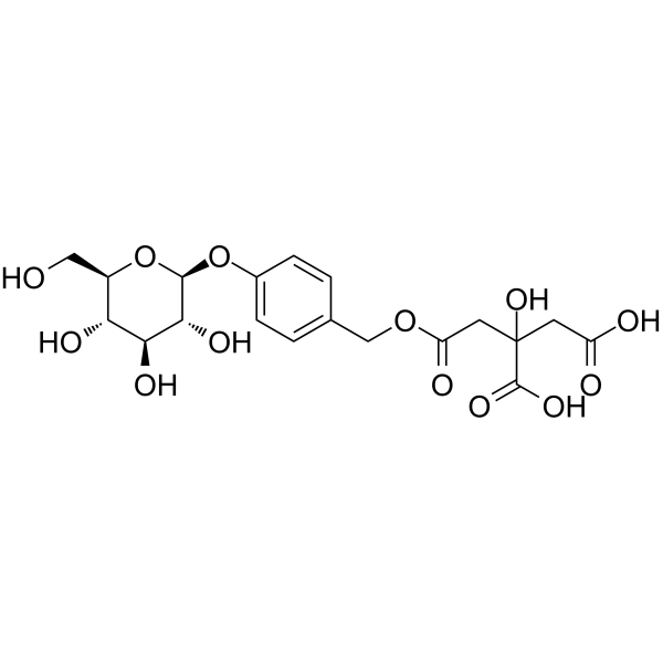
Parishin E is a modulator of hexokinase 2, lactate dehydrogenase A, and silent information regulator 6 (sirtuin 6), and is also a component present in Gastrodia elata Blume. Parishin E reduces the expression of hexokinase 2 and lactate dehydrogenase A, thereby regulating the process of cellular glycolysis. Parishin E regulates the deacetylation mediated by silent information regulator 6 (Sirtuin 6), and inhibits histone 3 lactylation modifications at the H3K18la and H3K27la sites. Parishin E inhibits macrophage polarization. Parishin E alleviates LPS-induced inflammatory responses and suppresses the expression of pro-inflammatory cytokines. Parishin E exerts ameliorating effects on rheumatoid arthritis. Parishin E can be used in studies related to rheumatoid arthritis.
For research use only. We do not sell to patients.
- Purity : 99.90%
- CAS No.: 952068-57-4
- Formula: C19H24O13
- Molecular Weight:460.39
-
Storage:
4°C, sealed storage, away from moisture and light
* In solvent : -80°C, 6 months; -20°C, 1 month (sealed storage, away from moisture and light)

To place orders, for customer services and technical support, please contact: MedChemExpress USA
Tel: 609-228-6898 E-mail: [email protected] [email protected]


-
 Biological Activity
-
Chemical Information
-
Solvent & Solubility
- Protocols
- Purity & Documentation
- References
-
 Help & FAQs





Biological Activity
Description
In Vitro
Parishin E (1-256 μM; 24 h) is tested for its effect on the viability of RAW264.7 cells[1].
Parishin E (32 μM; overnight) inhibits M1 polarization of LPS-stimulated RAW264.7 cells, as evidenced by decreased expression levels of CD86 and iNOS[1].
Parishin E (32 μM; 24 h) regulates glycolysis in LPS-stimulated RAW264.7 cells, thereby inhibiting inflammatory activation[1].
Parishin E (32 μM; 48 h) inhibits lactylation modifications of histone 3 at the H3K18la and H3K27la sites in RAW264.7 cells and reduces the expression of pro-inflammatory cytokines[1].
MedChemExpress (MCE) has not independently confirmed the accuracy of these methods. They are for reference only.
-
Cell Line:mouse macrophage RAW 264.7 cells
-
Concentration:1 μM; 2 μM; 4 μM; 8 μM; 16 μM; 32 μM; 64 μM; 128 μM; 256 μM
-
Incubation Time:24 h
-
Result:Assessed effects on RAW264.7 cell viability with no specific numerical viability values provided.
-
Cell Line:mouse macrophage RAW 264.7 cells
-
Concentration:32 μM
-
Incubation Time:overnight
-
Result:Reduced the fluorescence signals associated with CD86 and iNOS, markers of M1 macrophage polarization, in LPS-stimulated RAW264.7 cells.
In Vivo
-
Animal Model:Sprague Dawley (SD) (200 g; half male and half female)[3]
-
Dosage:72.5 mg/kg; 116 mg/kg; 220 mg/kg (parent parishin)
-
Administration:i.v.; single dose
-
Result:Was detected in rat plasma for up to 4 h after intravenous parishin administration.
Showed a poor correlation between systemic exposure level (AUC0-∞) and the dose of parent parishin, attributed to low plasma concentration and high biological sample variability.
Had a mean AUC-weighting coefficient (ω) of approximately 0.17% across the three parishin doses.


Chemical Information
-
CAS No. 952068-57-4
-
Appearance Solid
-
Molecular Weight 460.39
-
Formula C19H24O13
-
Color White to off-white
-
SMILES
O[C@H]([C@H]([C@@H]([C@@H](CO)O1)O)O)[C@@H]1OC2=CC=C(COC(CC(O)(C(O)=O)CC(O)=O)=O)C=C2
-
Structure Classification
-
Initial Source
-
Shipping
Room temperature in continental US; may vary elsewhere.
-
Storage
4°C, sealed storage, away from moisture and light
* In solvent : -80°C, 6 months; -20°C, 1 month (sealed storage, away from moisture and light)


Solvent & Solubility
In Vitro:
DMSO : 100 mg/mL (217.21 mM; Need ultrasonic; Hygroscopic DMSO has a significant impact on the solubility of product, please use newly opened DMSO)
Please refer to the solubility information to select the appropriate solvent. Once prepared, please aliquot and store the solution to prevent product inactivation from repeated freeze-thaw cycles.
Storage method and period of stock solution: -80°C, 6 months; -20°C, 1 month (sealed storage, away from moisture and light). When stored at -80°C, please use it within 6 months. When stored at -20°C, please use it within 1 month.
Please refer to the solubility information to select the appropriate solvent. Once prepared, please aliquot and store the solution to prevent product inactivation from repeated freeze-thaw cycles.
Storage method and period of stock solution: -80°C, 6 months; -20°C, 1 month (sealed storage, away from moisture and light). When stored at -80°C, please use it within 6 months. When stored at -20°C, please use it within 1 month.
Concentration (start) × Volume (start) = Concentration (final) × Volume (final)
In Vivo:
Select the appropriate dissolution method based on your experimental animal and administration route.
- For the following dissolution methods, please ensure to first prepare a clear stock solution using an In Vitro approach and then sequentially add co-solvents:
- To ensure reliable experimental results, the clarified stock solution can be appropriately stored based on storage conditions. As for the working solution for In Vivo experiments, it is recommended to prepare freshly and use it on the same day.
- The percentages shown for the solvents indicate their volumetric ratio in the final prepared solution. If precipitation or phase separation occurs during preparation, heat and/or sonication can be used to aid dissolution.
Add each solvent one by one: 10% DMSO 40% PEG300 5% Tween-80 45% Saline
Solubility: ≥ 2.5 mg/mL (5.43 mM); Clear solution
This protocol yields a clear solution of ≥ 2.5 mg/mL (saturation unknown).
Taking 1 mL working solution as an example, add 100 μL DMSO stock solution (25.0 mg/mL) to 400 μL PEG300, and mix evenly; then add 50 μL Tween-80 and mix evenly; then add 450 μL Saline to adjust the volume to 1 mL.
Preparation of Saline: Dissolve 0.9 g sodium chloride in ddH₂O and dilute to 100 mL to obtain a clear Saline solution.
Add each solvent one by one: 10% DMSO 90% (20% SBE-β-CD in Saline)
Solubility: ≥ 2.5 mg/mL (5.43 mM); Clear solution
This protocol yields a clear solution of ≥ 2.5 mg/mL (saturation unknown).
Taking 1 mL working solution as an example, add 100 μL DMSO stock solution (25.0 mg/mL) to 900 μL 20% SBE-β-CD in Saline, and mix evenly.
Preparation of 20% SBE-β-CD in Saline (4°C, storage for one week): 2 g SBE-β-CD powder is dissolved in 10 mL Saline, completely dissolve until clear.
In Vivo Dissolution Calculator
Please enter the basic information of animal experiments:
-
-
-
-
Recommended: Prepare an additional quantity of animals to account for potential losses during experiments.
Please enter your animal formula composition:
-
%DMSO +
Recommended: Keep the proportion of DMSO in working solution below 2% if your animal is weak.
-
%+
-
+%Tween-80 + +
-
%Saline +
The co-solvents required include: DMSO, . All of co-solvents are available by MedChemExpress (MCE). , Tween 80. All of co-solvents are available by MedChemExpress (MCE).
Working solution concentration: 0.22 mg/mL
Method for preparing stock solution: mg drug dissolved in μL DMSO. Stock solution concentration: mg/mL. * In solvent : -80°C, 6 months; -20°C, 1 month (sealed storage, away from moisture and light)
1. Take μL DMSO stock solution;
2. Add μL .
μL , mix evenly;
3. Then add μL Tween 80, mix evenly;
4. Then add μL
Please ensure that the stock solution in the first step is dissolved to a clear state, and add co-solvents in sequence. You can use ultrasonic heating (ultrasonic cleaner, recommended frequency 20-40 kHz), vortexing, etc. to assist dissolution.


Purity & Documentation
-
Data Sheet (273 KB)
-
SDS (393 KB)
- English - EN (393 KB)
- Français - FR (393 KB)
- Deutsch - DE (393 KB)
- Norwegian - NO (393 KB)
- Español - ES (393 KB)
- Swedish - SV (393 KB)
- Italian - IT (393 KB)
- Korean - KR (393 KB)
- Portuguese - PT (393 KB)
-
Handling Instructions (2659 KB)
References
[1]. Liu X, et al. Parishin E from ginger-processed Gastrodia elata Bl. alleviates rheumatoid arthritis by regulating histone 3 lactylation at H3K18la and H3K27la sites. Front Pharmacol. 2025 Oct 28;16:1682504. [Content Brief]
[2]. Liu J, et al. An Optimized and Sensitive Pharmacokinetic Quantitative Method of Investigating Gastrodin, Parishin, and Parishin B, C and E in Beagle Dog Plasma using LC-MS/MS after Intragastric Administration of Tall Gastrodia Capsules. Molecules. 2017 Nov 10;22(11):1938. [Content Brief]
[3]. Tang C, et al. Pharmacokinetic study of Gastrodia elata in rats. Analytical and bioanalytical chemistry. 2015 Nov;407(29):8903-10. [Content Brief]


Complete Stock Solution Preparation Table
Please refer to the solubility information to select the appropriate solvent. Once prepared, please aliquot and store the solution to prevent product inactivation from repeated freeze-thaw cycles.
Storage method and period of stock solution: -80°C, 6 months; -20°C, 1 month (sealed storage, away from moisture and light). When stored at -80°C, please use it within 6 months. When stored at -20°C, please use it within 1 month.
| Optional Solvent | Concentration Solvent Mass | 1 mg | 5 mg | 10 mg | 25 mg |
|---|---|---|---|---|---|
| DMSO | 1 mM | 2.1721 mL | 10.8604 mL | 21.7207 mL | 54.3018 mL |
| 5 mM | 0.4344 mL | 2.1721 mL | 4.3441 mL | 10.8604 mL | |
| 10 mM | 0.2172 mL | 1.0860 mL | 2.1721 mL | 5.4302 mL | |
| 15 mM | 0.1448 mL | 0.7240 mL | 1.4480 mL | 3.6201 mL | |
| 20 mM | 0.1086 mL | 0.5430 mL | 1.0860 mL | 2.7151 mL | |
| 25 mM | 0.0869 mL | 0.4344 mL | 0.8688 mL | 2.1721 mL | |
| 30 mM | 0.0724 mL | 0.3620 mL | 0.7240 mL | 1.8101 mL | |
| 40 mM | 0.0543 mL | 0.2715 mL | 0.5430 mL | 1.3575 mL | |
| 50 mM | 0.0434 mL | 0.2172 mL | 0.4344 mL | 1.0860 mL | |
| 60 mM | 0.0362 mL | 0.1810 mL | 0.3620 mL | 0.9050 mL | |
| 80 mM | 0.0272 mL | 0.1358 mL | 0.2715 mL | 0.6788 mL | |
| 100 mM | 0.0217 mL | 0.1086 mL | 0.2172 mL | 0.5430 mL |


Parishin E Related Classifications
HY-N2126 Related Classifications
 Powered by Bioz