



E260
Based on 3 publication(s) in Google Scholar
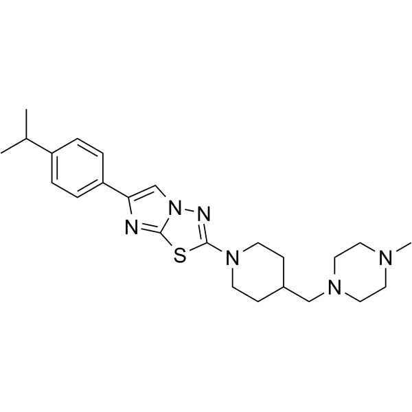
E260 is a Fer/FerT kinase inhibitor.
연구목적의 판매만을 진행합니다. 환자를 대상으로 한 판매는 하지 않습니다.
- Purity : 99.92%
- CAS No.: 1241537-79-0
- 화학식: C24H34N6S
- 분자량:438.63
-
보관:Powder -20°C, 3 years , 4°C, 2 years ; In solvent -80°C, 2 years , -20°C, 1 year

To place orders, for customer services and technical support, please contact: MedChemExpress USA
Tel: 609-228-6898 E-mail: [email protected] [email protected]


-
 Biological Activity
-
Chemical Information
-
용액&용해도
- 순도&문서
- References
-
 Help & FAQs


Publications Citing Use of MedChemExpress (MCE) E260
MoreAll EGFR Isoforms
More

Biological Activity
제품 설명
IC50 & Target
Fer/FerT kinase[1]
In Vitro
E260 is a Fer and FerT inhibitor, which selectively evokes metabolic stress in cancer cells by imposing mitochondrial dysfunction and deformation, and onset of energy-consuming autophagy which decreases the cellular ATP level. To demonstrate that E260 directly targets Fer and FerT, an in vitro kinase assay is performed using a purified kinase domain (KD)-containing fragment of these enzymes. This analysis demonstrates the direct inhibitory effect of E260 on this domain as reflected by the significantly decreased auto-phosphorylation level of the Fer/FerT KD when incubated with ATP and increasing concentrations of E260. Moreover, computational analysis of E260 docking in the modeled whole Fer protein reveals that the highest scored binding mode of E260 to Fer falls in the ATP-binding pocket of the enzyme’s KD. To measure the dissociation constant (Kd) of E260 from Fer/FerT KD, a microscale thermophoresis (MST) test is performed using ascending concentrations of E260. This analysis corroborates the direct binding of E260 to Fer/FerT KD and determines a Kd of 0.85 μM. To examine the effect of the E260 micellar formulation on Fer in malignant cells, the kinase is immunoprecipitated from untreated and from E260-treated SW620 CC cells. When applied to metastatic grade IV SW620 CC cells, which are serum starved for 16 h and treated with 3 mM H2O2 to activate Fer, E260 exhibits inhibitory effects on the Fer-kinase activity as is reflected by suppressed auto-phosphorylation activity of the enzyme. To characterize the effect of E260 on malignant cells, metastatic SW620 cells are treated with E260 followed by analysis of viability. Onset of death is observed in the E260-treated cells, with an EC50 value of 400 nM after 24 h of treatment and an EC50 of 300 nM after 48 h. E260 exhibits an EC50 of 3.2 μM after 72 h treatment of non-metastatic PANC-1 cells, which are derived from a primary pancreatic ductal carcinoma. Moreover, the maximum death level of these cells after 72 h of treatment with E260 is about 70% following treatment with 4 μM E260. In comparison, SU.86.86 which are metastatic ductal carcinoma cells, prove to be more susceptible to E260 with an EC50 of 1.1 μM after 72 h of treatment and 100% death level imposed by 2 μM E260[1].
MedChemExpress (MCE) has not independently confirmed the accuracy of these methods. They are for reference only.
In Vivo
MedChemExpress (MCE) has not independently confirmed the accuracy of these methods. They are for reference only.


Chemical Information
-
CAS No. 1241537-79-0
-
Appearance Solid
-
분자량 438.63
-
화학식 C24H34N6S
-
Color White to off-white
-
SMILES
CC(C)C1=CC=C(C2=CN3C(SC(N4CCC(CN5CCN(C)CC5)CC4)=N3)=N2)C=C1
-
선적
Room temperature in continental US; may vary elsewhere.
-
보관
Powder -20°C 3 years 4°C 2 years In solvent -80°C 2 years -20°C 1 year


Publications (3)
-
Journal Impact Factor
-
Most Recent
-
Nat Commun
Molecular landscape, subtypes, and therapeutic vulnerabilities of central nervous system solitary fibrous tumors. [Abstract]2025 Aug 23;16(1):7870. PMID: 40849425 -
Nat Commun
Spatiotemporal H2O2 flashes coordinate actin cytoskeletal remodeling and regulate cell migration and wound healing. [Abstract]2025 Jul 25;16(1):6868. PMID: 40715145 -
Biofactors
Lactate dehydrogenase A is implicated in the pathogenesis of B-cell lymphoma through regulation of the FER signaling pathway. [Abstract]2024 Sep-Oct;50(5):1024-1038. PMID: 38516823


용액&용해도
In Vitro:
Ethanol : 10 mg/mL (22.80 mM; Need ultrasonic)
DMSO : 1 mg/mL (2.28 mM; Need ultrasonic; Hygroscopic DMSO has a significant impact on the solubility of product, please use newly opened DMSO)
Please refer to the solubility information to select the appropriate solvent. Once prepared, please aliquot and store the solution to prevent product inactivation from repeated freeze-thaw cycles.
Storage method and period of stock solution: -80°C, 2 years; -20°C, 1 year. When stored at -80°C, please use it within 2 years. When stored at -20°C, please use it within 1 year.
Please refer to the solubility information to select the appropriate solvent. Once prepared, please aliquot and store the solution to prevent product inactivation from repeated freeze-thaw cycles.
Storage method and period of stock solution: -80°C, 2 years; -20°C, 1 year. When stored at -80°C, please use it within 2 years. When stored at -20°C, please use it within 1 year.
Concentration (start) × Volume (start) = Concentration (final) × Volume (final)
In Vivo:
Select the appropriate dissolution method based on your experimental animal and administration route.
- For the following dissolution methods, please ensure to first prepare a clear stock solution using an In Vitro approach and then sequentially add co-solvents:
- To ensure reliable experimental results, the clarified stock solution can be appropriately stored based on storage conditions. As for the working solution for In Vivo experiments, it is recommended to prepare freshly and use it on the same day.
- The percentages shown for the solvents indicate their volumetric ratio in the final prepared solution. If precipitation or phase separation occurs during preparation, heat and/or sonication can be used to aid dissolution.
Add each solvent one by one: 10% DMSO 40% PEG300 5% Tween-80 45% Saline
Solubility: ≥ 0.1 mg/mL (0.23 mM); Clear solution
This protocol yields a clear solution of ≥ 0.1 mg/mL (saturation unknown).
Taking 1 mL working solution as an example, add 100 μL DMSO stock solution (1.0 mg/mL) to 400 μL PEG300, and mix evenly; then add 50 μL Tween-80 and mix evenly; then add 450 μL Saline to adjust the volume to 1 mL.
Preparation of Saline: Dissolve 0.9 g sodium chloride in ddH₂O and dilute to 100 mL to obtain a clear Saline solution.
Add each solvent one by one: 10% DMSO 90% (20% SBE-β-CD in Saline)
Solubility: ≥ 0.1 mg/mL (0.23 mM); Clear solution
This protocol yields a clear solution of ≥ 0.1 mg/mL (saturation unknown).
Taking 1 mL working solution as an example, add 100 μL DMSO stock solution (1.0 mg/mL) to 900 μL 20% SBE-β-CD in Saline, and mix evenly.
Preparation of 20% SBE-β-CD in Saline (4°C, storage for one week): 2 g SBE-β-CD powder is dissolved in 10 mL Saline, completely dissolve until clear.
For the following dissolution methods, please prepare the working solution directly:
It is recommended to prepare fresh solutions and use them promptly within a short period of time.
The percentages shown for the solvents indicate their volumetric ratio in the final prepared solution. If precipitation or phase separation occurs during preparation, heat and/or sonication can be used to aid dissolution.
Add each solvent one by one: 0.5% CMC-Na/saline water
Solubility: 22 mg/mL (50.16 mM); Suspended solution; Need ultrasonic
In Vivo Dissolution Calculator
Please enter the basic information of animal experiments:
-
-
-
-
Recommended: Prepare an additional quantity of animals to account for potential losses during experiments.
Please enter your animal formula composition:
-
%DMSO +
Recommended: Keep the proportion of DMSO in working solution below 2% if your animal is weak.
-
%+
-
+%Tween-80 + +
-
%Saline +
The co-solvents required include: DMSO, . All of co-solvents are available by MedChemExpress (MCE). , Tween 80. All of co-solvents are available by MedChemExpress (MCE).
Working solution concentration: 0.22 mg/mL
Method for preparing stock solution: mg drug dissolved in μL DMSO. Stock solution concentration: mg/mL.
1. Take μL DMSO stock solution;
2. Add μL .
μL , mix evenly;
3. Then add μL Tween 80, mix evenly;
4. Then add μL
Please ensure that the stock solution in the first step is dissolved to a clear state, and add co-solvents in sequence. You can use ultrasonic heating (ultrasonic cleaner, recommended frequency 20-40 kHz), vortexing, etc. to assist dissolution.


순도&문서
-
Data Sheet (278 KB)
-
SDS (396 KB)
- English - EN (396 KB)
- Français - FR (396 KB)
- Deutsch - DE (396 KB)
- Norwegian - NO (396 KB)
- Español - ES (396 KB)
- Swedish - SV (396 KB)
- Italian - IT (396 KB)
- Korean - KR (396 KB)
- Portuguese - PT (396 KB)
-
Handling Instructions (2659 KB)
References


Complete Stock Solution Preparation Table
Please refer to the solubility information to select the appropriate solvent. Once prepared, please aliquot and store the solution to prevent product inactivation from repeated freeze-thaw cycles.
Storage method and period of stock solution: -80°C, 2 years; -20°C, 1 year. When stored at -80°C, please use it within 2 years. When stored at -20°C, please use it within 1 year.
| Optional Solvent | Concentration Solvent Mass | 1 mg | 5 mg | 10 mg | 25 mg |
|---|---|---|---|---|---|
| DMSO / Ethanol | 1 mM | 2.2798 mL | 11.3991 mL | 22.7983 mL | 56.9956 mL |
| Ethanol | 5 mM | 0.4560 mL | 2.2798 mL | 4.5597 mL | 11.3991 mL |
| 10 mM | 0.2280 mL | 1.1399 mL | 2.2798 mL | 5.6996 mL | |
| 15 mM | 0.1520 mL | 0.7599 mL | 1.5199 mL | 3.7997 mL | |
| 20 mM | 0.1140 mL | 0.5700 mL | 1.1399 mL | 2.8498 mL |


E260 Related Classifications
HY-112097 Related Classifications
 Powered by Bioz