



Leucic acid

Based on 1 Customer Validation
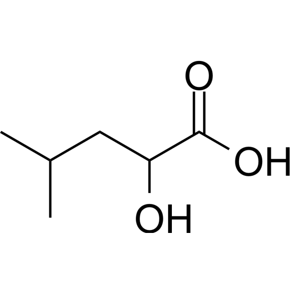
Leucic acid (α-Hydroxyisocaproic acid) is an orally active end-product of the microbial metabolism of leucine. Leucic acid can bind to HCAR2, alters AMPK and ERK1/2 phosphorylation status, suppresses lipid synthesis, promotes catabolism, reduces adiposity, enhances lean mass and exercise capacity. Leucic acid suppresses pro-inflammatory cytokine secretion, inflammation-related gene mRNA expression. Leucic acid decreases basal protein synthesis, attenuates myotube atrophy. Leucic acid can be used for the research of obesity.
For research use only. We do not sell to patients.
- Purity : 98.38%
- CAS No.: 498-36-2
- Formula: C6H12O3
- Molecular Weight:132.16
-
Storage:
Sealed storage, away from moisture.
Powder -80°C, 2 years , -20°C, 1 year* In solvent : -80°C, 6 months; -20°C, 1 month (sealed storage, away from moisture)

To place orders, for customer services and technical support, please contact: MedChemExpress USA
Tel: 609-228-6898 E-mail: [email protected] [email protected]


-
 Biological Activity
-
Chemical Information
-
Solvent & Solubility
- Protocols
- Purity & Documentation
- References
-
 Help & FAQs


-
HY-N0091
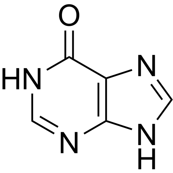
HypoxanthineEndogenous Metabolite -
HY-113381
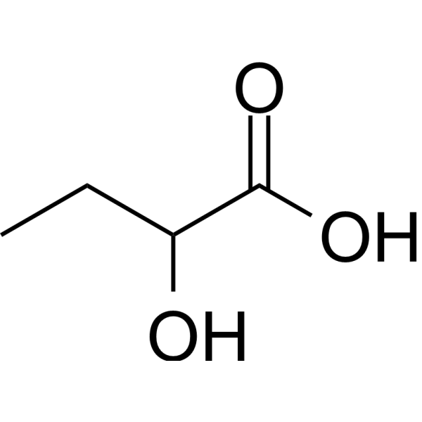
2-Hydroxybutyric acidEndogenous Metabolite -

HY-113218
Acetyl-L-carnitineNeuroprotective Agent -
HY-113126
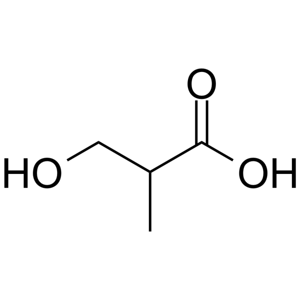
3-Hydroxyisobutyric acidEndogenous Metabolite -
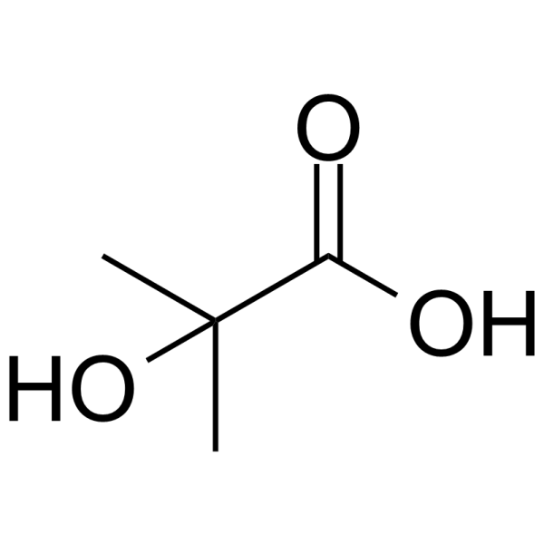
HY-W015924
2-Hydroxyisobutyric acidInsulin/IGF-1/p38 MAPK Regulator -
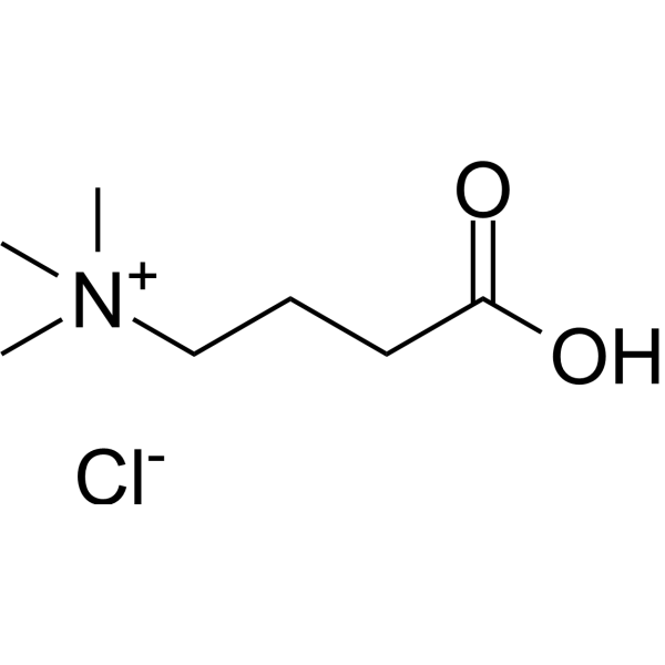
HY-113270A
Actinine chlorideEndogenous Metabolite -

HY-30216AR
Leucic acid (Standard)Endogenous Metabolite -
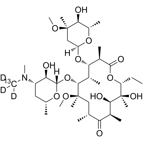
HY-17508S
Clarithromycin-13C,d3Stable Isotope -
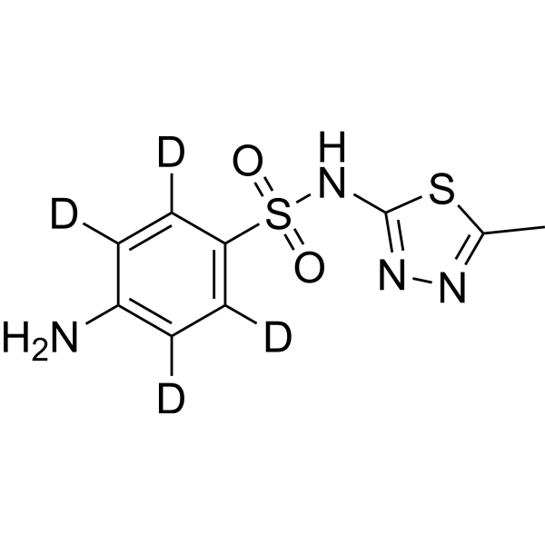
HY-B0333S2
Sulfamethizole-d4-1Stable Isotope -
HY-137911
SparsomycinMetabolite
All AMPK Isoforms
More

Biological Activity
Description
IC50 & Target
[1]|
HCAR2 |
ERK1 |
ERK2 |
In Vitro
Leucic acid (50 μM) targets HCAR2, which is required for leucic acid-mediated suppression of lipid storage and stimulation of oxygen consumption in vitro beige-like adipocytes[1].
Leucic acid (0.31-10 mM; 21 days) reduces lipid accumulation and adipokine levels in human adipose-derived mesenchymal stem cells[2].
Leucic acid modulates protein expression linked to immune and inflammatory signaling, including significant enrichment of the adipokine signaling pathway, in tumor necrosis factor-α-stimulated human adipose-derived mesenchymal stem cells[2].
Leucic acid (0.31-10 mM) suppresses pro-inflammatory cytokine secretion and inflammatory gene expression in tumor necrosis factor-α-stimulated human adipose-derived mesenchymal stem cells[2].
Leucic acid (1.67-15 mM; 30 min) activates AMPK and p38MAPK signaling, inhibits ERK1/2 signaling, and does not alter mTORC1 targets p70S6K or 4E-BP1 in fully differentiated murine C2C12 myotubes under normal conditions, indicating negative regulation of protein synthesis through multiple pathways[3].
Leucic acid (15 mM; 2 days (myotubes), 1 day (myoblasts)) significantly decelerates the fractional protein synthesis rate in murine C2C12 myotubes and myoblasts under normal conditions[3].
Leucic acid (15 mM; 2-day pretreatment) attenuates TNFα/IFNγ-induced myotube atrophy, reduces nitric oxide secretion under cachexic conditions, and increases myotube fusion index in murine C2C12 myotubes[3].
Leucic acid (15 mM; 2-day pretreatment) does not modify acute STAT3 or NFκB p65 signaling induced by TNFα/IFNγ co-exposure in murine C2C12 myotubes[3].
Leucic acid (15 mM; 2-day pretreatment) decreases fractional protein synthesis rate under both normal and TNFα/IFNγ-induced cachexic conditions, and attenuates cachexia-associated protein degradation, iNOS overexpression, and IL-6 production in murine C2C12 myotubes[3].
MedChemExpress (MCE) has not independently confirmed the accuracy of these methods. They are for reference only. Further protocols information, click here.
-
Cell Line:Human adipose-derived mesenchymal stem cells (hMSCs)
-
Concentration:0.31, 10 mM
-
Incubation Time:21 days
-
Result:Suppressed Oil Red O-positive lipid accumulation in a concentration-dependent manner, with significant reductions observed at all tested concentrations relative to control differentiated cells.
Reduced cellular triglyceride, glycerol, and free fatty acid levels significantly at tested concentrations relative to control differentiated cells.
In Vivo
Leucic acid (50 mM; p.o.; in drinking water; 12 weeks) attenuates HFD-induced weight gain in male Aida-/- mice, suppresses lipid synthesis in gWAT, and amplifies thermogenic lipid catabolism in iWAT via upregulation of UCP1 and related pathways[1].
Leucic acid (50 mM; p.o.; in drinking water; 13 weeks) relies on HCAR2 to drive iWAT fat mass reduction and browning in HFD-fed male C57BL/6J mice, as shown by attenuated effects in Hcar2-knockdown tissue[1].
MedChemExpress (MCE) has not independently confirmed the accuracy of these methods. They are for reference only.
-
Animal Model:C57BL/6J Aida-/- mice (male, 8 weeks old)[1]
-
Dosage:50 mM
-
Administration:p.o.; continuously in drinking water, refreshed every 5 days; up to 12 weeks
-
Result:Attenuated HFD-induced body weight gain. Improved forelimb grip strength.
Suppressed key lipogenic enzymes including DGAT2, MOGAT2, ACSS2, and FASN in gonadal white adipose tissue (gWAT).
Upregulated genes linked to muscle-like metabolic activation and thermogenesis in inguinal white adipose tissue (iWAT).
Increased UCP1 protein abundance in iWAT.
Amplified thermogenic and fatty acid degradation pathways in iWAT as shown by Gene Set Enrichment Analysis.


Chemical Information
-
CAS No. 498-36-2
-
Appearance Solid
-
Molecular Weight 132.16
-
Formula C6H12O3
-
Color White to off-white
-
SMILES
CC(C)CC(O)C(O)=O
-
Synonyms
α-Hydroxyisocaproic acid
-
Structure Classification
-
Initial Source
-
Shipping
Room temperature in continental US; may vary elsewhere.
-
Storage
Sealed storage, away from moisture
Powder -80°C 2 years -20°C 1 year * In solvent : -80°C, 6 months; -20°C, 1 month (sealed storage, away from moisture)


Solvent & Solubility
In Vitro:
DMSO : 175 mg/mL (1324.15 mM; Need ultrasonic; Hygroscopic DMSO has a significant impact on the solubility of product, please use newly opened DMSO)
H2O : 100 mg/mL (756.66 mM; Need ultrasonic)
Please refer to the solubility information to select the appropriate solvent. Once prepared, please aliquot and store the solution to prevent product inactivation from repeated freeze-thaw cycles.
Storage method and period of stock solution: -80°C, 6 months; -20°C, 1 month (sealed storage, away from moisture). When stored at -80°C, please use it within 6 months. When stored at -20°C, please use it within 1 month.
* Note: If you choose water as the stock solution, please dilute it to the working solution, then filter and sterilize it with a 0.22 μm filter before use.
Please refer to the solubility information to select the appropriate solvent. Once prepared, please aliquot and store the solution to prevent product inactivation from repeated freeze-thaw cycles.
Storage method and period of stock solution: -80°C, 6 months; -20°C, 1 month (sealed storage, away from moisture). When stored at -80°C, please use it within 6 months. When stored at -20°C, please use it within 1 month.
* Note: If you choose water as the stock solution, please dilute it to the working solution, then filter and sterilize it with a 0.22 μm filter before use.
Concentration (start) × Volume (start) = Concentration (final) × Volume (final)
In Vivo:
Select the appropriate dissolution method based on your experimental animal and administration route.
- For the following dissolution methods, please ensure to first prepare a clear stock solution using an In Vitro approach and then sequentially add co-solvents:
- To ensure reliable experimental results, the clarified stock solution can be appropriately stored based on storage conditions. As for the working solution for In Vivo experiments, it is recommended to prepare freshly and use it on the same day.
- The percentages shown for the solvents indicate their volumetric ratio in the final prepared solution. If precipitation or phase separation occurs during preparation, heat and/or sonication can be used to aid dissolution.
Add each solvent one by one: 10% DMSO 40% PEG300 5% Tween-80 45% Saline
Solubility: ≥ 5 mg/mL (37.83 mM); Clear solution
This protocol yields a clear solution of ≥ 5 mg/mL (saturation unknown).
Taking 1 mL working solution as an example, add 100 μL DMSO stock solution (50.0 mg/mL) to 400 μL PEG300, and mix evenly; then add 50 μL Tween-80 and mix evenly; then add 450 μL Saline to adjust the volume to 1 mL.
Preparation of Saline: Dissolve 0.9 g sodium chloride in ddH₂O and dilute to 100 mL to obtain a clear Saline solution.
Add each solvent one by one: 10% DMSO 90% (20% SBE-β-CD in Saline)
Solubility: ≥ 5 mg/mL (37.83 mM); Clear solution
This protocol yields a clear solution of ≥ 5 mg/mL (saturation unknown).
Taking 1 mL working solution as an example, add 100 μL DMSO stock solution (50.0 mg/mL) to 900 μL 20% SBE-β-CD in Saline, and mix evenly.
Preparation of 20% SBE-β-CD in Saline (4°C, storage for one week): 2 g SBE-β-CD powder is dissolved in 10 mL Saline, completely dissolve until clear.
In Vivo Dissolution Calculator
Please enter the basic information of animal experiments:
-
-
-
-
Recommended: Prepare an additional quantity of animals to account for potential losses during experiments.
Working solution concentration: 0.22 mg/mL
This product has good water solubility, please refer to the measured solubility data in water/PBS/Saline for details.

Protocols
-
Kinase activity and phosphorylation assays
Kinase activity assays measure the ability of kinases to transfer phosphate groups from ATP to specific substrates, while phosphorylation assays detect the presence and levels of phosphorylated proteins. Common methods include radiolabeled ATP incorporation (e. g. ,), ADP release detection via bioluminescence (e. g. ,[3]), enzyme-linked immunosorbent assays (ELISA) for phospho-specific epitopes (e. g. ,[6]), and microtiter-based formats for high-throughput screening (e. g. ,[8]). The ADP-Glo assay quantifies kinase activity by measuring ADP produced during phosphorylation using a luciferase-based system. Radiometric assays involve autoradiography or scintillation counting after incorporation of 32P-labeled ATP into substrate proteins. ELISA-based approaches rely on phospho-specific antibodies to detect activated kinases in cell lysates or purified samples.
-
RT-PCR
Reverse transcription technology uses RNA as a template to synthesize DNA. RT-PCR is simple, specific and sensitive, and can be used to detect gene expression levels and expression differences in cells; detect RNA virus content; clone cDNA sequences of specific genes.
-
Western Blot
Western blotting (WB) is a commonly used experimental method in molecular biology, biochemistry, and immunogenetics for identifying and quantifying target proteins. It combines gel electrophoresis with immunoassay, enabling researchers to analyze protein expression, post-translational modifications, and molecular weight.
-
RNA extraction experimental
By lysing cells, releasing RNA, and removing impurities such as proteins and DNA, high-purity RNA products are finally obtained. The commonly used traditional method is the guanidine isothiocyanate/phenol/chloroform method (Trizol), which is suitable for a variety of animal materials including animal tissues, microorganisms, cultured cells, etc., and most plant materials.
-
Protocol for Kinase activity and phosphorylation assays
Kinase activity assays measure transfer of phosphate from ATP to a protein or peptide substrate, generating phosphorylated substrate, ADP, or incorporated radiolabeled phosphate as the readout; phosphorylation assays measure site-specific phosphorylation in cells or tissues as a proxy for kinase-pathway activation, inhibition, or substrate regulation. Phosphorylation can be detected by phospho-specific Western blot, immunoprecipitation kinase assay, phospho-immunofluorescence, phospho-flow cytometry, luminescent ADP detection, radiolabeled ATP incorporation, or reporter-based pathway assays, and these readouts can be applied to cancer cells, primary neurons, mouse tumors, organoids, inflammatory macrophages, ferroptosis studies, and mitophagy studies when the kinase target is biologically relevant.
-
Research Protocol for Inflammation-related Diseases
The NLRP3 inflammasome is a cytosolic innate immune signaling platform that integrates priming signals and danger-signal activation to promote caspase-1 activation, maturation of IL-1β and IL-18, and gasdermin D-mediated pyroptotic cell death. The core experimental logic is to determine whether inflammatory disease phenotypes are driven by increased NLRP3 expression, ASC-containing inflammasome assembly, caspase-1 cleavage, GSDMD cleavage, and extracellular release of IL-1β/IL-18 rather than by nonspecific cell injury alone. The pathway is strongly linked to inflammation-related disease phenotypes because monosodium urate crystals activate NALP3/NLRP3 inflammasome signaling in gout-like crystal inflammation, cholesterol crystals activate NLRP3 inflammasomes in atherogenesis models, and DSS-induced intestinal inflammation has been reported to involve NLRP3 inflammasome activity. However, experimental colitis studies also show context-dependent protective effects of NLRP3 inflammasome co
-
Research Protocol for Metabolic Diseases
AMP-activated protein kinase, AMPK, is a conserved cellular energy sensor that responds to reduced cellular energy status and coordinates metabolism by increasing ATP-generating catabolic pathways while suppressing ATP-consuming anabolic processes. In metabolic disease research, the AMPK pathway is experimentally relevant because it regulates hepatic lipid synthesis, fatty acid oxidation, glucose production, skeletal-muscle glucose disposal, mTORC1-linked biosynthesis, autophagy, mitochondrial homeostasis, and whole-body energy balance. The central pathway logic is that energy stress, metformin, exercise-like stimulation, or direct AMPK activators increase AMPKα Thr172 phosphorylation and downstream substrate phosphorylation, including ACC and RAPTOR. Phosphorylation of ACC suppresses lipogenesis and supports fatty acid oxidation, whereas phosphorylation of RAPTOR suppresses mTORC1 signaling and links cellular energy status to growth and protein synthesis control. The pathway is linked
-
Real Time qPCR (Q-PCR)
Real-time quantitative PCR (qPCR) quantifies an amplifiable nucleic-acid target by monitoring fluorescence during PCR cycling rather than measuring product only after amplification. The increase in fluorescence tracks accumulation of PCR product, and the quantification cycle (Cq; historically also Ct/CP) is related to the initial amount of target: samples containing more starting target generally reach the defined fluorescence threshold in fewer cycles.


Purity & Documentation
-
Data Sheet (278 KB)
-
SDS (394 KB)
- English - EN (394 KB)
- Français - FR (394 KB)
- Deutsch - DE (394 KB)
- Norwegian - NO (394 KB)
- Español - ES (394 KB)
- Swedish - SV (394 KB)
- Italian - IT (394 KB)
- Korean - KR (394 KB)
- Portuguese - PT (394 KB)
-
Handling Instructions (2659 KB)
References
[2]. Lee M, et al. Metabolites of Kimchi Lactic Acid Bacteria, Indole-3-Lactic Acid, Phenyllactic Acid, and Leucic Acid, Inhibit Obesity-Related Inflammation in Human Mesenchymal Stem Cells. J Microbiol Biotechnol. 2024;34(2):306-313. [Content Brief]
[3]. Sumi K, et al. α-Hydroxyisocaproic Acid Decreases Protein Synthesis but Attenuates TNFα/IFNγ Co-Exposure-Induced Protein Degradation and Myotube Atrophy via Suppression of iNOS and IL-6 in Murine C2C12 Myotube. Nutrients. 2021 Jul 13;13(7):2391. [Content Brief]


Complete Stock Solution Preparation Table
Please refer to the solubility information to select the appropriate solvent. Once prepared, please aliquot and store the solution to prevent product inactivation from repeated freeze-thaw cycles.
Storage method and period of stock solution: -80°C, 6 months; -20°C, 1 month (sealed storage, away from moisture). When stored at -80°C, please use it within 6 months. When stored at -20°C, please use it within 1 month.
| Optional Solvent | Concentration Solvent Mass | 1 mg | 5 mg | 10 mg | 25 mg |
|---|---|---|---|---|---|
| H2O / DMSO | 1 mM | 7.5666 mL | 37.8329 mL | 75.6659 mL | 189.1646 mL |
| 5 mM | 1.5133 mL | 7.5666 mL | 15.1332 mL | 37.8329 mL | |
| 10 mM | 0.7567 mL | 3.7833 mL | 7.5666 mL | 18.9165 mL | |
| 15 mM | 0.5044 mL | 2.5222 mL | 5.0444 mL | 12.6110 mL | |
| 20 mM | 0.3783 mL | 1.8916 mL | 3.7833 mL | 9.4582 mL | |
| 25 mM | 0.3027 mL | 1.5133 mL | 3.0266 mL | 7.5666 mL | |
| 30 mM | 0.2522 mL | 1.2611 mL | 2.5222 mL | 6.3055 mL | |
| 40 mM | 0.1892 mL | 0.9458 mL | 1.8916 mL | 4.7291 mL | |
| 50 mM | 0.1513 mL | 0.7567 mL | 1.5133 mL | 3.7833 mL | |
| 60 mM | 0.1261 mL | 0.6305 mL | 1.2611 mL | 3.1527 mL | |
| 80 mM | 0.0946 mL | 0.4729 mL | 0.9458 mL | 2.3646 mL | |
| 100 mM | 0.0757 mL | 0.3783 mL | 0.7567 mL | 1.8916 mL |
* Note: If you choose water as the stock solution, please dilute it to the working solution, then filter and sterilize it with a 0.22 μm filter before use.


Leucic acid Related Classifications
HY-30216A Related Classifications
 Powered by Bioz