



Nafamostat hydrochloride
Based on 19 publication(s) in Google Scholar
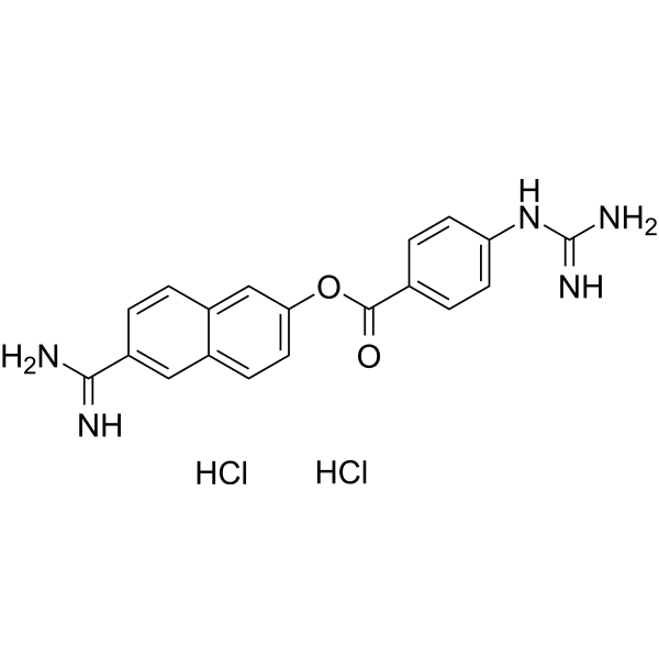
Nafamostat hydrochloride, an anticoagulant, is a synthetic serine protease inhibitor. Nafamostat hydrochloride has anticancer and antivirus effect. Nafamostat hydrochloride induces apoptosis by up-regulating the expression of tumor necrosis factor receptor-1 (TNFR1). Nafamostat hydrochloride can be used in the development of the pathological thickening of the arterial wall.
For research use only. We do not sell to patients.
- CAS No.: 80251-32-7
- Formula: C19H19Cl2N5O2
- Molecular Weight:420.29
-
Storage:
Please store the product under the recommended conditions in the Certificate of Analysis.

To place orders, for customer services and technical support, please contact: MedChemExpress USA
Tel: 609-228-6898 E-mail: [email protected] [email protected]


-
 Biological Activity
-
Chemical Information
- Protocols
- Purity & Documentation
- References
-
 Help & FAQs


-
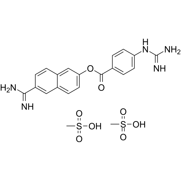
HY-B0190A
Nafamostat mesylateSerine Protease Inhibitor -
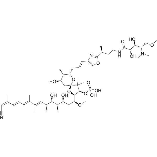
HY-18983
Calyculin APhosphatase Inhibitor -
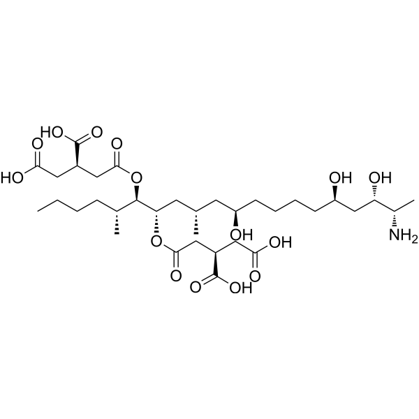
HY-N6719
Fumonisin B1Sphingosine N-acyltransferase Inhibitor -
HY-N0052A
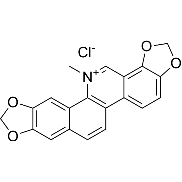
Sanguinarine chlorideApoptosis Inducer -
HY-18756
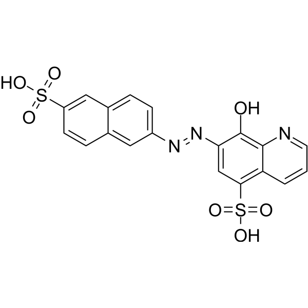
NSC-87877SHP PTP Inhibitor -
HY-B0190
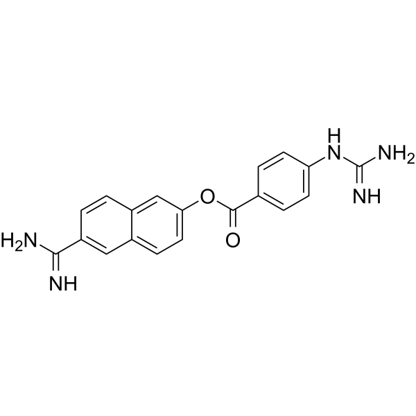
NafamostatSerine Protease Inhibitor -
HY-100034
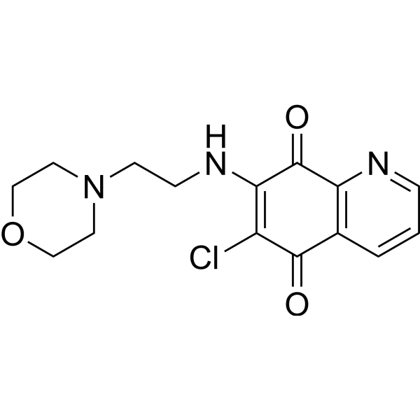
NSC 663284Cdc25 Inhibitor -
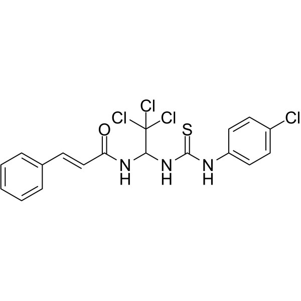
HY-15969
Sal003eIF2α Phophatase Inhibitor -
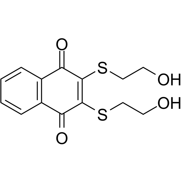
HY-108543
NSC 95397Cdc25 Phosphatase Inhibitor -
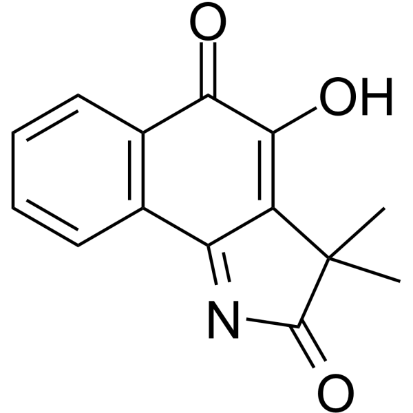
HY-100625
BVT948PTP Inhibitor -
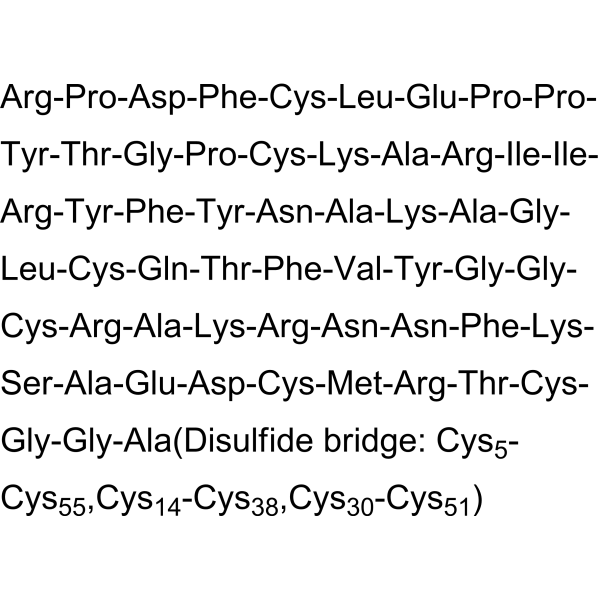
HY-P0017
AprotininBPTI Inhibitor -
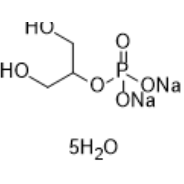
HY-D0886
β-Glycerophosphate disodium salt pentahydratePhosphatase Inhibitor
Publications Citing Use of MedChemExpress (MCE) Nafamostat hydrochloride
More- Cell Res. 2020 Mar;30(3):269-271. [Abstract]
- Nat Commun. 2024 Jun 27;15(1):5458. [Abstract]
- Exp Mol Med. 2024 Dec;56(12):2631-2641. [Abstract]
- Nat Chem Biol. 2022 Sep;18(9):963-971. [Abstract]
- Nucleic Acids Res. 2021 Jan 8;49(D1):D1113-D1121. [Abstract]
- Adv Healthc Mater. 2025 Aug 1:e02156. [Abstract]
- Cells. 2022 Jan 18;11(3):319. [Abstract]
- Cell Rep Methods. 2023 Oct 23;3(10):100599. [Abstract]
- Eur J Pharmacol. 2023 Jan 5:938:175394. [Abstract]
- Eur J Pharmacol. 2022 Mar 15:919:174795. [Abstract]
- Biomolecules. 2022 Jul 31;12(8):1063. [Abstract]
- Sci Rep. 2026 May 16. [Abstract]
- Antiviral Res. 2023 Jun:214:105606. [Abstract]
- Viruses. 2022 Sep 12;14(9):2017. [Abstract]
- ChemMedChem. 2026 Mar 31;21(7):e202501099.
- bioRxiv. 2026 Jan 24.
- Res Sq. 2025 Apr 07.
- bioRxiv. 2024 October 09.
- Patent. US20230210807A1.
 Customer Validation & Images

-
Bio/Physico-chemical Assay
-
Bio/Physico-chemical Assay
-
Bio/Physico-chemical Assay
-
IF
-
Bio/Physico-chemical Assay


Biological Activity
Description
IC50 & Target
|
I-kappaBalpha |
In Vitro
Nafamostat hydrochloride (10-80 μg/mL, 3-48 h) inhibits NF-κB activity by blocking IκBα phosphorylation in MDAPanc-28 cells[1].
Nafamostat hydrochloride (80 μg/mL, 24-48 h) induces apoptosis by up-regulating the expression of tumor necrosis factor receptor-1 (TNFR1) in MDAPanc-28 cells[1].
Nafamostat hydrochloride (0.1-10 μM, 24 h) has suppressive effect on invasiveness in Panc-1 cells[2].
MedChemExpress (MCE) has not independently confirmed the accuracy of these methods. They are for reference only. Further protocols information, click here.
-
Cell Line:MDAPanc-28 cells
-
Concentration:80 μg/mL
-
Incubation Time:24 h, 48 h
-
Result:Substantially reduced the cell viability of MDAPanc-28 cells at both 24 hours and 48 hours.
-
Cell Line:Panc-1 cells
-
Concentration:0.1 μM, 1 μM, 10 μM
-
Incubation Time:24 h
-
Result:Observed significant inhibition in Panc-1-Try clones at concentrations as low as 0.1 mM.
-
Cell Line:MDAPanc-28 cells
-
Concentration:10 μg/mL, 20 μg/mL ,40 μg/mL, 80 μg/mL
-
Incubation Time:3 h, 8 h, 24 h, 48 h
-
Result:Inhibited NF-κB DNA-binding activity and the degradation of IκBα in a dose-dependent manner as well as in a time-dependent manner.
Inhibited phosphorylation of IκBα in a time-dependent manner.
In Vivo
Nafamostat hydrochloride (0.5-2.0 mg/mL (dissolved in saline), Intraperitoneal injection, once a day for 7 consecutive days) has inhibitory effect on neointimal formation after balloon injury of the rat carotid wall[4].
MedChemExpress (MCE) has not independently confirmed the accuracy of these methods. They are for reference only.
-
Animal Model:A129 mice[3]
-
Dosage:10 mg/kg
-
Administration:Intraperitoneal injection (i.p.)
-
Result:Exhibit delayed lethality and improved survival (40%).
-
Animal Model:Balloon injury of the rat carotid wall [4]
-
Dosage:0.5 mg/mL, 1 mg/mL, 2 mg/mL (dissolved in saline)
-
Administration:Intraperitoneal injection (i.p.)
-
Result:Showed smaller ratios of the neointima/medial area.
Showed positive but reduced immunoreactivity of the cells in the neointimal.


Chemical Information
-
CAS No. 80251-32-7
-
Molecular Weight 420.29
-
Formula C19H19Cl2N5O2
-
SMILES
O=C(OC1=CC=C2C=C(C(N)=N)C=CC2=C1)C3=CC=C(NC(N)=N)C=C3.Cl.Cl
-
Shipping
Room temperature in continental US; may vary elsewhere.
-
Storage
Please store the product under the recommended conditions in the Certificate of Analysis.


Publications (19)
-
Journal Impact Factor
-
Most Recent
-
Cell Res
Remdesivir and chloroquine effectively inhibit the recently emerged novel coronavirus (2019-nCoV) in vitro. [Abstract]2020 Mar;30(3):269-271. PMID: 32020029 -
Nat Commun
Structural basis for potent neutralization of human respirovirus type 3 by protective single-domain camelid antibodies. [Abstract]2024 Jun 27;15(1):5458. PMID: 38937429 -
Exp Mol Med
Distribution and impact of p16INK4A+ senescent cells in elderly tissues: a focus on senescent immune cell and epithelial dysfunction. [Abstract]2024 Dec;56(12):2631-2641. PMID: 39617789 -
Nat Chem Biol
2022 Sep;18(9):963-971. PMID: 35676539 -
Nucleic Acids Res
COVID19 Drug Repository: text-mining the literature in search of putative COVID19 therapeutics. [Abstract]2021 Jan 8;49(D1):D1113-D1121. PMID: 33166390 -
Adv Healthc Mater
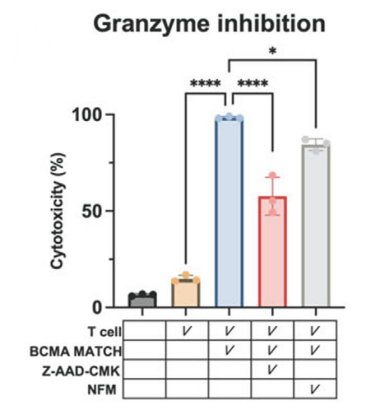
Self-Assembling Multi-Antigen T Cell Hybridizers for Precision Immunotherapy of Multiple Myeloma. [Abstract]2025 Aug 1:e02156. PMID: 40747784
Nafamostat hydrochloride purchased from MedChemExpress. Usage Cited in: Adv Healthc Mater. 2025 Aug 1:e02156. [Abstract]
Targeted inhibition of granzyme B with Z‐AAD‐CMK and granzyme A with Nafamostat mesylate (NFM) attenuates cell death.
-
Cells
Assessment of FDA-Approved Drugs as a Therapeutic Approach for Niemann-Pick Disease Type C1 Using Patient-Specific iPSC-Based Model Systems. [Abstract]2022 Jan 18;11(3):319. PMID: 35159129 -
Cell Rep Methods
RECOVER identifies synergistic drug combinations in vitro through sequential model optimization. [Abstract]2023 Oct 23;3(10):100599. PMID: 37797618 -
Eur J Pharmacol
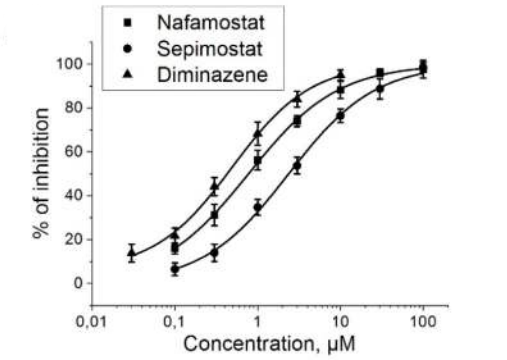
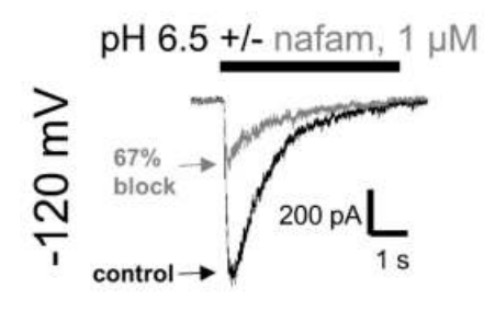
Mechanisms of acid-sensing ion channels inhibition by nafamostat, sepimostat and diminazene. [Abstract]2023 Jan 5:938:175394. PMID: 36403685
Nafamostat hydrochloride purchased from MedChemExpress. Usage Cited in: Eur J Pharmacol. 2023 Jan 5:938:175394. [Abstract]
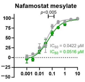
Concentration-inhibition curves for Nafamostat mesylate, sepimostat and diminazene aceturate.
Nafamostat hydrochloride purchased from MedChemExpress. Usage Cited in: Eur J Pharmacol. 2023 Jan 5:938:175394. [Abstract]
Representative examples of current inhibition by 1 μM Nafamostat mesylate.
-
Eur J Pharmacol
2022 Mar 15:919:174795. PMID: 35122868 -
Biomolecules
The Rabep1-Mediated Endocytosis and Activation of Trypsinogen to Promote Pancreatic Stellate Cell Activation. [Abstract]2022 Jul 31;12(8):1063. PMID: 36008957 -
Sci Rep
Production and characterization of gigastasin, a leech-derived inhibitor of complement and coagulation pathways. [Abstract]2026 May 16. PMID: 42143157 -
Antiviral Res
Omicsynin B4 potently blocks coronavirus infection by inhibiting host proteases cathepsin L and TMPRSS2. [Abstract]2023 Jun:214:105606. PMID: 37076089
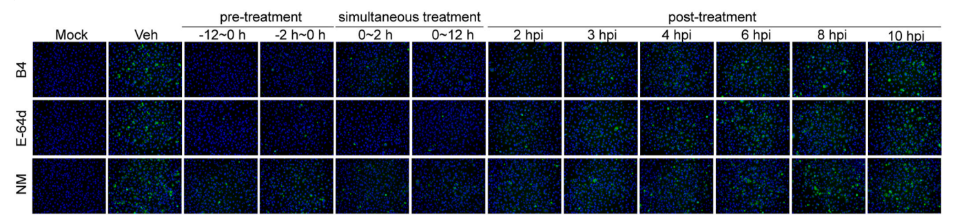
Nafamostat hydrochloride purchased from MedChemExpress. Usage Cited in: Antiviral Res. 2023 Jun:214:105606. [Abstract]
Huh7.5 cells was infected with HCoV-229E (MOI = 3.5) and treated with omicsynin B4 (10 μM), E−64d (1 μM) or nafamostat mesylate (40 μM) at the indicated times. Immunofluorescence staining of the dsRNA (green) were merged with the nuclear Hoechst 33342 dye (blue) using a fluorescence microscopy (200 × ). Mock, the cell control. Veh, the vehicle control. Cpd, compound. NH4Cl, ammonium chloride. B4, omicsynin B4. NM, nafamostat mesylate. hpi, hour post infection.
-
Viruses
Molecular Analyses of Clinical Isolates and Recombinant SARS-CoV-2 Carrying B.1 and B.1.617.2 Spike Mutations Suggest a Potential Role of Non-Spike Mutations in Infection Kinetics. [Abstract]2022 Sep 12;14(9):2017. PMID: 36146823
Nafamostat hydrochloride purchased from MedChemExpress. Usage Cited in: Viruses. 2022 Sep 12;14(9):2017. [Abstract]
Efficacy of small molecule inhibitors Remdesivir, Nafamostat mesylate, Nirmatrelvir, and Molnupiravir against B.1BAC-V (green), B.1.617.2BAC-V (orange), and corresponding clinical isolates (CI, grey).
-
-
-
-
-

Protocols
-
RNA extraction experimental
By lysing cells, releasing RNA, and removing impurities such as proteins and DNA, high-purity RNA products are finally obtained. The commonly used traditional method is the guanidine isothiocyanate/phenol/chloroform method (Trizol), which is suitable for a variety of animal materials including animal tissues, microorganisms, cultured cells, etc., and most plant materials.
-
Apoptosis
Apoptosis, also called programmed cell death, is generally characterized by distinct morphological characteristics.
-
TUNEL staining for apoptotic DNA fragmentation
TUNEL staining detects DNA strand breaks by using terminal deoxynucleotidyl transferase to add labeled nucleotides to exposed 3′-OH DNA termini, generating either microscopic staining in fixed cells or tissue sections, or fluorescence/cytometric signal in cell suspensions. TUNEL positivity reflects DNA fragmentation but should not be interpreted alone as definitive apoptosis, because TUNEL can also label necrotic, autolytic, mechanically damaged, or DNA-repair-associated DNA breaks.
-
Annexin V plus membrane-impermeant dye apoptosis staining
Annexin V-based apoptosis assays rely on the detection of phosphatidylserine (PS) externalization from the inner leaflet of the plasma membrane to the outer leaflet, an early biochemical hallmark of apoptosis. Fluorescently labeled Annexin V binds PS in a calcium-dependent manner, enabling identification of early apoptotic cells by flow cytometry or fluorescence microscopy. When combined with a membrane-impermeant DNA-binding dye (e. g. , propidium iodide), this approach allows discrimination between viable (Annexin V−/dye−), early apoptotic (Annexin V+/dye−), and late apoptotic or necrotic (Annexin V+/dye+) cell populations by assessing membrane integrity and PS exposure.
-
Apoptosis Solutions
Apoptosis is a regulated, generally non-lytic cell-death pathway that removes unwanted, damaged, infected, or abnormal cells through coordinated morphological changes, caspase activation, DNA fragmentation, and membrane remodeling. The intrinsic apoptosis pathway is controlled mainly by mitochondrial outer membrane permeabilization, BCL-2 family proteins, cytochrome c release, apoptosome formation, caspase-9 activation, and downstream executioner caspase-3/7 activation. The extrinsic apoptosis pathway is initiated by death receptors such as Fas, TNFR, and TRAIL receptors, which recruit adaptor proteins and activate caspase-8 before engaging executioner caspases or mitochondrial amplification through BID cleavage. Apoptosis is linked to many phenotypes, including cancer cell killing, tissue homeostasis, immune regulation, neurodegeneration, infection response, and treatment-induced cytotoxicity; unresolved questions include how apoptosis interacts with necroptosis, pyroptosis, ferroptos


Purity & Documentation
References
[1]. Uwagawa T, et al. Mechanisms of synthetic serine protease inhibitor (FUT‐175)‐mediated cell death [J]. Cancer: Interdisciplinary International Journal of the American Cancer Society, 2007, 109(10): 2142-2153. [Content Brief]
[2]. Tajima H, et al. Enhanced invasiveness of pancreatic adenocarcinoma cells stably transfected with cationic trypsinogen cDNA [J]. International journal of cancer, 2001, 94(5): 699-704. [Content Brief]
[3]. Yan Y, et al. Nafamostat mesylate as a broad-spectrum candidate for the treatment of flavivirus infections by targeting envelope proteins [J]. Antiviral research, 2022, 202: 105325. [Content Brief]
[4]. Sawada M, et al. Prevention of neointimal formation by a serine protease inhibitor, FUT-175, after carotid balloon injury in rats [J]. Stroke, 1999, 30(3): 644-650. [Content Brief]

Calculators
Concentration (start) × Volume (start) = Concentration (final) × Volume (final)


Nafamostat hydrochloride Related Classifications
HY-B0190B Related Classifications
 Powered by Bioz