



Patulin
Based on 4 publication(s) in Google Scholar
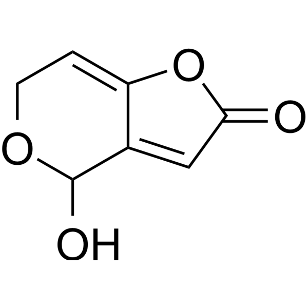
Patulin (Terinin) is a mycotoxin produced by fungi including the Aspergillus, Penicillium, and Byssochlamys species, causes chromosome breakage, mutation, teratogenic and cytotoxic. Patulin induces autophagy-dependent apoptosis through lysosomal-mitochondrial axis, and causes DNA damage.
For research use only. We do not sell to patients.
- Purity : 99.94%
- CAS No.: 149-29-1
- Formula: C7H6O4
- Molecular Weight:154.12
-
Storage:
-20°C, protect from light
* In solvent : -80°C, 6 months; -20°C, 1 month (protect from light)

To place orders, for customer services and technical support, please contact: MedChemExpress USA
Tel: 609-228-6898 E-mail: [email protected] [email protected]


-
 Biological Activity
-
Chemical Information
-
Solvent & Solubility
- Protocols
- Purity & Documentation
- References
-
 Help & FAQs


-
HY-N6719
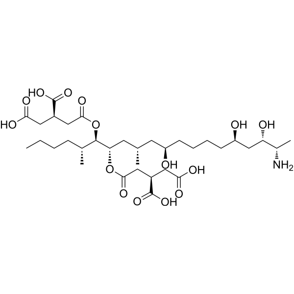
Fumonisin B1Sphingosine N-acyltransferase Inhibitor -

HY-15763
ErastinFerroptosis Inducer -
HY-B0332
MenadioneEndogenous Metabolite -
HY-N6684
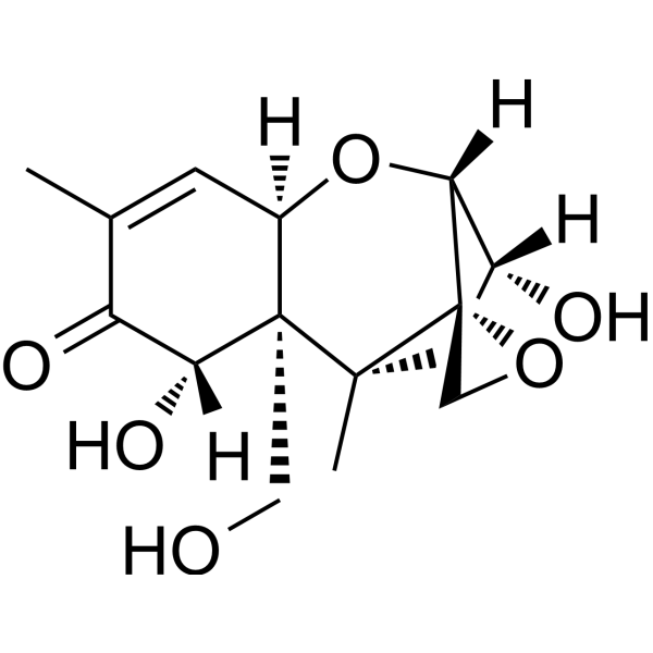
DeoxynivalenolMycotoxin -
HY-50856
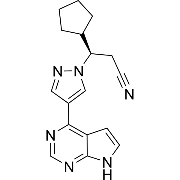
RuxolitinibJAK1/2 Inhibitor -
HY-112037
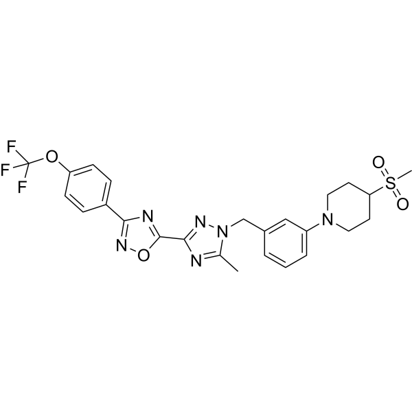
IACS-010759OXPHOS Inhibitor -
HY-103447
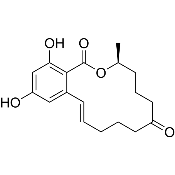
ZearalenoneMycotoxin Compound -

HY-N6779R
Patulin (Standard)Mycotoxin -
HY-N6723
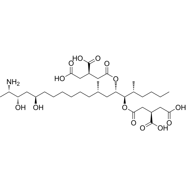
Fumonisin B2Ceramide Synthase Inhibitor
Publications Citing Use of MedChemExpress (MCE) Patulin
MoreAll Antibiotic Isoforms
More

Biological Activity
Description
In Vitro
Patulin (2 μM, 6-24 h) induces LMP and reduces Cathepsin B release in an autophagy-dependent manner in HepG2 cells[3].
Patulin (2 μM, 6-24 h) increases cytochrome c protein level and leads apoptosis induction in an autophagy-dependent manner in HepG2 cells[3].
Patulin (2 μM, 6-24 h) impairs mitophagy in HepG2 cells[3].
MedChemExpress (MCE) has not independently confirmed the accuracy of these methods. They are for reference only. Further protocols information, click here.
-
Cell Line:HepG2 cells
-
Concentration:2 μM
-
Incubation Time:6-24 h
-
Result:Increased the levels of active cleaved caspase-3 at 12 or 24 h.
-
Cell Line:HepG2 cells
-
Concentration:2 μM
-
Incubation Time:3-24 h
-
Result:Increases the LC3-II levels.
-
Cell Line:HepG2 cells
-
Concentration:2 μM
-
Incubation Time:6-24 h
-
Result:Increased the numbers of swollen mitochondria with scarce or disordered cristae.
In Vivo
MedChemExpress (MCE) has not independently confirmed the accuracy of these methods. They are for reference only.
-
Animal Model:Male CF-1 mice[4]
-
Dosage:1-3.75 mg/kg
-
Administration:i.p., once time
-
Result:Increased the damage index and TBARS production in the brain, liver and kidneys of mice in a dose-dependently manner (2.5-3.75 mg/kg).
Reduced the concentration of the reduced form of intracellular glutathione in the brain, liver and kidneys of mice at a dose of 2.5-3.75 mg/kg.
Decreased the levels of GSH.
Increased lipid peroxidation.


Chemical Information
-
CAS No. 149-29-1
-
Appearance Solid
-
Molecular Weight 154.12
-
Formula C7H6O4
-
Color White to off-white
-
SMILES
O=C1OC2=CCOC(C2=C1)O
-
Synonyms
Terinin
-
Structure Classification
-
Initial Source
Penicillium and Aspergillus
-
Shipping
Room temperature in continental US; may vary elsewhere.
-
Storage
-20°C, protect from light
* In solvent : -80°C, 6 months; -20°C, 1 month (protect from light)


Publications (4)
-
Journal Impact Factor
-
Most Recent
-
Antioxidants (Basel)
Patulin Ameliorates Hypertrophied Lipid Accumulation and Lipopolysaccharide-Induced Inflammatory Response by Modulating Mitochondrial Respiration. [Abstract]2023 Sep 11;12(9):1750. PMID: 37760053 -
Anal Chem
Highly Sensitive Chemiluminescent Immunoassay of Mycotoxins Using ZIF-8-Derived Yolk-Shell Co Single-Atom Site Catalysts as Superior Fenton-like Probes. [Abstract]2022 Feb 22;94(7):3400-3407. PMID: 35138805 -
Anal Chim Acta
CoN4-supported Co2N metal clusters for developing sensitive chemiluminescent immunochromatographic assays. [Abstract]2022 Nov 1:1232:340478. PMID: 36257748 -


Solvent & Solubility
In Vitro:
DMSO : 100 mg/mL (648.85 mM; Need ultrasonic; Hygroscopic DMSO has a significant impact on the solubility of product, please use newly opened DMSO)
H2O : 50 mg/mL (324.42 mM; Need ultrasonic and warming)
Please refer to the solubility information to select the appropriate solvent. Once prepared, please aliquot and store the solution to prevent product inactivation from repeated freeze-thaw cycles.
Storage method and period of stock solution: -80°C, 6 months; -20°C, 1 month (protect from light). When stored at -80°C, please use it within 6 months. When stored at -20°C, please use it within 1 month.
* Note: If you choose water as the stock solution, please dilute it to the working solution, then filter and sterilize it with a 0.22 μm filter before use.
Please refer to the solubility information to select the appropriate solvent. Once prepared, please aliquot and store the solution to prevent product inactivation from repeated freeze-thaw cycles.
Storage method and period of stock solution: -80°C, 6 months; -20°C, 1 month (protect from light). When stored at -80°C, please use it within 6 months. When stored at -20°C, please use it within 1 month.
* Note: If you choose water as the stock solution, please dilute it to the working solution, then filter and sterilize it with a 0.22 μm filter before use.
Concentration (start) × Volume (start) = Concentration (final) × Volume (final)
In Vivo:
Select the appropriate dissolution method based on your experimental animal and administration route.
- For the following dissolution methods, please ensure to first prepare a clear stock solution using an In Vitro approach and then sequentially add co-solvents:
- To ensure reliable experimental results, the clarified stock solution can be appropriately stored based on storage conditions. As for the working solution for In Vivo experiments, it is recommended to prepare freshly and use it on the same day.
- The percentages shown for the solvents indicate their volumetric ratio in the final prepared solution. If precipitation or phase separation occurs during preparation, heat and/or sonication can be used to aid dissolution.
Add each solvent one by one: 10% DMSO 40% PEG300 5% Tween-80 45% Saline
Solubility: 2.5 mg/mL (16.22 mM); Suspended solution; Need ultrasonic
This protocol yields a suspended solution of 2.5 mg/mL. Suspended solution can be used for oral and intraperitoneal injection.
Taking 1 mL working solution as an example, add 100 μL DMSO stock solution (25.0 mg/mL) to 400 μL PEG300, and mix evenly; then add 50 μL Tween-80 and mix evenly; then add 450 μL Saline to adjust the volume to 1 mL.
Preparation of Saline: Dissolve 0.9 g sodium chloride in ddH₂O and dilute to 100 mL to obtain a clear Saline solution.
Add each solvent one by one: 10% DMSO 90% (20% SBE-β-CD in Saline)
Solubility: ≥ 2.5 mg/mL (16.22 mM); Clear solution
This protocol yields a clear solution of ≥ 2.5 mg/mL (saturation unknown).
Taking 1 mL working solution as an example, add 100 μL DMSO stock solution (25.0 mg/mL) to 900 μL 20% SBE-β-CD in Saline, and mix evenly.
Preparation of 20% SBE-β-CD in Saline (4°C, storage for one week): 2 g SBE-β-CD powder is dissolved in 10 mL Saline, completely dissolve until clear.
In Vivo Dissolution Calculator
Please enter the basic information of animal experiments:
-
-
-
-
Recommended: Prepare an additional quantity of animals to account for potential losses during experiments.
Working solution concentration: 0.22 mg/mL
This product has good water solubility, please refer to the measured solubility data in water/PBS/Saline for details.

Protocols
-
Apoptosis
Apoptosis, also called programmed cell death, is generally characterized by distinct morphological characteristics.
-
TUNEL staining for apoptotic DNA fragmentation
TUNEL staining detects DNA strand breaks by using terminal deoxynucleotidyl transferase to add labeled nucleotides to exposed 3′-OH DNA termini, generating either microscopic staining in fixed cells or tissue sections, or fluorescence/cytometric signal in cell suspensions. TUNEL positivity reflects DNA fragmentation but should not be interpreted alone as definitive apoptosis, because TUNEL can also label necrotic, autolytic, mechanically damaged, or DNA-repair-associated DNA breaks.
-
Annexin V plus membrane-impermeant dye apoptosis staining
Annexin V-based apoptosis assays rely on the detection of phosphatidylserine (PS) externalization from the inner leaflet of the plasma membrane to the outer leaflet, an early biochemical hallmark of apoptosis. Fluorescently labeled Annexin V binds PS in a calcium-dependent manner, enabling identification of early apoptotic cells by flow cytometry or fluorescence microscopy. When combined with a membrane-impermeant DNA-binding dye (e. g. , propidium iodide), this approach allows discrimination between viable (Annexin V−/dye−), early apoptotic (Annexin V+/dye−), and late apoptotic or necrotic (Annexin V+/dye+) cell populations by assessing membrane integrity and PS exposure.
-
Autophagy
Autophagy is a process in which eukaryotic cells use lysosomes to degrade their own cytoplasmic proteins and damaged organelles under the regulation of autophagy related gene (Atg). Microtubule-associated proteins light chain 3 (LC3) is recognized as autophagy marker, which transfers from cytoplasmic LC3 (LC3-I) to membrane type (LC3-II). LC3-II/I ratio could be detected by Western Blot and fluorescence microscopy.
-
Apoptosis Solutions
Apoptosis is a regulated, generally non-lytic cell-death pathway that removes unwanted, damaged, infected, or abnormal cells through coordinated morphological changes, caspase activation, DNA fragmentation, and membrane remodeling. The intrinsic apoptosis pathway is controlled mainly by mitochondrial outer membrane permeabilization, BCL-2 family proteins, cytochrome c release, apoptosome formation, caspase-9 activation, and downstream executioner caspase-3/7 activation. The extrinsic apoptosis pathway is initiated by death receptors such as Fas, TNFR, and TRAIL receptors, which recruit adaptor proteins and activate caspase-8 before engaging executioner caspases or mitochondrial amplification through BID cleavage. Apoptosis is linked to many phenotypes, including cancer cell killing, tissue homeostasis, immune regulation, neurodegeneration, infection response, and treatment-induced cytotoxicity; unresolved questions include how apoptosis interacts with necroptosis, pyroptosis, ferroptos
-
Lysosome and acidic-vesicle live-cell staining
Lysosome and acidic-vesicle live-cell staining detects acidic intracellular compartments by using membrane-permeant acidotropic probes that accumulate in low-pH vesicles, including lysosomes, late endosomes, autolysosomes, and acidic phagosomes. LysoTracker staining is commonly used as an intensity-based readout of acidic lysosomal compartment abundance or enlargement, while acridine orange produces green fluorescence in less concentrated compartments and red fluorescence after concentration-dependent accumulation in acidic vesicular organelles. Loss or reduction of acridine-orange red signal can be used as a readout of lysosomal membrane permeabilization or reduced acidic-vesicle integrity. This protocol is designed for live cultured cells and can be adapted for fluorescence microscopy, high-content imaging, plate-reader readout, or flow cytometry when the selected literature supports the readout. Because these dyes report acidotropic accumulation rather than lysosome identity alone,
-
Macroautophagy Solutions
Macroautophagy is a conserved lysosome-dependent degradation pathway in which cytoplasmic material is sequestered into double-membrane autophagosomes and delivered to lysosomes for degradation and recycling. The pathway supports cellular homeostasis during nutrient limitation, organelle stress, protein-aggregate accumulation, infection, differentiation, and tissue remodeling by coupling cargo sequestration, autophagosome maturation, lysosomal fusion, and degradation of cargo-derived macromolecules. The core molecular sequence includes initiation by nutrient- and stress-regulated autophagy machinery, autophagosome nucleation, LC3/ATG8-family conjugation to autophagosomal membranes, cargo selection through receptors such as SQSTM1/p62, autophagosome-lysosome fusion, and lysosomal degradation. LC3 was identified as a mammalian homolog of yeast Atg8 that localizes to autophagosomal membranes after processing, and p62/SQSTM1 was shown to connect ubiquitinated cargo with autophagic degradati
-
Genotoxicity/Mutagenicity Study
The bacterial reverse mutation assay detects point mutations that restore amino-acid prototrophy in auxotrophic Salmonella typhimurium or Escherichia coli tester strains; after exposure to a test article, mutagenic activity is read out as an increased number of revertant colonies on minimal agar compared with the vehicle control. The assay uses tester strains with different mutation targets so that base-substitution and frameshift mutagens can be detected, and testing is performed with and without exogenous mammalian metabolic activation because some chemicals require biotransformation to become mutagenic.
-
Cell Viability Determination by MTT Colorimetric Assay
The following protocol uses the MTT colorimetric assay as a classic literature-established method for assessing cell viability/metabolic activity in cultured mammalian cells. MTT[3-(4,5-dimethylthiazol-2-yl)-2,5-diphenyltetrazolium bromide] is reduced by metabolically active cells to a colored formazan product; the amount of formazan is quantified spectrophotometrically and provides an indirect measure of metabolically active viable cells. Importantly, MTT reduction reflects cellular oxidoreductase/metabolic activity rather than an absolute direct count of living cells, so changes in cellular metabolism can alter the signal independently of cell number.


Purity & Documentation
-
Data Sheet (280 KB)
-
SDS (419 KB)
- English - EN (419 KB)
- Français - FR (419 KB)
- Deutsch - DE (419 KB)
- Norwegian - NO (419 KB)
- Español - ES (419 KB)
- Swedish - SV (419 KB)
- Italian - IT (419 KB)
- Korean - KR (419 KB)
- Portuguese - PT (419 KB)
-
Handling Instructions (2659 KB)
References
[1]. Puel O, et al. Biosynthesis and toxicological effects of patulin. Toxins (Basel). 2010 Apr;2(4):613-31. [Content Brief]
[2]. Glaser N, et al. Patulin: Mechanism of genotoxicity. Food Chem Toxicol. 2012 May;50(5):1796-801. [Content Brief]
[3]. Sun M, et al. Patulin Induces Autophagy-Dependent Apoptosis through Lysosomal-Mitochondrial Axis and Impaired Mitophagy in HepG2 Cells. J Agric Food Chem. 2018 Nov 21;66(46):12376-12384. [Content Brief]
[4]. Glaser N, et al. Patulin: Mechanism of genotoxicity. Food Chem Toxicol. 2012 May;50(5):1796-801. [Content Brief]


Complete Stock Solution Preparation Table
Please refer to the solubility information to select the appropriate solvent. Once prepared, please aliquot and store the solution to prevent product inactivation from repeated freeze-thaw cycles.
Storage method and period of stock solution: -80°C, 6 months; -20°C, 1 month (protect from light). When stored at -80°C, please use it within 6 months. When stored at -20°C, please use it within 1 month.
| Optional Solvent | Concentration Solvent Mass | 1 mg | 5 mg | 10 mg | 25 mg |
|---|---|---|---|---|---|
| H2O / DMSO | 1 mM | 6.4885 mL | 32.4423 mL | 64.8845 mL | 162.2113 mL |
| 5 mM | 1.2977 mL | 6.4885 mL | 12.9769 mL | 32.4423 mL | |
| 10 mM | 0.6488 mL | 3.2442 mL | 6.4885 mL | 16.2211 mL | |
| 15 mM | 0.4326 mL | 2.1628 mL | 4.3256 mL | 10.8141 mL | |
| 20 mM | 0.3244 mL | 1.6221 mL | 3.2442 mL | 8.1106 mL | |
| 25 mM | 0.2595 mL | 1.2977 mL | 2.5954 mL | 6.4885 mL | |
| 30 mM | 0.2163 mL | 1.0814 mL | 2.1628 mL | 5.4070 mL | |
| 40 mM | 0.1622 mL | 0.8111 mL | 1.6221 mL | 4.0553 mL | |
| 50 mM | 0.1298 mL | 0.6488 mL | 1.2977 mL | 3.2442 mL | |
| 60 mM | 0.1081 mL | 0.5407 mL | 1.0814 mL | 2.7035 mL | |
| 80 mM | 0.0811 mL | 0.4055 mL | 0.8111 mL | 2.0276 mL | |
| 100 mM | 0.0649 mL | 0.3244 mL | 0.6488 mL | 1.6221 mL |
* Note: If you choose water as the stock solution, please dilute it to the working solution, then filter and sterilize it with a 0.22 μm filter before use.


Patulin Related Classifications
HY-N6779 Related Classifications
 Powered by Bioz