



PROTAC Axl Degrader 2

Based on 1 Customer Validation
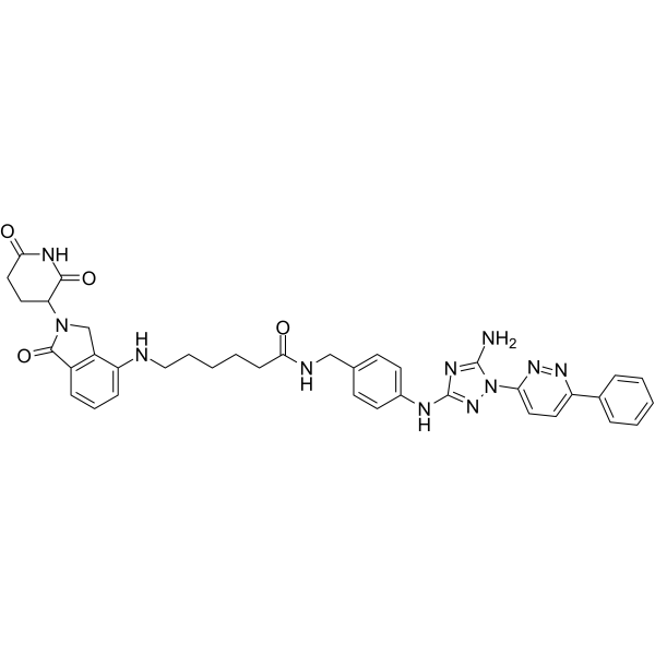
PROTAC Axl Degrader 2 is an orally active and selective AXL PROTAC degrader with dual activities of AXL kinase inhibition and AXL protein degradation. PROTAC Axl Degrader 2 induces cytoplasmic vacuolization, excessive macropinosome production, oncosis, H-Ras activation, and Rac1-dependent non-apoptotic cell death. PROTAC Axl Degrader 2 inhibits the proliferation and migration of cancer cells, and also suppresses the growth of tumor cell xenografts under in vivo conditions. PROTAC Axl Degrader 2 can be used in research related to breast cancer.
(Pink: Axl ligand (HY-184184); Blue: Cereblon ligand (HY-A0003); Black: linker (HY-W041970)).
For research use only. We do not sell to patients.
- Purity : 96.07%
- CAS No.: 3033401-45-2
- Formula: C38H39N11O4
- Molecular Weight:713.79
-
Storage:
-20°C, protect from light, stored under nitrogen
* In solvent : -80°C, 6 months; -20°C, 1 month (protect from light, stored under nitrogen)

To place orders, for customer services and technical support, please contact: MedChemExpress USA
Tel: 609-228-6898 E-mail: [email protected] [email protected]


-
 Biological Activity
-
Chemical Information
-
Solvent & Solubility
- Protocols
- Purity & Documentation
- References
-
 Help & FAQs


All PROTACs Isoforms
More

Biological Activity
Description
IC50 & Target
[1]|
Axl 1.61 μM (IC50) |
H-Ras |
Cellular Effect
|
Cell Line

|
Type | Value | Description | References |
|---|---|---|---|---|
| MDA-MB-231 | IC50 |
6.23 μM
|
Antiproliferative activity against human MDA-MB-231 breast cancer cells assessed as reduction in cell viability incubated for 72 hrs by MTT assay.
Antiproliferative activity against human MDA-MB-231 breast cancer cells assessed as reduction in cell viability incubated for 72 hrs by MTT assay.
|
35279611 |
| 4T1 | IC50 |
2.06 μM
|
Antiproliferative activity against mouse 4T1 breast cancer cells assessed as reduction in cell viability incubated for 72 hrs by MTT assay.
Antiproliferative activity against mouse 4T1 breast cancer cells assessed as reduction in cell viability incubated for 72 hrs by MTT assay.
|
35279611 |
| MCF-10A | IC50 |
>60 μM
|
Cytotoxicity against non-tumorigenic human MCF-10A breast epithelial cells assessed as reduction in cell viability incubated for 72 hrs by MTT assay.
Cytotoxicity against non-tumorigenic human MCF-10A breast epithelial cells assessed as reduction in cell viability incubated for 72 hrs by MTT assay.
|
35279611 |
| GES1 | IC50 |
>60 μM
|
Cytotoxicity against human GES-1 gastric epithelial cells assessed as reduction in cell viability incubated for 72 hrs by MTT assay.
Cytotoxicity against human GES-1 gastric epithelial cells assessed as reduction in cell viability incubated for 72 hrs by MTT assay.
|
35279611 |
In Vitro
PROTAC Axl Degrader 2 (compound 20) potently inhibits recombinant AXL kinase activity with an IC50 of 1.61 μM; it suppresses the proliferation of MDA-MB-231 and 4T1 breast cancer cells with IC50 values of 6.23 μM and 2.06 μM, respectively, and shows no cytotoxicity in non-tumorigenic MCF-10A and GES-1 cells[1].
PROTAC Axl Degrader 2 exhibits high selectivity for AXL kinase, with an inhibition rate of over 90% at a concentration of 5 μM, while the inhibition rates against all other tested human kinases do not exceed 90%[1].
PROTAC Axl Degrader 2 (0.5-2 μM; 24-48 h) induces proteasome-dependent degradation of AXL protein in MDA-MB-231 cells[1].
PROTAC Axl Degrader 2 (0.0001-100 μM; 30 min) forms a ternary complex with AXL and CRBN proteins, and exhibits maximum activity at a concentration of 1 μM[1].
PROTAC Axl Degrader 2 (1-10 μM; 48 h) inhibits the migration of MDA-MB-231 and 4T1 breast cancer cells[1].
PROTAC Axl Degrader 2 (1 μM; 24 h) induces Rac1-dependent, non-apoptotic oncosis via macropinocytosis in MDA-MB-231 and 4T1 breast cancer cells, which is characterized by large cytoplasmic vacuoles not originating from mitochondria or lysosomes[1].
MedChemExpress (MCE) has not independently confirmed the accuracy of these methods. They are for reference only. Further protocols information, click here.
-
Cell Line:MDA-MB-231 human breast cancer cells
-
Concentration:0.5, 1, 2 μM
-
Incubation Time:24 h, 48 h
-
Result:Reduced AXL protein levels after 24h of treatment.
Showed a more pronounced decrease in AXL protein levels after 48h at both 0.5 μM and 2 μM.
Reversed AXL protein degradation was observed with co-treatment with the proteasome inhibitor Epoxymycin (EPO).
-
Cell Line:MDA-MB-231 human breast cancer cells, 4T1 mouse breast cancer cells
-
Concentration:1, 10 μM
-
Incubation Time:48 h
-
Result:Inhibited migration of both cell lines
-
Cell Line:MDA-MB-231 human breast cancer cells, 4T1 mouse breast cancer cells
-
Concentration:2 μM
-
Incubation Time:48 h
-
Result:Inhibited migration of 4T1 cells more effectively than R428 at 2 μM.
Inhibited MDA-MB-231 cell migration to an equivalent extent as R428 at 2 μM.
Parmacokinetics
In Vivo
MedChemExpress (MCE) has not independently confirmed the accuracy of these methods. They are for reference only.


Chemical Information
-
CAS No. 3033401-45-2
-
Appearance Solid
-
Molecular Weight 713.79
-
Formula C38H39N11O4
-
Color White to light yellow
-
SMILES
NC1=NC(NC2=CC=C(C=C2)CNC(CCCCCNC3=CC=CC4=C3CN(C4=O)C5CCC(NC5=O)=O)=O)=NN1C6=CC=C(N=N6)C7=CC=CC=C7
-
Shipping
Room temperature in continental US; may vary elsewhere.
-
Storage
-20°C, protect from light, stored under nitrogen
* In solvent : -80°C, 6 months; -20°C, 1 month (protect from light, stored under nitrogen)


Solvent & Solubility
In Vitro:
DMSO : 100 mg/mL (140.10 mM; Need ultrasonic; Hygroscopic DMSO has a significant impact on the solubility of product, please use newly opened DMSO)
Please refer to the solubility information to select the appropriate solvent. Once prepared, please aliquot and store the solution to prevent product inactivation from repeated freeze-thaw cycles.
Storage method and period of stock solution: -80°C, 6 months; -20°C, 1 month (protect from light, stored under nitrogen). When stored at -80°C, please use it within 6 months. When stored at -20°C, please use it within 1 month.
Please refer to the solubility information to select the appropriate solvent. Once prepared, please aliquot and store the solution to prevent product inactivation from repeated freeze-thaw cycles.
Storage method and period of stock solution: -80°C, 6 months; -20°C, 1 month (protect from light, stored under nitrogen). When stored at -80°C, please use it within 6 months. When stored at -20°C, please use it within 1 month.
Concentration (start) × Volume (start) = Concentration (final) × Volume (final)

Protocols
-
Apoptosis
Apoptosis, also called programmed cell death, is generally characterized by distinct morphological characteristics.
-
Cell Cytotoxicity Assay
Cytotoxicity assays are usually based on the assessment of cell membrane damage, which can also be indirectly detected by measuring cell viability. Detection methods include MTT assay, CKK-8 assay, LDH assay and ATP assay, etc.
-
Mammalian live/dead viability and cytotoxicity staining
Live/dead viability and cytotoxicity staining assays are based on the simultaneous detection of intracellular esterase activity in metabolically active (viable) cells and membrane integrity loss in non-viable cells. In commonly used dual-staining approaches, membrane-permeant fluorogenic substrates are converted by intracellular esterases into fluorescent products in live cells, while impermeant DNA-binding dyes selectively enter cells with compromised plasma membranes and label nucleic acids in dead or dying cells, enabling discrimination between viable and non-viable populations by fluorescence microscopy or flow cytometry.
-
Cell migration
Cell migration is a method that plays an important role in wound healing, cell differentiation, embryonic development, etc.
-
Apoptosis Solutions
Apoptosis is a regulated, generally non-lytic cell-death pathway that removes unwanted, damaged, infected, or abnormal cells through coordinated morphological changes, caspase activation, DNA fragmentation, and membrane remodeling. The intrinsic apoptosis pathway is controlled mainly by mitochondrial outer membrane permeabilization, BCL-2 family proteins, cytochrome c release, apoptosome formation, caspase-9 activation, and downstream executioner caspase-3/7 activation. The extrinsic apoptosis pathway is initiated by death receptors such as Fas, TNFR, and TRAIL receptors, which recruit adaptor proteins and activate caspase-8 before engaging executioner caspases or mitochondrial amplification through BID cleavage. Apoptosis is linked to many phenotypes, including cancer cell killing, tissue homeostasis, immune regulation, neurodegeneration, infection response, and treatment-induced cytotoxicity; unresolved questions include how apoptosis interacts with necroptosis, pyroptosis, ferroptos
-
Breast Cancer Modeling
Breast cancer is a heterogeneous cancer, and it has been distinguished into four subtypes: luminal A, luminal B, HER2-positive and basal-like. Molecular mutations, epigenetic alterations, hormone exposure and immune microenvironment are related to the progression of breast cancer.
-
Cell Viability Determination by MTT Colorimetric Assay
The following protocol uses the MTT colorimetric assay as a classic literature-established method for assessing cell viability/metabolic activity in cultured mammalian cells. MTT[3-(4,5-dimethylthiazol-2-yl)-2,5-diphenyltetrazolium bromide] is reduced by metabolically active cells to a colored formazan product; the amount of formazan is quantified spectrophotometrically and provides an indirect measure of metabolically active viable cells. Importantly, MTT reduction reflects cellular oxidoreductase/metabolic activity rather than an absolute direct count of living cells, so changes in cellular metabolism can alter the signal independently of cell number.


Purity & Documentation
-
Data Sheet (280 KB)
-
SDS (252 KB)
- English - EN (252 KB)
- Français - FR (252 KB)
- Deutsch - DE (252 KB)
- Norwegian - NO (252 KB)
- Español - ES (252 KB)
- Swedish - SV (252 KB)
- Italian - IT (252 KB)
- Korean - KR (252 KB)
- Portuguese - PT (252 KB)
-
Handling Instructions (2659 KB)
References


Complete Stock Solution Preparation Table
Please refer to the solubility information to select the appropriate solvent. Once prepared, please aliquot and store the solution to prevent product inactivation from repeated freeze-thaw cycles.
Storage method and period of stock solution: -80°C, 6 months; -20°C, 1 month (protect from light, stored under nitrogen). When stored at -80°C, please use it within 6 months. When stored at -20°C, please use it within 1 month.
| Optional Solvent | Concentration Solvent Mass | 1 mg | 5 mg | 10 mg | 25 mg |
|---|---|---|---|---|---|
| DMSO | 1 mM | 1.4010 mL | 7.0049 mL | 14.0097 mL | 35.0243 mL |
| 5 mM | 0.2802 mL | 1.4010 mL | 2.8019 mL | 7.0049 mL | |
| 10 mM | 0.1401 mL | 0.7005 mL | 1.4010 mL | 3.5024 mL | |
| 15 mM | 0.0934 mL | 0.4670 mL | 0.9340 mL | 2.3350 mL | |
| 20 mM | 0.0700 mL | 0.3502 mL | 0.7005 mL | 1.7512 mL | |
| 25 mM | 0.0560 mL | 0.2802 mL | 0.5604 mL | 1.4010 mL | |
| 30 mM | 0.0467 mL | 0.2335 mL | 0.4670 mL | 1.1675 mL | |
| 40 mM | 0.0350 mL | 0.1751 mL | 0.3502 mL | 0.8756 mL | |
| 50 mM | 0.0280 mL | 0.1401 mL | 0.2802 mL | 0.7005 mL | |
| 60 mM | 0.0233 mL | 0.1167 mL | 0.2335 mL | 0.5837 mL | |
| 80 mM | 0.0175 mL | 0.0876 mL | 0.1751 mL | 0.4378 mL | |
| 100 mM | 0.0140 mL | 0.0700 mL | 0.1401 mL | 0.3502 mL |


PROTAC Axl Degrader 2 Related Classifications
HY-144627 Related Classifications
 Powered by Bioz