



PROTAC EZH2 Degrader-2
Based on 1 publication(s) in Google Scholar
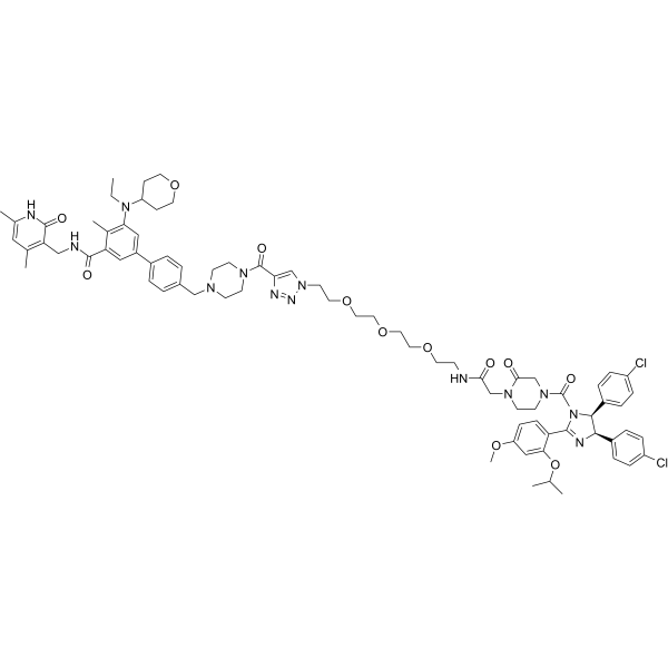
PROTAC EZH2 Degrader-2 is a PROTAC EZH2 inhibitor. PROTAC EZH2 Degrader-2 degrades EZH2 in SU-DHL-6 cells in a dose-dependent manner. PROTAC EZH2 Degrader-2 induces apoptosis and reduces mitochondrial membrane potential in SU-DHL-6 cells. PROTAC EZH2 Degrader-2 has anti-cancer and anti-proliferative activity.
(Pink: EZH2 ligand (HY-179351); Blue: MDM2 ligand (HY-128836); Black: linker (HY-W021401)).
Para uso exclusivo en investigación. No vendemos a pacientes.
- Pureza : 98.77%
- No. CAS: 3093642-32-8
- Fòrmula: C77H93Cl2N13O12
- Peso molecular:1463.55
-
Almacenamiento:
4°C, stored under nitrogen
* In solvent : -80°C, 6 months; -20°C, 1 month (stored under nitrogen)

To place orders, for customer services and technical support, please contact: MedChemExpress USA
Tel: 609-228-6898 E-mail: [email protected] [email protected]


-
 Actividad biológica
-
Chemical Information
-
Solvente y solubilidad
- Protocolo
- Pureza y Documentación
- Referencias
-
 Help & FAQs


Publications Citing Use of MedChemExpress (MCE) PROTAC EZH2 Degrader-2
MoreVer todos los productos específicos de isoformas PROTACs
MoreVer todos los productos específicos de isoformas Histone Methyltransferase
More

Actividad biológica
Descripciòn
IC50 & Target
[1]|
EZH2 |
MDM2 |
In Vitro
PROTAC EZH2 Degrader-2 (Compound E-3P-MDM2) (0.037-3 μM, 24-72 h) degrades EZH2, EED, and SUZ12 proteins of PRC2 complex in a concentration and time-dependent manner in SU-DHL-6 cells[1].
PROTAC EZH2 Degrader-2 (0.037-3 μM, 48-72 h) exhibits antiproliferative activity and induces apoptosis in SU-DHL-6 cells[1].
PROTAC EZH2 Degrader-2 (3 μM, 72 h) upregulates the expression of H3K27me3-related genes (ADRB2 and CDKN2) in SU-DHL-6 cells[1].
PROTAC EZH2 Degrader-2 (0.37-30 μM, 48 h) shows more potent inhibitory effect on cell proliferation in SU-DHL-6 (EZH2Y614N) cells than in HBL-1 (EZH2WT) cells[1].
MedChemExpress (MCE) has not independently confirmed the accuracy of these methods. They are for reference only. Further protocols information, click here.
-
Cell Line:SU-DHL-6 cells
-
Concentration:0.037, 0.11, 0.33, 1, 3 μM
-
Incubation Time:48, 72 h
-
Result:Reduced mitochondrial membrane potential.
Upregulated Bax, cytochrome c, cleaved-caspase 9, cleaved-caspase 3.
Disrupted Bax/Bcl-2 balance.


Chemical Information
-
No. CAS 3093642-32-8
-
Appearance Solid
-
Peso molecular 1463.55
-
Fòrmula C77H93Cl2N13O12
-
Color White to off-white
-
SMILES
CC1=CC(C)=C(CNC(C2=CC(C3=CC=C(CN4CCN(C(C5=CN(CCOCCOCCOCCNC(CN6CCN(C(N7C(C8=CC=C(OC)C=C8OC(C)C)=N[C@H](C9=CC=C(Cl)C=C9)[C@@H]7C%10=CC=C(Cl)C=C%10)=O)CC6=O)=O)N=N5)=O)CC4)C=C3)=CC(N(C%11CCOCC%11)CC)=C2C)=O)C(N1)=O
-
Envío
Room temperature in continental US; may vary elsewhere.
-
Almacenamiento
4°C, stored under nitrogen
* In solvent : -80°C, 6 months; -20°C, 1 month (stored under nitrogen)


Publications (1)
-
Journal Impact Factor
-
Most Recent
-
bioRxiv
Dual EZH1/2 inhibition enhances DNMT inhibitor efficacy in colon cancer through targeting H3K27me1. [Abstract]2025 Sep 18:2025.09.16.676613. PMID: 41000734


Solvente y solubilidad
In Vitro:
DMSO : 100 mg/mL (68.33 mM; Need ultrasonic; Hygroscopic DMSO has a significant impact on the solubility of product, please use newly opened DMSO)
Please refer to the solubility information to select the appropriate solvent. Once prepared, please aliquot and store the solution to prevent product inactivation from repeated freeze-thaw cycles.
Storage method and period of stock solution: -80°C, 6 months; -20°C, 1 month (stored under nitrogen). When stored at -80°C, please use it within 6 months. When stored at -20°C, please use it within 1 month.
Please refer to the solubility information to select the appropriate solvent. Once prepared, please aliquot and store the solution to prevent product inactivation from repeated freeze-thaw cycles.
Storage method and period of stock solution: -80°C, 6 months; -20°C, 1 month (stored under nitrogen). When stored at -80°C, please use it within 6 months. When stored at -20°C, please use it within 1 month.
Concentration (start) × Volume (start) = Concentration (final) × Volume (final)
In Vivo:
Select the appropriate dissolution method based on your experimental animal and administration route.
- For the following dissolution methods, please ensure to first prepare a clear stock solution using an In Vitro approach and then sequentially add co-solvents:
- To ensure reliable experimental results, the clarified stock solution can be appropriately stored based on storage conditions. As for the working solution for In Vivo experiments, it is recommended to prepare freshly and use it on the same day.
- The percentages shown for the solvents indicate their volumetric ratio in the final prepared solution. If precipitation or phase separation occurs during preparation, heat and/or sonication can be used to aid dissolution.
In Vivo Dissolution Calculator
Please enter the basic information of animal experiments:
-
-
-
-
Recommended: Prepare an additional quantity of animals to account for potential losses during experiments.
Please enter your animal formula composition:
-
%DMSO +
Recommended: Keep the proportion of DMSO in working solution below 2% if your animal is weak.
-
%+
-
+%Tween-80 + +
-
%Saline +
The co-solvents required include: DMSO, . All of co-solvents are available by MedChemExpress (MCE). , Tween 80. All of co-solvents are available by MedChemExpress (MCE).
Working solution concentration: 0.22 mg/mL
Method for preparing stock solution: mg drug dissolved in μL DMSO. Stock solution concentration: mg/mL. * In solvent : -80°C, 6 months; -20°C, 1 month (stored under nitrogen)
1. Take μL DMSO stock solution;
2. Add μL .
μL , mix evenly;
3. Then add μL Tween 80, mix evenly;
4. Then add μL
Please ensure that the stock solution in the first step is dissolved to a clear state, and add co-solvents in sequence. You can use ultrasonic heating (ultrasonic cleaner, recommended frequency 20-40 kHz), vortexing, etc. to assist dissolution.

Protocolo
-
Apoptosis
Apoptosis, also called programmed cell death, is generally characterized by distinct morphological characteristics.
-
TUNEL staining for apoptotic DNA fragmentation
TUNEL staining detects DNA strand breaks by using terminal deoxynucleotidyl transferase to add labeled nucleotides to exposed 3′-OH DNA termini, generating either microscopic staining in fixed cells or tissue sections, or fluorescence/cytometric signal in cell suspensions. TUNEL positivity reflects DNA fragmentation but should not be interpreted alone as definitive apoptosis, because TUNEL can also label necrotic, autolytic, mechanically damaged, or DNA-repair-associated DNA breaks.
-
Mitochondrial membrane-potential fluorescent assay
Mitochondrial membrane potential fluorescent assays estimate ΔΨm in living cells using lipophilic cationic dyes such as TMRM, TMRE, rhodamine 123, and JC-1, which accumulate in mitochondria according to membrane polarization; loss of signal after FCCP or CCCP treatment is interpreted as mitochondrial depolarization. TMRM/TMRE and rhodamine 123 are commonly used for semi-quantitative live-cell microscopy or flow cytometry, while JC-1 can report a shift from red aggregate fluorescence to green monomer fluorescence during depolarization; interpretation requires controls because dye concentration, quenching mode, cell type, dye efflux, and mitochondrial mass can affect fluorescence independently of ΔΨm.
-
Annexin V plus membrane-impermeant dye apoptosis staining
Annexin V-based apoptosis assays rely on the detection of phosphatidylserine (PS) externalization from the inner leaflet of the plasma membrane to the outer leaflet, an early biochemical hallmark of apoptosis. Fluorescently labeled Annexin V binds PS in a calcium-dependent manner, enabling identification of early apoptotic cells by flow cytometry or fluorescence microscopy. When combined with a membrane-impermeant DNA-binding dye (e. g. , propidium iodide), this approach allows discrimination between viable (Annexin V−/dye−), early apoptotic (Annexin V+/dye−), and late apoptotic or necrotic (Annexin V+/dye+) cell populations by assessing membrane integrity and PS exposure.
-
Fluorescent plasma-membrane potential dye assay
Fluorescent plasma-membrane potential dye assays measure changes in cell membrane potential using voltage-sensitive dyes whose fluorescence changes when cells depolarize or hyperpolarize. Anionic bis-oxonol dyes such as DiBAC4(3) enter depolarized cells more readily and show increased fluorescence after intracellular binding, while hyperpolarization reduces dye accumulation and fluorescence. FMP/FLIPR membrane-potential dyes are used for faster, homogeneous microplate assays of ion-channel or receptor-mediated membrane-potential changes.
-
Mitochondrial membrane-potential and mitochondrial mass staining
Mitochondrial membrane potential staining measures the electrochemical polarization across the mitochondrial inner membrane in live cells using lipophilic cationic fluorescent probes; early rhodamine-based work showed that selective mitochondrial dye accumulation is lost when the mitochondrial transmembrane potential is dissipated. JC-1 reports mitochondrial polarization by shifting from green monomer fluorescence to red J-aggregate fluorescence as dye concentration increases within energized mitochondria; therefore, the red/green fluorescence ratio is used as a relative readout of mitochondrial membrane potential. TMRE or TMRM staining provides a single-channel relative readout because these cationic rhodamine esters accumulate in polarized mitochondria, and lower fluorescence indicates reduced mitochondrial polarization when acquisition and dye-loading conditions are controlled. Mitochondrial mass staining is commonly performed with MitoTracker Green FM or related MitoTracker dyes as
-
Apoptosis Solutions
Apoptosis is a regulated, generally non-lytic cell-death pathway that removes unwanted, damaged, infected, or abnormal cells through coordinated morphological changes, caspase activation, DNA fragmentation, and membrane remodeling. The intrinsic apoptosis pathway is controlled mainly by mitochondrial outer membrane permeabilization, BCL-2 family proteins, cytochrome c release, apoptosome formation, caspase-9 activation, and downstream executioner caspase-3/7 activation. The extrinsic apoptosis pathway is initiated by death receptors such as Fas, TNFR, and TRAIL receptors, which recruit adaptor proteins and activate caspase-8 before engaging executioner caspases or mitochondrial amplification through BID cleavage. Apoptosis is linked to many phenotypes, including cancer cell killing, tissue homeostasis, immune regulation, neurodegeneration, infection response, and treatment-induced cytotoxicity; unresolved questions include how apoptosis interacts with necroptosis, pyroptosis, ferroptos
-
Cell Viability Determination by MTT Colorimetric Assay
The following protocol uses the MTT colorimetric assay as a classic literature-established method for assessing cell viability/metabolic activity in cultured mammalian cells. MTT[3-(4,5-dimethylthiazol-2-yl)-2,5-diphenyltetrazolium bromide] is reduced by metabolically active cells to a colored formazan product; the amount of formazan is quantified spectrophotometrically and provides an indirect measure of metabolically active viable cells. Importantly, MTT reduction reflects cellular oxidoreductase/metabolic activity rather than an absolute direct count of living cells, so changes in cellular metabolism can alter the signal independently of cell number.


Pureza y Documentación
-
Ficha de datos (272 KB)
-
SDS (252 KB)
- English - EN (252 KB)
- Français - FR (252 KB)
- Deutsch - DE (252 KB)
- Norwegian - NO (252 KB)
- Español - ES (252 KB)
- Swedish - SV (252 KB)
- Italian - IT (252 KB)
- Korean - KR (252 KB)
- Portuguese - PT (252 KB)
-
Instrucciones de manejo (2659 KB)
Referencias


Complete Stock Solution Preparation Table
Please refer to the solubility information to select the appropriate solvent. Once prepared, please aliquot and store the solution to prevent product inactivation from repeated freeze-thaw cycles.
Storage method and period of stock solution: -80°C, 6 months; -20°C, 1 month (stored under nitrogen). When stored at -80°C, please use it within 6 months. When stored at -20°C, please use it within 1 month.
| Optional Solvent | Concentration Solvent Mass | 1 mg | 5 mg | 10 mg | 25 mg |
|---|---|---|---|---|---|
| DMSO | 1 mM | 0.6833 mL | 3.4164 mL | 6.8327 mL | 17.0818 mL |
| 5 mM | 0.1367 mL | 0.6833 mL | 1.3665 mL | 3.4164 mL | |
| 10 mM | 0.0683 mL | 0.3416 mL | 0.6833 mL | 1.7082 mL | |
| 15 mM | 0.0456 mL | 0.2278 mL | 0.4555 mL | 1.1388 mL | |
| 20 mM | 0.0342 mL | 0.1708 mL | 0.3416 mL | 0.8541 mL | |
| 25 mM | 0.0273 mL | 0.1367 mL | 0.2733 mL | 0.6833 mL | |
| 30 mM | 0.0228 mL | 0.1139 mL | 0.2278 mL | 0.5694 mL | |
| 40 mM | 0.0171 mL | 0.0854 mL | 0.1708 mL | 0.4270 mL | |
| 50 mM | 0.0137 mL | 0.0683 mL | 0.1367 mL | 0.3416 mL | |
| 60 mM | 0.0114 mL | 0.0569 mL | 0.1139 mL | 0.2847 mL |


PROTAC EZH2 Degrader-2 Related Classifications
HY-157164 Related Classifications
 Powered by Bioz