



Zovegalisib
Based on 1 publication(s) in Google Scholar
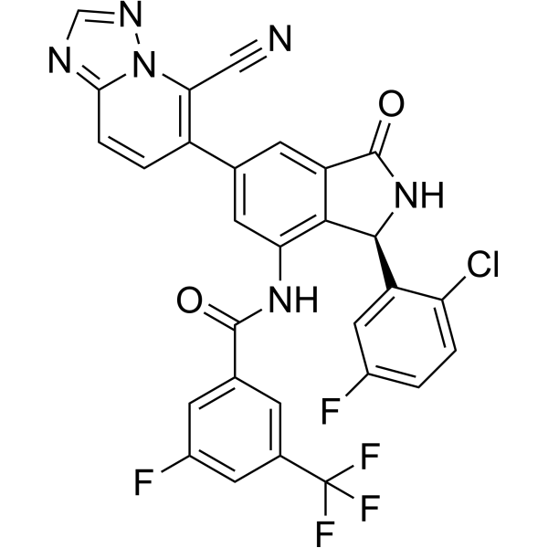
Zovegalisib (RLY-2608) is an orally active first-in-class allosteric mutant-selective inhibitor of PI3Ka with anti-tumor activity. Zovegalisib inhibits tumor growth in PIK3CA-mutant xenograft mice models with minimal impact on insulin.
Nos produits utilisent uniquement pour la recherche. Nous ne vendons pas aux patients.
- Pureté : 99.79%
- CAS No.: 2733573-94-7
- Formule: C29H14ClF5N6O2
- Masse moléculaire:608.91
-
Stockage:Powder -20°C, 3 years , 4°C, 2 years ; In solvent -80°C, 6 months , -20°C, 1 month

To place orders, for customer services and technical support, please contact: MedChemExpress USA
Tel: 609-228-6898 E-mail: [email protected] [email protected]


-
 Activité biologique
-
Chemical Information
-
Solvant et solubilité
- Protocole
- Pureté et documentation
- Références
-
 Help & FAQs


-
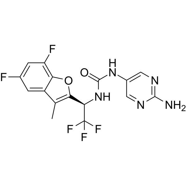
HY-156681
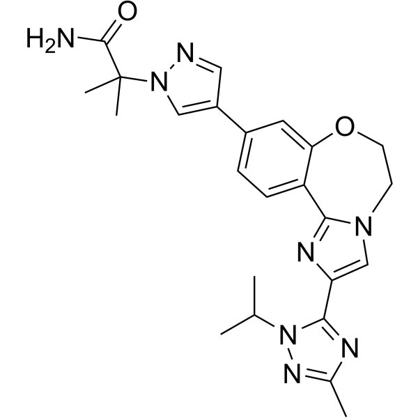
TersolisibPI3Kα Inhibitor -
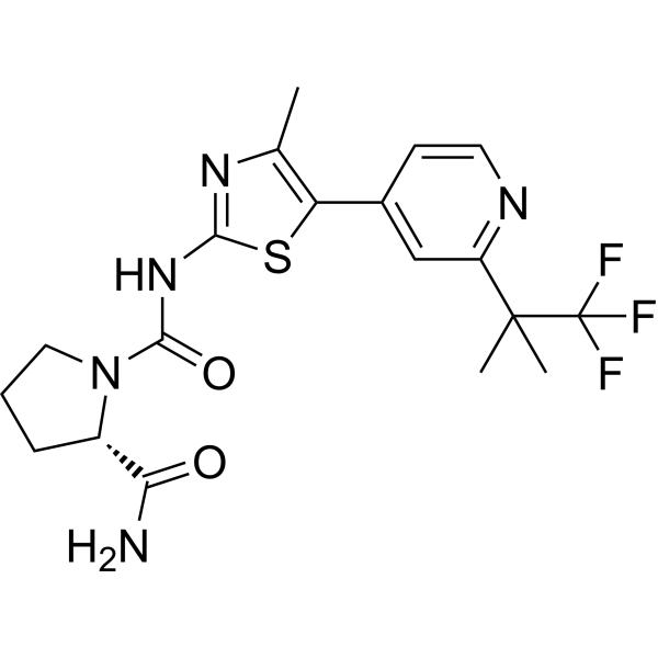
HY-15244
AlpelisibPI3Kα Inhibitor -
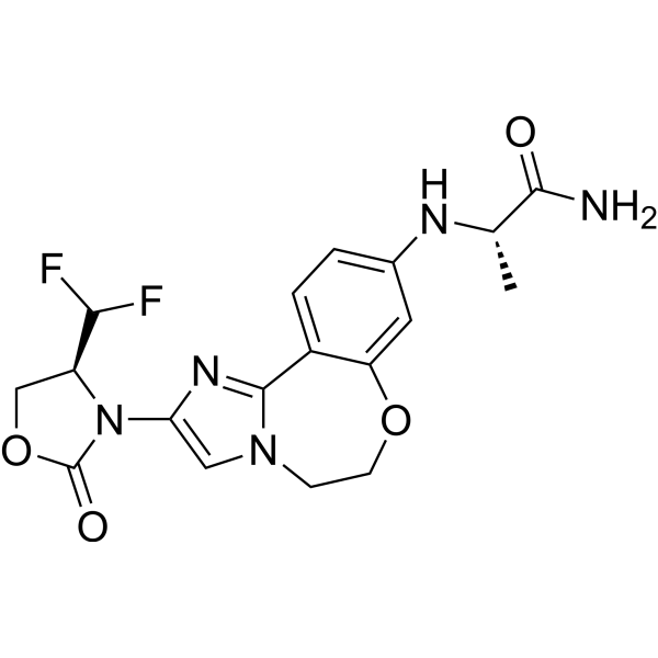
HY-101562
InavolisibPI3Kα Inhibitor -
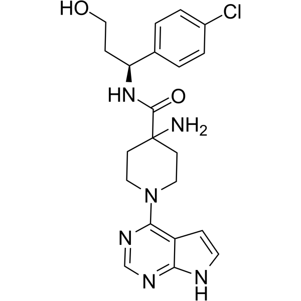
HY-15431
CapivasertibAkt Inhibitor -
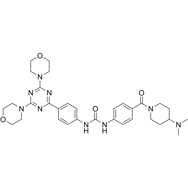
HY-10681
GedatolisibPI3K/mTOR Inhibitor -
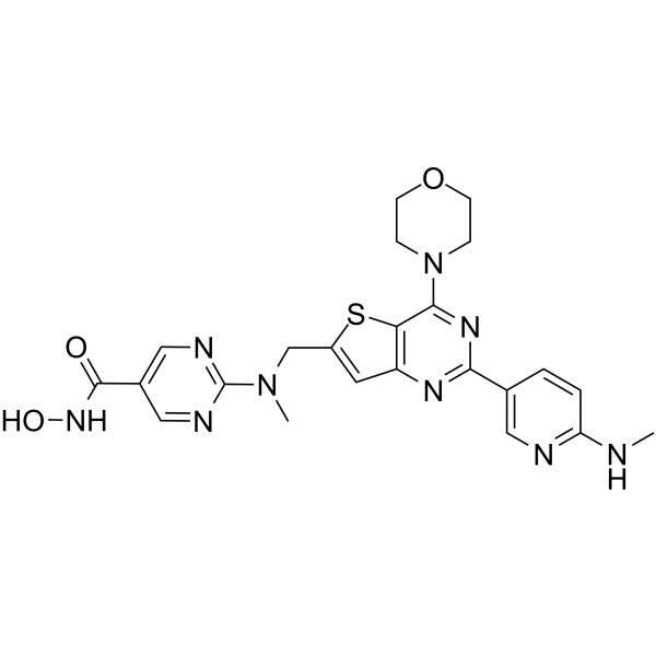
HY-19763
IfupinostatHDAC/PI3K Inhibitor -
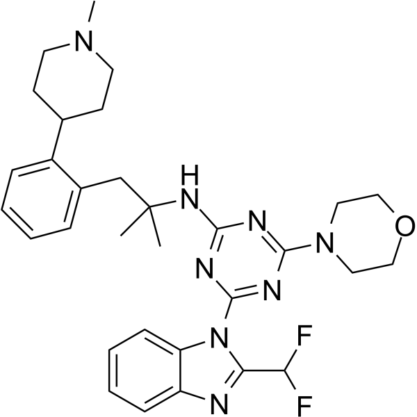
HY-109198
ZandelisibPI3K Inhibitor -
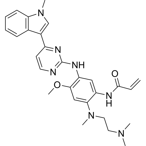
HY-15772
OsimertinibMutant-Selective EGFR Inhibitor -
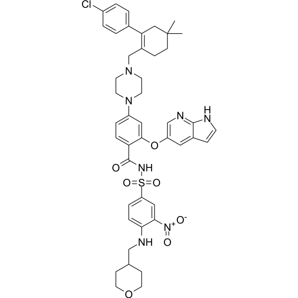
HY-15531
VenetoclaxBCL-2 Inhibitor -
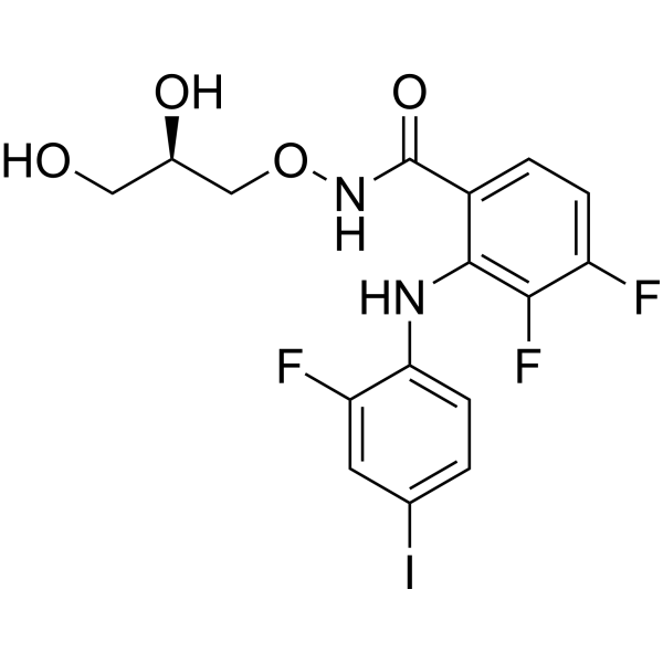
HY-10254
MirdametinibMEK Inhibitor -
HY-18749
SC79Akt Activator -
HY-13898
TaselisibPIK3CA Inhibitor
Publications Citing Use of MedChemExpress (MCE) Zovegalisib
More Customer Validation & Images

-
Cell Proliferation/Viability Assay


Activité biologique
Description
In Vitro
In Vivo
Zovegalisib (12.5-100 mg/kg, p.o., a single dose for 4 days) demonstrates potent tumor growth inhibition in Balb/c nude female mice bearing HSC-2 tumors[2].
MedChemExpress (MCE) has not independently confirmed the accuracy of these methods. They are for reference only.
-
Animal Model:Tumor xenograft mice models[1]
-
Dosage:25-100 mg/kg
-
Administration:p.o., twice a day for 50 days
-
Result:Potentiated tumor regression in tumor xenograft mice models.
Essai clinique
| NCT Number | Sponsor | Condition | Start Date |
Phase

|
|---|---|---|---|---|
| NCT01329991 | 2011-05 | PHASE1 |


Chemical Information
-
CAS No. 2733573-94-7
-
Appearance Solid
-
Masse moléculaire 608.91
-
Formule C29H14ClF5N6O2
-
Color White to yellow
-
SMILES
O=C1N[C@@H](C2=CC(F)=CC=C2Cl)C(C1=C3)=C(NC(C4=CC(F)=CC(C(F)(F)F)=C4)=O)C=C3C5=C(C#N)N6C(C=C5)=NC=N6
-
Synonyms
RLY-2608
-
Livraison
Room temperature in continental US; may vary elsewhere.
-
Stockage
Powder -20°C 3 years 4°C 2 years In solvent -80°C 6 months -20°C 1 month


Publications (1)
-
Journal Impact Factor
-
Most Recent
-
Br J Cancer
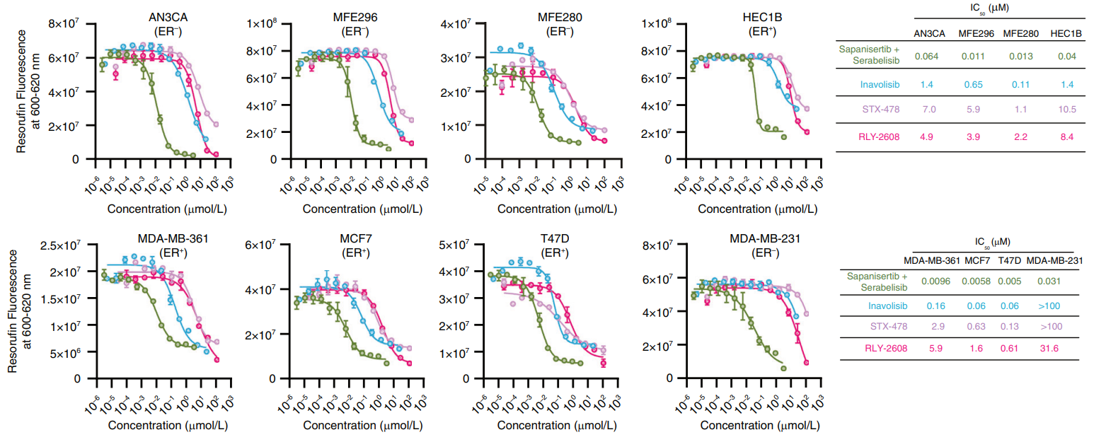
Multi-node inhibition targeting mTORC1, mTORC2 and PI3Kα potently inhibits the PI3K/AKT/mTOR pathway in endometrial and breast cancer models. [Abstract]2025 Aug;133(2):144-154. PMID: 40360883
Zovegalisib purchased from MedChemExpress. Usage Cited in: Br J Cancer. 2025 Aug;133(2):144-154. [Abstract]
Compared to mutant-specific PI3Kα inhibitors (RLY-2608 (Zovegalisib) & STX-478) and PI3Kα inhibitor/degrader inavolisib, the sapanisertib plus serabelisib combination had a substantially lower mean IC50 i.e., 32.0 nM versus 0.89 μM (inavolisib), 6.13 μM (STX-478), 4.85 μM (RLY-2608, Zovegalisib).


Solvant et solubilité
In Vitro:
DMSO : 100 mg/mL (164.23 mM; ultrasonic and warming and heat to 60°C; Hygroscopic DMSO has a significant impact on the solubility of product, please use newly opened DMSO)
Please refer to the solubility information to select the appropriate solvent. Once prepared, please aliquot and store the solution to prevent product inactivation from repeated freeze-thaw cycles.
Storage method and period of stock solution: -80°C, 6 months; -20°C, 1 month. When stored at -80°C, please use it within 6 months. When stored at -20°C, please use it within 1 month.
Please refer to the solubility information to select the appropriate solvent. Once prepared, please aliquot and store the solution to prevent product inactivation from repeated freeze-thaw cycles.
Storage method and period of stock solution: -80°C, 6 months; -20°C, 1 month. When stored at -80°C, please use it within 6 months. When stored at -20°C, please use it within 1 month.
Concentration (start) × Volume (start) = Concentration (final) × Volume (final)
In Vivo:
Select the appropriate dissolution method based on your experimental animal and administration route.
- For the following dissolution methods, please ensure to first prepare a clear stock solution using an In Vitro approach and then sequentially add co-solvents:
- To ensure reliable experimental results, the clarified stock solution can be appropriately stored based on storage conditions. As for the working solution for In Vivo experiments, it is recommended to prepare freshly and use it on the same day.
- The percentages shown for the solvents indicate their volumetric ratio in the final prepared solution. If precipitation or phase separation occurs during preparation, heat and/or sonication can be used to aid dissolution.
Add each solvent one by one: 10% DMSO 40% PEG300 5% Tween-80 45% Saline
Solubility: ≥ 5 mg/mL (8.21 mM); Clear solution
This protocol yields a clear solution of ≥ 5 mg/mL (saturation unknown).
Taking 1 mL working solution as an example, add 100 μL DMSO stock solution (50.0 mg/mL) to 400 μL PEG300, and mix evenly; then add 50 μL Tween-80 and mix evenly; then add 450 μL Saline to adjust the volume to 1 mL.
Preparation of Saline: Dissolve 0.9 g sodium chloride in ddH₂O and dilute to 100 mL to obtain a clear Saline solution.
In Vivo Dissolution Calculator
Please enter the basic information of animal experiments:
-
-
-
-
Recommended: Prepare an additional quantity of animals to account for potential losses during experiments.
Please enter your animal formula composition:
-
%DMSO +
Recommended: Keep the proportion of DMSO in working solution below 2% if your animal is weak.
-
%+
-
+%Tween-80 + +
-
%Saline +
The co-solvents required include: DMSO, . All of co-solvents are available by MedChemExpress (MCE). , Tween 80. All of co-solvents are available by MedChemExpress (MCE).
Working solution concentration: 0.22 mg/mL
Method for preparing stock solution: mg drug dissolved in μL DMSO. Stock solution concentration: mg/mL.
1. Take μL DMSO stock solution;
2. Add μL .
μL , mix evenly;
3. Then add μL Tween 80, mix evenly;
4. Then add μL
Please ensure that the stock solution in the first step is dissolved to a clear state, and add co-solvents in sequence. You can use ultrasonic heating (ultrasonic cleaner, recommended frequency 20-40 kHz), vortexing, etc. to assist dissolution.


Pureté et documentation
-
Fiche technique (283 KB)
-
SDS (254 KB)
- English - EN (254 KB)
- Français - FR (254 KB)
- Deutsch - DE (254 KB)
- Norwegian - NO (254 KB)
- Español - ES (254 KB)
- Swedish - SV (254 KB)
- Italian - IT (254 KB)
- Korean - KR (254 KB)
- Portuguese - PT (254 KB)
-
Instruction de manipulation (2659 KB)
Références


Complete Stock Solution Preparation Table
Please refer to the solubility information to select the appropriate solvent. Once prepared, please aliquot and store the solution to prevent product inactivation from repeated freeze-thaw cycles.
Storage method and period of stock solution: -80°C, 6 months; -20°C, 1 month. When stored at -80°C, please use it within 6 months. When stored at -20°C, please use it within 1 month.
| Optional Solvent | Concentration Solvent Mass | 1 mg | 5 mg | 10 mg | 25 mg |
|---|---|---|---|---|---|
| DMSO | 1 mM | 1.6423 mL | 8.2114 mL | 16.4228 mL | 41.0570 mL |
| 5 mM | 0.3285 mL | 1.6423 mL | 3.2846 mL | 8.2114 mL | |
| 10 mM | 0.1642 mL | 0.8211 mL | 1.6423 mL | 4.1057 mL | |
| 15 mM | 0.1095 mL | 0.5474 mL | 1.0949 mL | 2.7371 mL | |
| 20 mM | 0.0821 mL | 0.4106 mL | 0.8211 mL | 2.0528 mL | |
| 25 mM | 0.0657 mL | 0.3285 mL | 0.6569 mL | 1.6423 mL | |
| 30 mM | 0.0547 mL | 0.2737 mL | 0.5474 mL | 1.3686 mL | |
| 40 mM | 0.0411 mL | 0.2053 mL | 0.4106 mL | 1.0264 mL | |
| 50 mM | 0.0328 mL | 0.1642 mL | 0.3285 mL | 0.8211 mL | |
| 60 mM | 0.0274 mL | 0.1369 mL | 0.2737 mL | 0.6843 mL | |
| 80 mM | 0.0205 mL | 0.1026 mL | 0.2053 mL | 0.5132 mL | |
| 100 mM | 0.0164 mL | 0.0821 mL | 0.1642 mL | 0.4106 mL |


Zovegalisib Related Classifications
HY-153306 Related Classifications
 Powered by Bioz