



S-Acetylglutathione

Based on 1 Customer Validation
S-Acetylglutathione is a glutathione derivative and intracellular glutathione supplement that bypasses glutathione synthetase deficiency through intracellular thioesterase conversion, exhibits higher blood stability than glutathione, and selectively induces apoptosis in lymphoma and malignant cells by inducing oxidative stress (apoptosis). S-Acetylglutathione can be used in research on glutathione synthetase deficiency, herpes simplex virus type 1 infection, murine AIDS, lymphoma, and malignant tumors.
연구목적의 판매만을 진행합니다. 환자를 대상으로 한 판매는 하지 않습니다.
- Purity : 98.57%
- CAS No.: 3054-47-5
- 화학식: C12H19N3O7S
- 분자량:349.36
-
보관:Powder -20°C, 3 years , 4°C, 2 years ; In solvent -80°C, 6 months , -20°C, 1 month


To place orders, for customer services and technical support, please contact: MedChemExpress USA
Tel: 609-228-6898 E-mail: [email protected] [email protected]


-
 Biological Activity
-
Chemical Information
-
용액&용해도
- Protocol
- 순도&문서
- References
-
 Help & FAQs


-
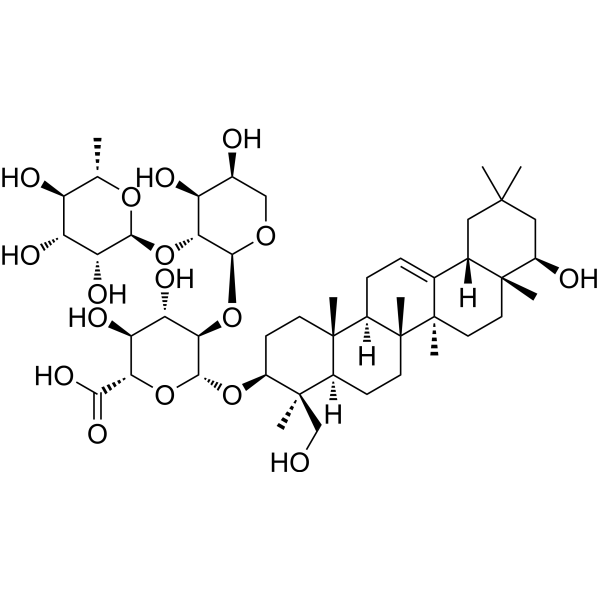
HY-122920
Soyasaponin IICMV Inhibitor -
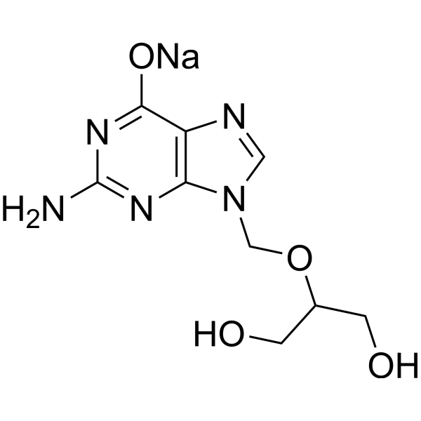
HY-13637A
Ganciclovir sodiumAntiviral Agent -
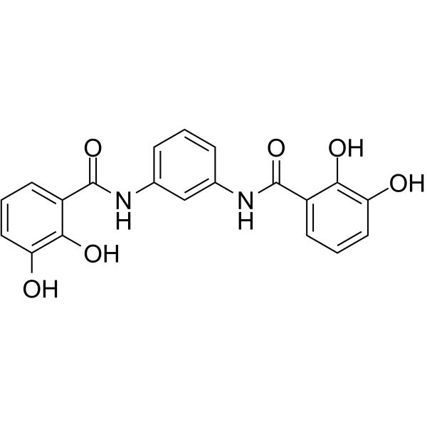
HY-120145
MST-312Telomerase Inhibitor -
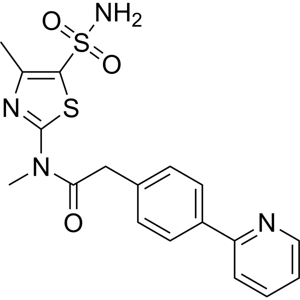
HY-15303
PritelivirHSV Inhibitor -

HY-W012311
3′-DeoxythymidineDNA Polymerase Inhibtior -
HY-145010
SN-011STING Inhibitor -
HY-136490
PsychosineCytotoxic Lipid -
HY-125118
GSK-A1PI4KIIIα Inhibitor -

HY-N11224
Pregnanetriol17α-Hydroxyprogesterone Metabolite -
HY-18944
FIT-039CDK9 Inhibitor -
HY-108137
Z-LVG-CHN2Cysteine Proteinase Inhibitor -
HY-N3848
ErmaniniNOS/COX-2 Inhibitor


Biological Activity
제품 설명
In Vitro
S-Acetylglutathione (50 μmol/L; 5 days) restores intracellular glutathione content to normal in cultured fibroblasts from patients with glutathione synthetase deficiency by recovering severely depleted GSH levels to the normal control range[1].
S-Acetylglutathione (5-10 mM; 120 h) does not affect the viability of confluent human foreskin fibroblasts at concentrations up to 10 mM, maintaining over 95% cell viability[2].
S-Acetylglutathione (10 mM; 24 h) is a potent GSH supplement in human foreskin fibroblasts, and at a concentration of 10 mM, it increases intracellular GSH levels by 7.4-fold in uninfected cells and 6.1-fold in HSV-1-infected cells[2].
S-acetyl-glutathione (5 mM; 24 h) selectively induces apoptosis in the human lymphoma cell lines Jurkat, Daudi, and Raji, but shows no such effect in Hut-78 cells or normal BT lymphocytes[4].
S-Acetylglutathione (2 mM; 5 days) induces extensive cell lysis in UKF-NB-3 neuroblastoma cells[5].
S-Acetylglutathione (0.001-20 mM; 14 days) induces cytotoxic effects in H69 small cell lung cancer cells[5].
S-acetyl-glutathione (0.2-5 mM; 24-96 h) induces apoptosis in Jurkat, Daudi, and Raji cell lines in a dose- and time-dependent manner, with significant effects starting at 1 mM, while showing no significant effect on Hut-78 or normal BT lymphocytes[4].
S-acetyl-glutathione (5 mM; 24 h for Jurkat and Raji, 48 h for Daudi) induces DNA fragmentation, confirming that apoptosis is the predominant mode of cell death in Jurkat, Daudi, and Raji cells[4].
S-Acetylglutathione (2 mM; 10 days) induces apoptosis in MOLT 4 cells, reducing the viable cell population from 91.8% to 52.7%[5].
S-acetyl-glutathione (5 mM; 1-24 h) induces a significant decrease in intracellular GSH in the lymphoma cell lines Jurkat, Daudi, and Raji, but has no such effect in the resistant Hut-78 or BTL cells, indicating that Sag-induced apoptosis is not mediated by an increase in GSH[4].
S-acetyl-glutathione (5 mM; 1-24 h) does not induce apoptosis in any of the cell lines tested (Jurkat, Daudi, Raji, Hut-78, BTL), although it increases intracellular GSH levels in some cell lines[4].
S-Acetylglutathione (1-2 mM; 14 days) induces apoptotic DNA fragmentation in H69 cells[5].
S-Acetylglutathione (1-2 mM; 14 days) induces apoptosis in MOLT 4 and UKF-NB-3 cells[5].
MedChemExpress (MCE) has not independently confirmed the accuracy of these methods. They are for reference only. Further protocols information, click here.
-
Cell Line:Human foreskin fibroblasts (HFF)
-
Concentration:5, 10 mM
-
Incubation Time:120 h
-
Result:Maintained 95.2% cell viability at 5 mM and 95.1% at 10 mM, compared to 88.6% viability in the untreated control.
-
Cell Line:Jurkat, Daudi, Raji, Hut-78, BT lymphocytes
-
Concentration:5 mM
-
Incubation Time:24 h
-
Result:Induced a strong increase in the whole Annexin-V positive population in Jurkat, Daudi, and Raji cells.
Did not affect Hut-78 cells or normal BT lymphocytes.
-
Cell Line:Jurkat, Daudi, Raji, Hut-78, BT lymphocytes
-
Concentration:0.2, 0.5, 1, 2, 5 mM (Dose-response); 5 mM (Time-course)
-
Incubation Time:24 h (Dose-response); 24, 48, 96 h (Time-course)
-
Result:Induced significant cell death at 1 mM for Daudi and Raji cells and at 2 mM for Jurkat cells.
Induced 100% apoptosis of Daudi and Raji cells at 2-5 mM.
Showed a significant but not maximal induction of cell death after 24 h, with a plateau reached after 48 h for sensitive cell lines.
Did not affect Hut-78 cells even at the highest concentration or after 96 h.
Caused a weak increase in the total apoptotic population in normal BT lymphocytes after 48 h.
-
Cell Line:Jurkat, Daudi, Raji
-
Concentration:5 mM
-
Incubation Time:24 h (Jurkat, Raji); 48 h (Daudi)
-
Result:Induced DNA fragmentation, confirming apoptosis in Jurkat, Daudi, and Raji cells.
-
Cell Line:Jurkat, Daudi, Raji, Hut-78, BT lymphocytes
-
Concentration:5 mM
-
Incubation Time:0-24 h
-
Result:Did not induce apoptosis in any of the tested cell lines (Jurkat, Daudi, Raji, Hut-78, BTL).
-
Cell Line:MOLT 4
-
Concentration:2 mM
-
Incubation Time:10 days
-
Result:Reduced the viable cell population from 91.8% to 52.7%.
Increased the early apoptotic population to 28.5% and the late apoptotic or dead cell population to 19%.
-
Cell Line:UKF-NB-3
-
Concentration:2 mM
-
Incubation Time:5 days
-
Result:Induced extensive cellular lysis over the 5-day period.
-
Cell Line:H69
-
Concentration:1, 2 mM
-
Incubation Time:14 days
-
Result:Induced cytotoxic effects.
-
Cell Line:H69
-
Concentration:1, 2 mM
-
Incubation Time:14 days
-
Result:Induced apoptotic DNA fragmentation, as evidenced by a DNA-laddering pattern with fragments of 340-1130 bp.
-
Cell Line:MOLT 4, UKF-NB-3
-
Concentration:1, 2 mM
-
Incubation Time:14 days
-
Result:Induced apoptosis as confirmed by DNA-laddering.
In Vivo
S-Acetylglutathione (14-143 μmol/mouse; intramuscular injection; five consecutive days per week; 10 weeks) shows antiviral efficacy in a mouse AIDS model by inhibiting viral DNA content in target organs, with a 62-77% reduction in the spleen, without affecting organ weight, immunoglobulin levels, or lymphocyte proliferation[3].
MedChemExpress (MCE) has not independently confirmed the accuracy of these methods. They are for reference only.
-
Animal Model:hr/hr (outbreed hairless mice, 11 weeks old, weight about 20 g; induced by intraperitoneal inoculation of a lethal dose of HSV-1 strain KM, 1×10⁴ PFU in 500 μl MEM)[2]
-
Dosage:3.125, 6.25, 12.5, 25 μg/g body weight per day
-
Administration:i.m.; once per day; five consecutive days for 2 weeks
-
Result:Delayed virus-induced animal mortality with a mean day of death of 18.5 days at 6.25 μg/g body weight per day, compared to 8.2 days for the virus control group.
Achieved 50% survival (7 out of 14 mice) on day 26 post-infection at 6.25 μg/g body weight per day.
Resulted in a mean day of death of 9.8 days with 4/14 survivors at 12.5 μg/g body weight per day.
Resulted in a mean day of death of 10.9 days with 2/14 survivors at 25 μg/g body weight per day.
Resulted in a mean day of death of 8.1 days with 0/14 survivors at 3.125 μg/g body weight per day.
-
Animal Model:C57BL/6 mice (Female, 4 weeks old, induced by intraperitoneal inoculation with the LP-BM5 viral complex)[3]
-
Dosage:14 μmol/mouse (0.23 g/kg); 72 μmol/mouse (1.15 g/kg); 143 μmol/mouse (2.3 g/kg)
-
Administration:i.m.; five consecutive days weekly; 10 weeks
-
Result:Reduced spleen BM5d DNA content by 62-77% at the 14 and 143 μmol/mouse doses.
Reduced lymph node BM5d DNA content by 35-65% at the 72 μmol/mouse dose.
Did not significantly reduce splenomegaly or lymphadenopathy (p = 0.096 and p = 0.073, respectively).
Did not influence serum IgG levels.
Did not restore the proliferative index of T or B lymphocytes.


Chemical Information
-
CAS No. 3054-47-5
-
Appearance Solid
-
분자량 349.36
-
화학식 C12H19N3O7S
-
Color White to light yellow
-
SMILES
O=C(O)CNC([C@H](CSC(C)=O)NC(CC[C@@H](C(O)=O)N)=O)=O
-
선적
Room temperature in continental US; may vary elsewhere.
-
보관
Powder -20°C 3 years 4°C 2 years In solvent -80°C 6 months -20°C 1 month


용액&용해도
In Vitro:
DMSO : 43.75 mg/mL (125.23 mM; Need ultrasonic; Hygroscopic DMSO has a significant impact on the solubility of product, please use newly opened DMSO)
Please refer to the solubility information to select the appropriate solvent. Once prepared, please aliquot and store the solution to prevent product inactivation from repeated freeze-thaw cycles.
Storage method and period of stock solution: -80°C, 6 months; -20°C, 1 month. When stored at -80°C, please use it within 6 months. When stored at -20°C, please use it within 1 month.
Please refer to the solubility information to select the appropriate solvent. Once prepared, please aliquot and store the solution to prevent product inactivation from repeated freeze-thaw cycles.
Storage method and period of stock solution: -80°C, 6 months; -20°C, 1 month. When stored at -80°C, please use it within 6 months. When stored at -20°C, please use it within 1 month.
Concentration (start) × Volume (start) = Concentration (final) × Volume (final)
In Vivo:
Select the appropriate dissolution method based on your experimental animal and administration route.
- For the following dissolution methods, please ensure to first prepare a clear stock solution using an In Vitro approach and then sequentially add co-solvents:
- To ensure reliable experimental results, the clarified stock solution can be appropriately stored based on storage conditions. As for the working solution for In Vivo experiments, it is recommended to prepare freshly and use it on the same day.
- The percentages shown for the solvents indicate their volumetric ratio in the final prepared solution. If precipitation or phase separation occurs during preparation, heat and/or sonication can be used to aid dissolution.
Add each solvent one by one: 10% DMSO 40% PEG300 5% Tween-80 45% Saline
Solubility: ≥ 5 mg/mL (14.31 mM); Clear solution
This protocol yields a clear solution of ≥ 5 mg/mL (saturation unknown).
Taking 1 mL working solution as an example, add 100 μL DMSO stock solution (50.0 mg/mL) to 400 μL PEG300, and mix evenly; then add 50 μL Tween-80 and mix evenly; then add 450 μL Saline to adjust the volume to 1 mL.
Preparation of Saline: Dissolve 0.9 g sodium chloride in ddH₂O and dilute to 100 mL to obtain a clear Saline solution.
Add each solvent one by one: 10% DMSO 90% (20% SBE-β-CD in Saline)
Solubility: ≥ 5 mg/mL (14.31 mM); Clear solution
This protocol yields a clear solution of ≥ 5 mg/mL (saturation unknown).
Taking 1 mL working solution as an example, add 100 μL DMSO stock solution (50.0 mg/mL) to 900 μL 20% SBE-β-CD in Saline, and mix evenly.
Preparation of 20% SBE-β-CD in Saline (4°C, storage for one week): 2 g SBE-β-CD powder is dissolved in 10 mL Saline, completely dissolve until clear.
In Vivo Dissolution Calculator
Please enter the basic information of animal experiments:
-
-
-
-
Recommended: Prepare an additional quantity of animals to account for potential losses during experiments.
Please enter your animal formula composition:
-
%DMSO +
Recommended: Keep the proportion of DMSO in working solution below 2% if your animal is weak.
-
%+
-
+%Tween-80 + +
-
%Saline +
The co-solvents required include: DMSO, . All of co-solvents are available by MedChemExpress (MCE). , Tween 80. All of co-solvents are available by MedChemExpress (MCE).
Working solution concentration: 0.22 mg/mL
Method for preparing stock solution: mg drug dissolved in μL DMSO. Stock solution concentration: mg/mL.
1. Take μL DMSO stock solution;
2. Add μL .
μL , mix evenly;
3. Then add μL Tween 80, mix evenly;
4. Then add μL
Please ensure that the stock solution in the first step is dissolved to a clear state, and add co-solvents in sequence. You can use ultrasonic heating (ultrasonic cleaner, recommended frequency 20-40 kHz), vortexing, etc. to assist dissolution.

Protocol
-
Research Protocol for Infectious Diseases
Infectious-disease experiments test how pathogens interact with host barriers, innate immune receptors, inflammatory signaling, pathogen replication, and tissue injury; pattern-recognition receptors such as TLRs, RIG-I-like receptors, NOD-like receptors, and inflammasomes detect microbial molecules and activate NF-κB, interferon, and cytokine responses. The central hypothesis is that infection severity reflects the balance between pathogen burden and host response: protective inflammation restricts pathogen growth, whereas excessive or mislocalized inflammation contributes to tissue damage and disease phenotype. Unresolved questions include which host pathways are protective versus pathogenic, why some infection models fail to translate to human disease, and which combined readouts best predict clinically relevant infection outcomes.
-
Apoptosis
Apoptosis, also called programmed cell death, is generally characterized by distinct morphological characteristics.
-
TUNEL staining for apoptotic DNA fragmentation
TUNEL staining detects DNA strand breaks by using terminal deoxynucleotidyl transferase to add labeled nucleotides to exposed 3′-OH DNA termini, generating either microscopic staining in fixed cells or tissue sections, or fluorescence/cytometric signal in cell suspensions. TUNEL positivity reflects DNA fragmentation but should not be interpreted alone as definitive apoptosis, because TUNEL can also label necrotic, autolytic, mechanically damaged, or DNA-repair-associated DNA breaks.
-
Annexin V plus membrane-impermeant dye apoptosis staining
Annexin V-based apoptosis assays rely on the detection of phosphatidylserine (PS) externalization from the inner leaflet of the plasma membrane to the outer leaflet, an early biochemical hallmark of apoptosis. Fluorescently labeled Annexin V binds PS in a calcium-dependent manner, enabling identification of early apoptotic cells by flow cytometry or fluorescence microscopy. When combined with a membrane-impermeant DNA-binding dye (e. g. , propidium iodide), this approach allows discrimination between viable (Annexin V−/dye−), early apoptotic (Annexin V+/dye−), and late apoptotic or necrotic (Annexin V+/dye+) cell populations by assessing membrane integrity and PS exposure.
-
ROS/oxidative-stress fluorescent staining
ROS/oxidative-stress fluorescent staining uses cell-permeant fluorogenic probes that become fluorescent after oxidation inside cells or tissues; commonly used examples include DCFH-DA/DCFDA for broad cellular oxidant detection, DHE for superoxide-related signal detection, MitoSOX for mitochondrial superoxide-related signal detection, and CellROX probes for oxidative-stress-associated fluorescence readouts. The assay detects probe oxidation rather than a single ROS species unless the probe and analysis method have been chemically validated for that species. DCFH-DA enters cells, is deacetylated by intracellular esterases to DCFH, and produces fluorescent DCF after oxidation, so the readout is used as an operational measure of total cellular oxidative stress rather than a species-specific ROS measurement. DHE and MitoSOX can report superoxide-related oxidation, but red fluorescence alone can include non-specific ethidium-like oxidation products; HPLC or optimized spectral approaches are
-
Apoptosis Solutions
Apoptosis is a regulated, generally non-lytic cell-death pathway that removes unwanted, damaged, infected, or abnormal cells through coordinated morphological changes, caspase activation, DNA fragmentation, and membrane remodeling. The intrinsic apoptosis pathway is controlled mainly by mitochondrial outer membrane permeabilization, BCL-2 family proteins, cytochrome c release, apoptosome formation, caspase-9 activation, and downstream executioner caspase-3/7 activation. The extrinsic apoptosis pathway is initiated by death receptors such as Fas, TNFR, and TRAIL receptors, which recruit adaptor proteins and activate caspase-8 before engaging executioner caspases or mitochondrial amplification through BID cleavage. Apoptosis is linked to many phenotypes, including cancer cell killing, tissue homeostasis, immune regulation, neurodegeneration, infection response, and treatment-induced cytotoxicity; unresolved questions include how apoptosis interacts with necroptosis, pyroptosis, ferroptos


순도&문서
-
Data Sheet (294 KB)
-
SDS (393 KB)
- English - EN (393 KB)
- Français - FR (393 KB)
- Deutsch - DE (393 KB)
- Norwegian - NO (393 KB)
- Español - ES (393 KB)
- Swedish - SV (393 KB)
- Italian - IT (393 KB)
- Korean - KR (393 KB)
- Portuguese - PT (393 KB)
-
Handling Instructions (2659 KB)
References
[1]. Okun JG, et al. S-Acetylglutathione normalizes intracellular glutathione content in cultured fibroblasts from patients with glutathione synthetase deficiency. Journal of inherited metabolic disease. 2004;27(6):783-6. [Content Brief]
[2]. Vogel JU, et al. Effects of S-acetylglutathione in cell and animal model of herpes simplex virus type 1 infection. Medical microbiology and immunology. 2005 Jan;194(1-2):55-9. [Content Brief]
[3]. Fraternale A, et al. Inhibition of murine AIDS by pro-glutathione (GSH) molecules. Antiviral research. 2008 Feb;77(2):120-7. [Content Brief]
[4]. Locigno R, et al. S-acetyl-glutathione selectively induces apoptosis in human lymphoma cells through a GSH-independent mechanism. International journal of oncology. 2002 Jan;20(1):69-75. [Content Brief]
[5]. Donnerstag B, et al. Reduced glutathione and S-acetylglutathione as selective apoptosis-inducing agents in cancer therapy. Cancer letters. 1996 Dec 20;110(1-2):63-70. [Content Brief]


Complete Stock Solution Preparation Table
Please refer to the solubility information to select the appropriate solvent. Once prepared, please aliquot and store the solution to prevent product inactivation from repeated freeze-thaw cycles.
Storage method and period of stock solution: -80°C, 6 months; -20°C, 1 month. When stored at -80°C, please use it within 6 months. When stored at -20°C, please use it within 1 month.
| Optional Solvent | Concentration Solvent Mass | 1 mg | 5 mg | 10 mg | 25 mg |
|---|---|---|---|---|---|
| DMSO | 1 mM | 2.8624 mL | 14.3119 mL | 28.6238 mL | 71.5594 mL |
| 5 mM | 0.5725 mL | 2.8624 mL | 5.7248 mL | 14.3119 mL | |
| 10 mM | 0.2862 mL | 1.4312 mL | 2.8624 mL | 7.1559 mL | |
| 15 mM | 0.1908 mL | 0.9541 mL | 1.9083 mL | 4.7706 mL | |
| 20 mM | 0.1431 mL | 0.7156 mL | 1.4312 mL | 3.5780 mL | |
| 25 mM | 0.1145 mL | 0.5725 mL | 1.1450 mL | 2.8624 mL | |
| 30 mM | 0.0954 mL | 0.4771 mL | 0.9541 mL | 2.3853 mL | |
| 40 mM | 0.0716 mL | 0.3578 mL | 0.7156 mL | 1.7890 mL | |
| 50 mM | 0.0572 mL | 0.2862 mL | 0.5725 mL | 1.4312 mL | |
| 60 mM | 0.0477 mL | 0.2385 mL | 0.4771 mL | 1.1927 mL | |
| 80 mM | 0.0358 mL | 0.1789 mL | 0.3578 mL | 0.8945 mL | |
| 100 mM | 0.0286 mL | 0.1431 mL | 0.2862 mL | 0.7156 mL |


S-Acetylglutathione Related Classifications
HY-131944 Related Classifications
 Powered by Bioz