



Galeterone
Based on 3 publication(s) in Google Scholar
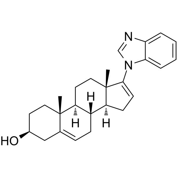
Galeterone (TOK-001) is a potent, orally active molecular glue degrader, which degrades androgen receptor (AR) and its splice variants (AR-Vs) and MAP kinase-interacting serine/threonine protein kinase Mnk1/2. Galeterone also functions as a CYP17 inhibitor (IC50 = 47 nM). Galeterone induces cell apoptosis. Galeterone inhibits tumor growth in human prostate cancer xenograft mouse models. Galeterone can be used for castration resistant prostate cancer (CRPC) and pancreatic ductal adenocarcinoma (PDAC) research[1][2].
For research use only. We do not sell to patients.
- Purity : 99.87%
- CAS No.: 851983-85-2
- Formula: C26H32N2O
- Molecular Weight:388.55
-
Storage:Powder -20°C, 3 years , 4°C, 2 years ; In solvent -80°C, 2 years , -20°C, 1 year

To place orders, for customer services and technical support, please contact: MedChemExpress USA
Tel: 609-228-6898 E-mail: [email protected] [email protected]


-
 Biological Activity
-
Chemical Information
-
Solvent & Solubility
- Protocols
- Purity & Documentation
- References
-
 Help & FAQs


-
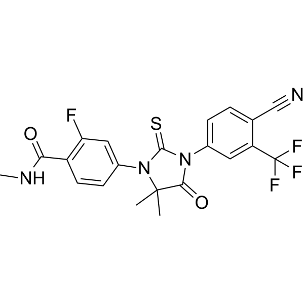
HY-70002
EnzalutamideAndrogen Receptor Antagonist -
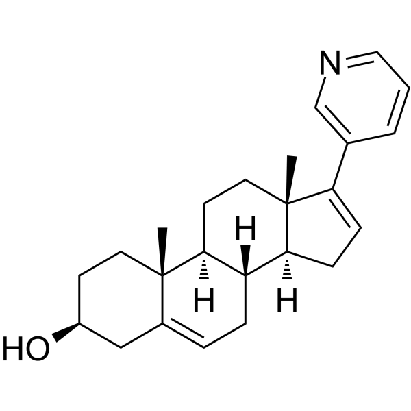
HY-70013
AbirateroneCYP17A1 Inhibitor -
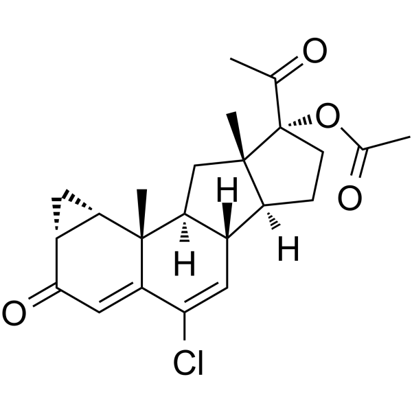
HY-13604
Cyproterone acetateAndrogen Receptor Antagonist -
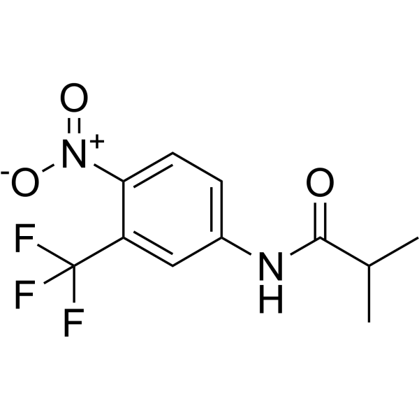
HY-B0022
FlutamideAntiandrogen Agent -
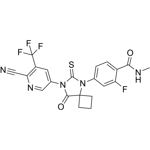
HY-16060
ApalutamideAR Antagonist -
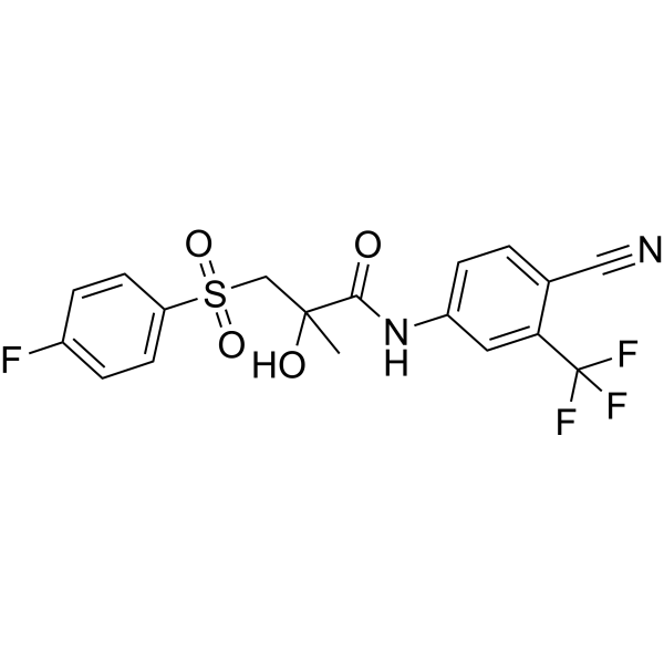
HY-14249
BicalutamideAndrogen Receptor Inhibitor -
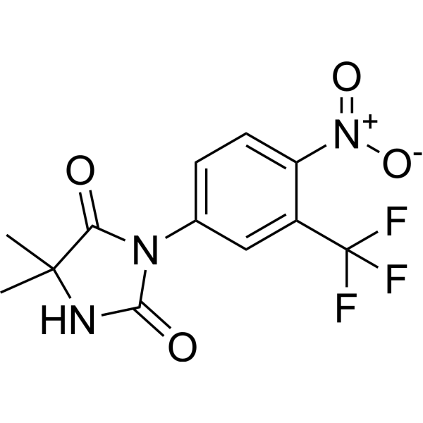
HY-13702
NilutamideAndrogen Receptor Antagonist -
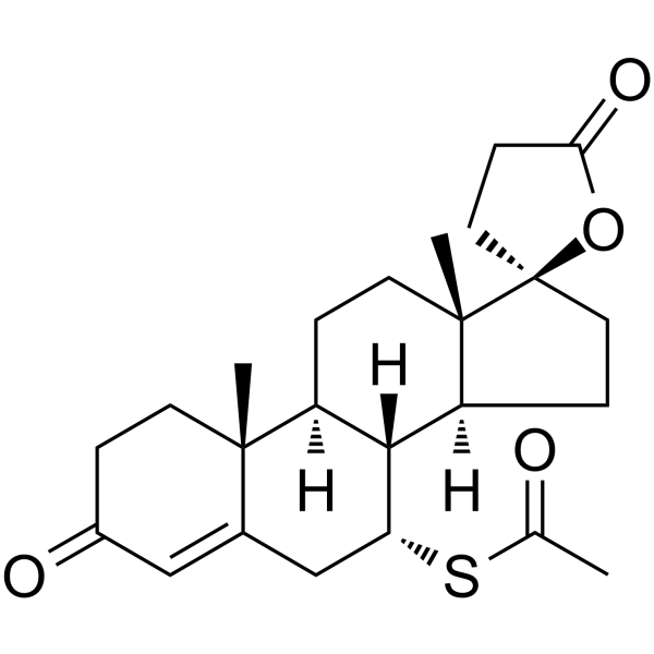
HY-B0561
SpironolactoneAldosterone/Corticoid Receptor/Androgen Antagonists -

HY-13604R
Cyproterone acetate (Standard)Androgen Receptor Antagonist -

HY-70002R
Enzalutamide (Standard)Androgen Receptor Antagonist -
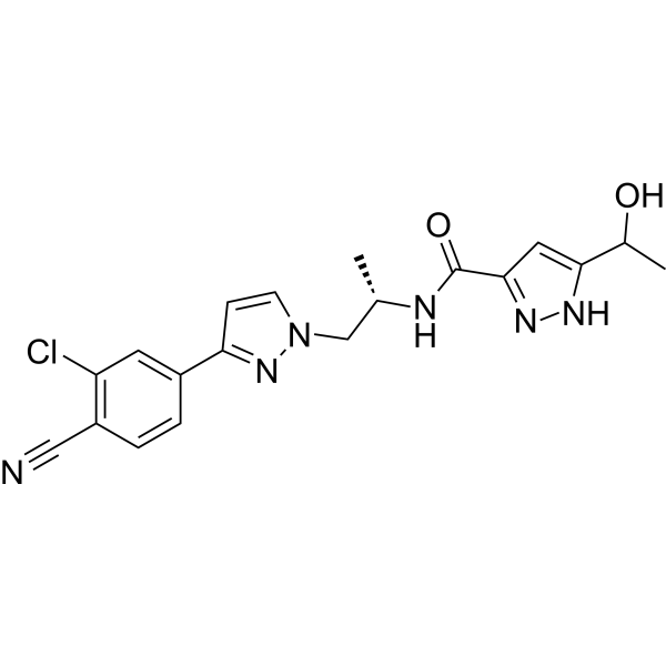
HY-16985
DarolutamideAndrogen Receptor Antagonist -
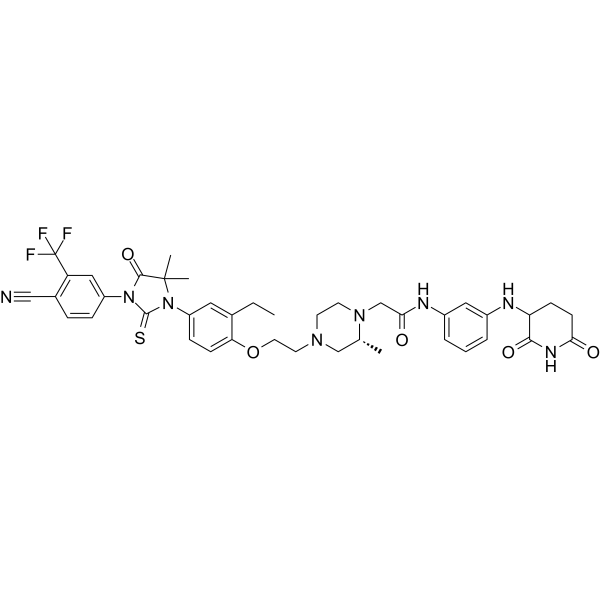
HY-158101
BMS-986365PROTAC AR Degrader
Publications Citing Use of MedChemExpress (MCE) Galeterone
More

Biological Activity
Description
IC50 & Target
|
CYP17 47 nM (IC50) |
Cellular Effect
|
Cell Line

|
Type | Value | Description | References |
|---|---|---|---|---|
| A549 | GI50 |
3.9 μM
Compound: Galeterone
|
Antiproliferative activity against human A549 cells after 48 hrs by SRB assay
Antiproliferative activity against human A549 cells after 48 hrs by SRB assay
|
[PMID: 29172080] |
| Cancer cell lines | IC50 |
2.9 μM
Compound: 33, VN/124-1, galeterone, TOK-001
|
Growth inhibition of human castration-resistant prostate cancer cells
Growth inhibition of human castration-resistant prostate cancer cells
|
[PMID: 25591066] |
| CWR22R | GI50 |
37.8 μM
Compound: Galeterone
|
Antiproliferative activity against AR-positive human 22Rv1 cells harboring ARE14 construct assessed as growth inhibition after 72 hrs by resazurin dye based fluorescence assay
Antiproliferative activity against AR-positive human 22Rv1 cells harboring ARE14 construct assessed as growth inhibition after 72 hrs by resazurin dye based fluorescence assay
|
[PMID: 31271960] |
| CWR22R | IC50 |
11.6 μM
Compound: Galeterone
|
Antiproliferative activity against AR/AR-V7 positive human 22Rv1 cells assessed as inhibition of cell growth incubated for 48 hrs by CCK-8 assay
Antiproliferative activity against AR/AR-V7 positive human 22Rv1 cells assessed as inhibition of cell growth incubated for 48 hrs by CCK-8 assay
|
[PMID: 38512060] |
| CWR22R | IC50 |
8.91 μM
Compound: 5; Galeterone
|
Antiproliferative activity against human CWR22Rv1 cells after 7 days by MTT assay
Antiproliferative activity against human CWR22Rv1 cells after 7 days by MTT assay
|
[PMID: 27437082] |
| HBL-100 | GI50 |
10 μM
Compound: Galeterone
|
Antiproliferative activity against human HBL100 cells after 48 hrs by SRB assay
Antiproliferative activity against human HBL100 cells after 48 hrs by SRB assay
|
[PMID: 29172080] |
| HeLa | GI50 |
5.3 μM
Compound: Galeterone
|
Antiproliferative activity against human HeLa cells after 48 hrs by SRB assay
Antiproliferative activity against human HeLa cells after 48 hrs by SRB assay
|
[PMID: 29172080] |
| hTERT-BJ | GI50 |
5 μM
Compound: Galeterone
|
Cytotoxicity against human hTERT-BJ cells assessed as cell growth inhibition after 48 hrs SRB assay
Cytotoxicity against human hTERT-BJ cells assessed as cell growth inhibition after 48 hrs SRB assay
|
[PMID: 29172080] |
| L02 | IC50 |
>100 μM
Compound: Galeterone
|
Antiproliferative activity against human L02 cells assessed as inhibition of cell growth incubated for 48 hrs by CCK-8 assay
Antiproliferative activity against human L02 cells assessed as inhibition of cell growth incubated for 48 hrs by CCK-8 assay
|
[PMID: 38512060] |
| LNCaP | EC50 |
670 nM
Compound: 5, TOK-001, Galeterone
|
Displacement of [3H]R1881 from AR in human LNCaP cells after 2 hrs by scintillation counting analysis
Displacement of [3H]R1881 from AR in human LNCaP cells after 2 hrs by scintillation counting analysis
|
[PMID: 23713567] |
| LNCaP | EC50 |
845 nM
Compound: 33, VN/124-1, galeterone, TOK-001
|
Displacement of [3H]R1881 from androgen receptor in human LNCAP cells
Displacement of [3H]R1881 from androgen receptor in human LNCAP cells
|
[PMID: 25591066] |
| LNCaP | GI50 |
3.35 μM
Compound: 5, TOK-001, Galeterone
|
Cytotoxicity against human LNCAP cells after 7 days by MTT assay
Cytotoxicity against human LNCAP cells after 7 days by MTT assay
|
[PMID: 23713567] |
| LNCaP | IC50 |
2 μM
Compound: Galeterone
|
Antiproliferative activity against human LNCaP cells assessed as inhibition of cell growth incubated for 48 hrs by CCK-8 assay
Antiproliferative activity against human LNCaP cells assessed as inhibition of cell growth incubated for 48 hrs by CCK-8 assay
|
[PMID: 38512060] |
| LNCaP | IC50 |
3.93 μM
Compound: 5; Galeterone
|
Antiproliferative activity against human LNCAP cells after 7 days by MTT assay
Antiproliferative activity against human LNCAP cells after 7 days by MTT assay
|
[PMID: 27437082] |
| LNCaP | IC50 |
3548 nM
Compound: 6; TOK-001, VN/124-1
|
Antiproliferative activity against human LNCAP cells assessed as reduction in cell viability measured after 6 days by CellTiter-Glo assay
Antiproliferative activity against human LNCAP cells assessed as reduction in cell viability measured after 6 days by CellTiter-Glo assay
|
[PMID: 36070471] |
| LNCaP C4-2 | GI50 |
>40 μM
Compound: Galeterone
|
Antiproliferative activity against AR-positive human C4-2 cells assessed as growth inhibition after 72 hrs by resazurin dye based fluorescence assay
Antiproliferative activity against AR-positive human C4-2 cells assessed as growth inhibition after 72 hrs by resazurin dye based fluorescence assay
|
[PMID: 31271960] |
| PC-3 | EC50 |
405 nM
Compound: 33, VN/124-1, galeterone, TOK-001
|
Displacement of [3H]R1881 from androgen receptor in human PC3 cells
Displacement of [3H]R1881 from androgen receptor in human PC3 cells
|
[PMID: 25591066] |
| PC-3 | GI50 |
37.7 μM
Compound: Galeterone
|
Antiproliferative activity against AR-negative human PC3 cells assessed as growth inhibition after 72 hrs by resazurin dye based fluorescence assay
Antiproliferative activity against AR-negative human PC3 cells assessed as growth inhibition after 72 hrs by resazurin dye based fluorescence assay
|
[PMID: 31271960] |
| SW1573 | GI50 |
3.9 μM
Compound: Galeterone
|
Antiproliferative activity against human SW1573 cells after 48 hrs by SRB assay
Antiproliferative activity against human SW1573 cells after 48 hrs by SRB assay
|
[PMID: 29172080] |
| T47D | GI50 |
2.1 μM
Compound: Galeterone
|
Antiproliferative activity against human T47D cells after 48 hrs by SRB assay
Antiproliferative activity against human T47D cells after 48 hrs by SRB assay
|
[PMID: 29172080] |
| VCaP | GI50 |
>40 μM
Compound: Galeterone
|
Antiproliferative activity against AR-positive human VCaP cells assessed as growth inhibition after 72 hrs by resazurin dye based fluorescence assay
Antiproliferative activity against AR-positive human VCaP cells assessed as growth inhibition after 72 hrs by resazurin dye based fluorescence assay
|
[PMID: 31271960] |
| WiDr | GI50 |
2.7 μM
Compound: Galeterone
|
Antiproliferative activity against human WiDr cells after 48 hrs by SRB assay
Antiproliferative activity against human WiDr cells after 48 hrs by SRB assay
|
[PMID: 29172080] |
In Vitro
Galeterone (compound 1) possess potent antiproliferative activities with GI50 of 2.43 μM, 2.20 μM, 3.80 μM against LNCaP (androgen-sensitive), C42B (androgen-insensitive) and CWR22Rv1 (castration-resistant), respectively[1].
Galeterone (5-20 μM; 24 h) can degrade AR/AR-V7 and Mnk1/2 with consequent inhibition of AR signaling and depletion of peIF4E, respectively, and modulation of the downstream molecular targets in human CWR22Rv1 prostate cancer cell[1].
Galeterone (compound 5) (1-15 μM, 24 h) causes AR down-regulation in LNCaP cells, with nearly complete (>95%) AR down-regulation at 15 μM[2].
MedChemExpress (MCE) has not independently confirmed the accuracy of these methods. They are for reference only.
-
Cell Line:CWR22Rv1 prostate cancer cells
-
Concentration:5, 10 and 20 μM
-
Incubation Time:24 h
-
Result:Significantly and dose-dependently reduced the expressions of Mnk1, and peIF4E.
Caused significant depletion of the downstream target, cyclin D1, and induction of apoptosis via significant downregulation of antiapoptotic Bcl-2 and upregulation of proapoptotic Bax.
-
Cell Line:LNCaP cells
-
Concentration:1, 5, 10 and 15 μM
-
Incubation Time:24 h
-
Result:Almost completed AR down-regulation at 15 μM.
In Vivo
Galeterone (compound 5) (0.15 mmol/kg; s.c.; b.i.d. for 31 days) shows antitumor activity in a LAPC-4 xenograft mouse model[2].
MedChemExpress (MCE) has not independently confirmed the accuracy of these methods. They are for reference only.
-
Animal Model:Male NRG mice (5-6 weeks) bearing CWR22Rv1 tumors[1]
-
Dosage:100 mg/kg
-
Administration:p.o.; b.i.d.; 5 days per/week; for 16 days
-
Result:Exhibited statistically significant robust inhibition of CWR22Rv1 tumor growth at the tested doses.
Inhibited tumor growth with a TGI of 64%.
-
Animal Model:Male SCID mice (4-6 weeks) subcutaneously injected with LAPC-4 tumors[2]
-
Dosage:0.15 mmol/kg
-
Administration:s.c.; b.i.d. for 31 days
-
Result:Significantly reduced the growth rate of tumor growth compared to control.
- Detection of Apoptosis
- TUNEL staining for apoptotic DNA fragmentation
- Annexin V plus membrane-impermeant dye apoptosis staining
- Research Protocol for Endocrine Diseases
- Patient-Derived Xenograft (PDX)
- Apoptosis Solutions
- Patient-Derived Orthotopic Xenograft (PDOX)
- Splenic/Portal-Vein Liver Metastasis Xenograft
- Subcutaneous Cell-Line-Derived Xenograft
- Orthotopic Cell-Line Xenograft
- Intraperitoneal/Peritoneal Dissemination Xenograft
Clinical Trial
| NCT Number | Sponsor | Condition | Start Date |
Phase
|
|---|---|---|---|---|
| NCT01329991 | Plexxikon| | 2011-05 | PHASE1 |


Chemical Information
-
CAS No. 851983-85-2
-
Appearance Solid
-
Molecular Weight 388.55
-
Formula C26H32N2O
-
Color White to off-white
-
SMILES
C[C@@]12C(N3C=NC4=CC=CC=C34)=CC[C@]1([C@@]5(CC=C6[C@@](C)([C@]5(CC2)[H])CC[C@@H](C6)O)[H])[H]
-
Synonyms
TOK-001; VN-124-1
-
Shipping
Room temperature in continental US; may vary elsewhere.
-
Storage
Powder -20°C 3 years 4°C 2 years In solvent -80°C 2 years -20°C 1 year


Publications (3)
-
Journal Impact Factor
-
Most Recent
-
Nature
2012 Jan 22;482(7383):116-9. PMID: 22266943 -
Drug Metab Dispos
Structural and Functional Evaluation of Clinically Relevant Inhibitors of Steroidogenic Cytochrome P450 17A1. [Abstract]2017 Jun;45(6):635-645. PMID: 28373265 -


Solvent & Solubility
In Vitro:
DMSO : 25 mg/mL (64.34 mM; Need ultrasonic; Hygroscopic DMSO has a significant impact on the solubility of product, please use newly opened DMSO)
Please refer to the solubility information to select the appropriate solvent. Once prepared, please aliquot and store the solution to prevent product inactivation from repeated freeze-thaw cycles.
Storage method and period of stock solution: -80°C, 2 years; -20°C, 1 year. When stored at -80°C, please use it within 2 years. When stored at -20°C, please use it within 1 year.
Please refer to the solubility information to select the appropriate solvent. Once prepared, please aliquot and store the solution to prevent product inactivation from repeated freeze-thaw cycles.
Storage method and period of stock solution: -80°C, 2 years; -20°C, 1 year. When stored at -80°C, please use it within 2 years. When stored at -20°C, please use it within 1 year.
Concentration (start) × Volume (start) = Concentration (final) × Volume (final)
In Vivo:
Select the appropriate dissolution method based on your experimental animal and administration route.
- For the following dissolution methods, please ensure to first prepare a clear stock solution using an In Vitro approach and then sequentially add co-solvents:
- To ensure reliable experimental results, the clarified stock solution can be appropriately stored based on storage conditions. As for the working solution for In Vivo experiments, it is recommended to prepare freshly and use it on the same day.
- The percentages shown for the solvents indicate their volumetric ratio in the final prepared solution. If precipitation or phase separation occurs during preparation, heat and/or sonication can be used to aid dissolution.
Add each solvent one by one: 10% DMSO 40% PEG300 5% Tween-80 45% Saline
Solubility: ≥ 2.5 mg/mL (6.43 mM); Clear solution
This protocol yields a clear solution of ≥ 2.5 mg/mL (saturation unknown).
Taking 1 mL working solution as an example, add 100 μL DMSO stock solution (25.0 mg/mL) to 400 μL PEG300, and mix evenly; then add 50 μL Tween-80 and mix evenly; then add 450 μL Saline to adjust the volume to 1 mL.
Preparation of Saline: Dissolve 0.9 g sodium chloride in ddH₂O and dilute to 100 mL to obtain a clear Saline solution.
Add each solvent one by one: 10% DMSO 90% (20% SBE-β-CD in Saline)
Solubility: ≥ 2.5 mg/mL (6.43 mM); Clear solution
This protocol yields a clear solution of ≥ 2.5 mg/mL (saturation unknown).
Taking 1 mL working solution as an example, add 100 μL DMSO stock solution (25.0 mg/mL) to 900 μL 20% SBE-β-CD in Saline, and mix evenly.
Preparation of 20% SBE-β-CD in Saline (4°C, storage for one week): 2 g SBE-β-CD powder is dissolved in 10 mL Saline, completely dissolve until clear.
In Vivo Dissolution Calculator
Please enter the basic information of animal experiments:
-
-
-
-
Recommended: Prepare an additional quantity of animals to account for potential losses during experiments.
Please enter your animal formula composition:
-
%DMSO +
Recommended: Keep the proportion of DMSO in working solution below 2% if your animal is weak.
-
%+
-
+%Tween-80 + +
-
%Saline +
The co-solvents required include: DMSO, . All of co-solvents are available by MedChemExpress (MCE). , Tween 80. All of co-solvents are available by MedChemExpress (MCE).
Working solution concentration: 0.22 mg/mL
Method for preparing stock solution: mg drug dissolved in μL DMSO. Stock solution concentration: mg/mL.
1. Take μL DMSO stock solution;
2. Add μL .
μL , mix evenly;
3. Then add μL Tween 80, mix evenly;
4. Then add μL
Please ensure that the stock solution in the first step is dissolved to a clear state, and add co-solvents in sequence. You can use ultrasonic heating (ultrasonic cleaner, recommended frequency 20-40 kHz), vortexing, etc. to assist dissolution.


Purity & Documentation
-
Data Sheet (282 KB)
-
SDS (419 KB)
- English - EN (419 KB)
- Français - FR (419 KB)
- Deutsch - DE (419 KB)
- Norwegian - NO (419 KB)
- Español - ES (419 KB)
- Swedish - SV (419 KB)
- Italian - IT (419 KB)
- Korean - KR (419 KB)
- Portuguese - PT (419 KB)
-
Handling Instructions (2659 KB)
References
[1]. Thankan RS, et al. Salinization Dramatically Enhance the Anti-Prostate Cancer Efficacies of AR/AR-V7 and Mnk1/2 Molecular Glue Degraders, Galeterone and VNPP433-3β Which Outperform Docetaxel and Enzalutamide in CRPC CWR22Rv1 Xenograft Mouse Model. Bioorg Chem. 2023 Oct;139:106700. [Content Brief]
[2]. Bruno RD, et al. Synthesis and biological evaluations of putative metabolically stable analogs of VN/124-1 (TOK-001): head to head anti-tumor efficacy evaluation of VN/124-1 (TOK-001) and abiraterone in LAPC-4 human prostate cancer xenograft model.Steroid [Content Brief]


Complete Stock Solution Preparation Table
Please refer to the solubility information to select the appropriate solvent. Once prepared, please aliquot and store the solution to prevent product inactivation from repeated freeze-thaw cycles.
Storage method and period of stock solution: -80°C, 2 years; -20°C, 1 year. When stored at -80°C, please use it within 2 years. When stored at -20°C, please use it within 1 year.
| Optional Solvent | Concentration Solvent Mass | 1 mg | 5 mg | 10 mg | 25 mg |
|---|---|---|---|---|---|
| DMSO | 1 mM | 2.5737 mL | 12.8684 mL | 25.7367 mL | 64.3418 mL |
| 5 mM | 0.5147 mL | 2.5737 mL | 5.1473 mL | 12.8684 mL | |
| 10 mM | 0.2574 mL | 1.2868 mL | 2.5737 mL | 6.4342 mL | |
| 15 mM | 0.1716 mL | 0.8579 mL | 1.7158 mL | 4.2895 mL | |
| 20 mM | 0.1287 mL | 0.6434 mL | 1.2868 mL | 3.2171 mL | |
| 25 mM | 0.1029 mL | 0.5147 mL | 1.0295 mL | 2.5737 mL | |
| 30 mM | 0.0858 mL | 0.4289 mL | 0.8579 mL | 2.1447 mL | |
| 40 mM | 0.0643 mL | 0.3217 mL | 0.6434 mL | 1.6085 mL | |
| 50 mM | 0.0515 mL | 0.2574 mL | 0.5147 mL | 1.2868 mL | |
| 60 mM | 0.0429 mL | 0.2145 mL | 0.4289 mL | 1.0724 mL |


Galeterone Related Classifications
HY-70006 Related Classifications
 Powered by Bioz