



Soyasaponin II

Based on 1 Customer Validation
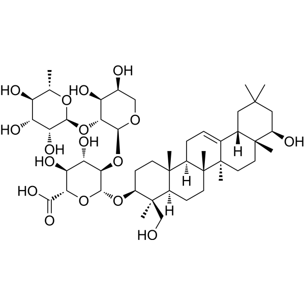
Soyasaponin II is a saponin with antiviral activity. Soyasaponin II inhibits the replication of HSV-1, HCMV, influenza virus, and HIV-1. Soyasaponin II shows potent inhibition on HSV-1 replication. Soyasaponin II serves as a inhibitor for YB-1 phosphorylation and NLRP3 inflammasome priming and could protect mice against LPS/GalN induced acute liver failure.
For research use only. We do not sell to patients.
- Purity : 99.79%
- CAS No.: 55319-36-3
- Formula: C47H76O17
- Molecular Weight:913.10
-
Storage:
-20°C, protect from light
* In solvent : -80°C, 6 months; -20°C, 1 month (protect from light)

To place orders, for customer services and technical support, please contact: MedChemExpress USA
Tel: 609-228-6898 E-mail: [email protected] [email protected]


-
 Biological Activity
-
Chemical Information
-
Solvent & Solubility
- Protocols
- Purity & Documentation
- References
-
 Help & FAQs


-
HY-N0310
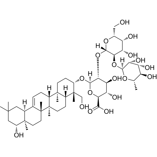
Soyasaponin BbAldose Reductase Inhibitor -
HY-13637A
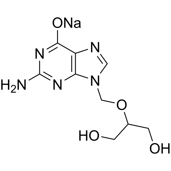
Ganciclovir sodiumAntiviral Agent -

HY-131944
S-AcetylglutathioneGlutathione Replenisher -
HY-120145
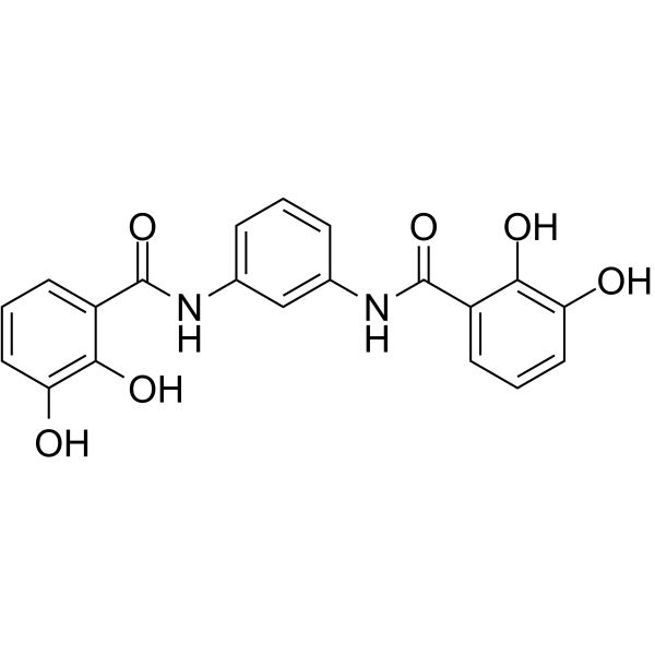
MST-312Telomerase Inhibitor -
HY-B0217
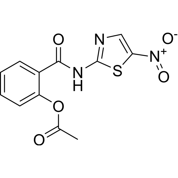
NitazoxanideAnthelmintic Agent -

HY-13637AR
Ganciclovir sodium (Standard)Antiviral Agent -
HY-13637
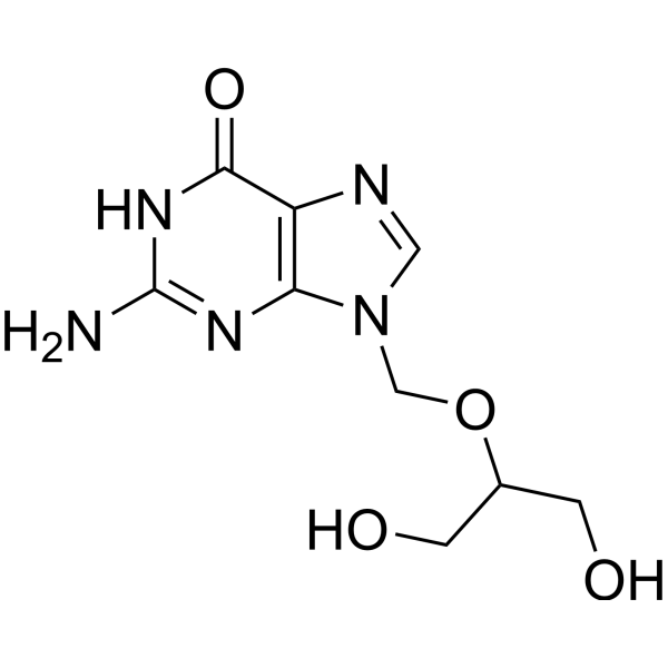
GanciclovirAntiviral Agent -
HY-100381
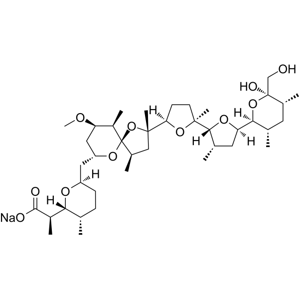
Nigericin sodium saltNLRP3 Activator -
HY-12815A
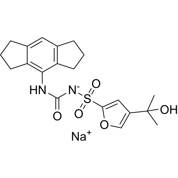
MCC950 sodiumNLRP3 Inhibitor -
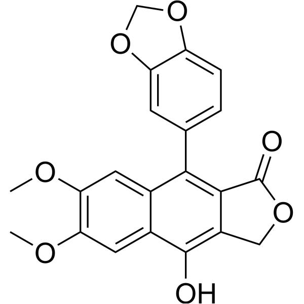
HY-N2532
DiphyllinV-ATPase/HIV-1 Inhibitor -
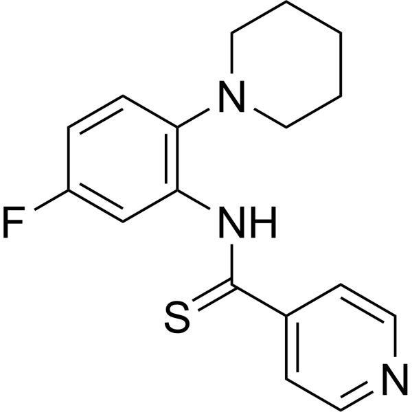
HY-18944
FIT-039CDK9 Inhibitor -
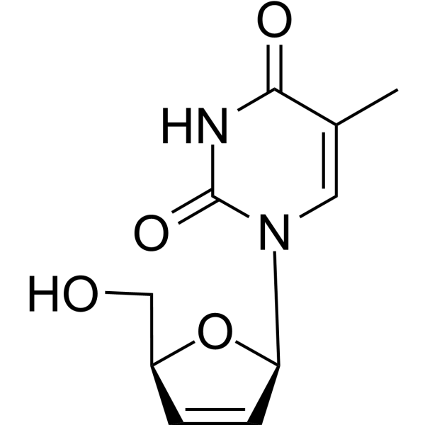
HY-B0116
StavudineNucleoside Reverse Transcriptase Inhibitor


Biological Activity
Description
|
HSV-1 54 μM (IC50) |
HIV-1 112 μM (IC50) |
influenza virus 88 μM (IC50) |
HCMV 104 μM (IC50) |
NLRP3 inflammasome |
In Vitro
Soyasaponin II inhibits the replication of HSV-1 (IC50=54μM), HCMV (104μM), influenza virus (88μM), and HIV-1 (112 μM)[1].
MedChemExpress (MCE) has not independently confirmed the accuracy of these methods. They are for reference only. Further protocols information, click here.
In Vivo


Chemical Information
-
CAS No. 55319-36-3
-
Appearance Solid
-
Molecular Weight 913.10
-
Formula C47H76O17
-
Color White to off-white
-
SMILES
C[C@]12[C@@](CC=C3[C@]2(CC[C@@]4([C@@]3([H])CC(C)(C[C@H]4O)C)C)C)([H])[C@@]5([C@@]([C@](C)([C@H](CC5)O[C@H]6[C@@H]([C@H]([C@@H]([C@H](O6)C(O)=O)O)O)O[C@H]7[C@@H]([C@H]([C@H](CO7)O)O)O[C@H]8[C@@H]([C@@H]([C@H]([C@@H](O8)C)O)O)O)CO)([H])CC1)C
-
Structure Classification
-
Initial Source
-
Shipping
Room temperature in continental US; may vary elsewhere.
-
Storage
-20°C, protect from light
* In solvent : -80°C, 6 months; -20°C, 1 month (protect from light)


Solvent & Solubility
In Vitro:
DMSO : 100 mg/mL (109.52 mM; Need ultrasonic; Hygroscopic DMSO has a significant impact on the solubility of product, please use newly opened DMSO)
Please refer to the solubility information to select the appropriate solvent. Once prepared, please aliquot and store the solution to prevent product inactivation from repeated freeze-thaw cycles.
Storage method and period of stock solution: -80°C, 6 months; -20°C, 1 month (protect from light). When stored at -80°C, please use it within 6 months. When stored at -20°C, please use it within 1 month.
Please refer to the solubility information to select the appropriate solvent. Once prepared, please aliquot and store the solution to prevent product inactivation from repeated freeze-thaw cycles.
Storage method and period of stock solution: -80°C, 6 months; -20°C, 1 month (protect from light). When stored at -80°C, please use it within 6 months. When stored at -20°C, please use it within 1 month.
Concentration (start) × Volume (start) = Concentration (final) × Volume (final)
In Vivo:
Select the appropriate dissolution method based on your experimental animal and administration route.
- For the following dissolution methods, please ensure to first prepare a clear stock solution using an In Vitro approach and then sequentially add co-solvents:
- To ensure reliable experimental results, the clarified stock solution can be appropriately stored based on storage conditions. As for the working solution for In Vivo experiments, it is recommended to prepare freshly and use it on the same day.
- The percentages shown for the solvents indicate their volumetric ratio in the final prepared solution. If precipitation or phase separation occurs during preparation, heat and/or sonication can be used to aid dissolution.
Add each solvent one by one: 10% DMSO 40% PEG300 5% Tween-80 45% Saline
Solubility: ≥ 2.5 mg/mL (2.74 mM); Clear solution
This protocol yields a clear solution of ≥ 2.5 mg/mL (saturation unknown).
Taking 1 mL working solution as an example, add 100 μL DMSO stock solution (25.0 mg/mL) to 400 μL PEG300, and mix evenly; then add 50 μL Tween-80 and mix evenly; then add 450 μL Saline to adjust the volume to 1 mL.
Preparation of Saline: Dissolve 0.9 g sodium chloride in ddH₂O and dilute to 100 mL to obtain a clear Saline solution.
Add each solvent one by one: 10% DMSO 90% (20% SBE-β-CD in Saline)
Solubility: ≥ 2.5 mg/mL (2.74 mM); Clear solution
This protocol yields a clear solution of ≥ 2.5 mg/mL (saturation unknown).
Taking 1 mL working solution as an example, add 100 μL DMSO stock solution (25.0 mg/mL) to 900 μL 20% SBE-β-CD in Saline, and mix evenly.
Preparation of 20% SBE-β-CD in Saline (4°C, storage for one week): 2 g SBE-β-CD powder is dissolved in 10 mL Saline, completely dissolve until clear.
In Vivo Dissolution Calculator
Please enter the basic information of animal experiments:
-
-
-
-
Recommended: Prepare an additional quantity of animals to account for potential losses during experiments.
Please enter your animal formula composition:
-
%DMSO +
Recommended: Keep the proportion of DMSO in working solution below 2% if your animal is weak.
-
%+
-
+%Tween-80 + +
-
%Saline +
The co-solvents required include: DMSO, . All of co-solvents are available by MedChemExpress (MCE). , Tween 80. All of co-solvents are available by MedChemExpress (MCE).
Working solution concentration: 0.22 mg/mL
Method for preparing stock solution: mg drug dissolved in μL DMSO. Stock solution concentration: mg/mL. * In solvent : -80°C, 6 months; -20°C, 1 month (protect from light)
1. Take μL DMSO stock solution;
2. Add μL .
μL , mix evenly;
3. Then add μL Tween 80, mix evenly;
4. Then add μL
Please ensure that the stock solution in the first step is dissolved to a clear state, and add co-solvents in sequence. You can use ultrasonic heating (ultrasonic cleaner, recommended frequency 20-40 kHz), vortexing, etc. to assist dissolution.

Protocols
-
Kinase activity and phosphorylation assays
Kinase activity assays measure the ability of kinases to transfer phosphate groups from ATP to specific substrates, while phosphorylation assays detect the presence and levels of phosphorylated proteins. Common methods include radiolabeled ATP incorporation (e. g. ,), ADP release detection via bioluminescence (e. g. ,[3]), enzyme-linked immunosorbent assays (ELISA) for phospho-specific epitopes (e. g. ,[6]), and microtiter-based formats for high-throughput screening (e. g. ,[8]). The ADP-Glo assay quantifies kinase activity by measuring ADP produced during phosphorylation using a luciferase-based system. Radiometric assays involve autoradiography or scintillation counting after incorporation of 32P-labeled ATP into substrate proteins. ELISA-based approaches rely on phospho-specific antibodies to detect activated kinases in cell lysates or purified samples.
-
Research Protocol for Infectious Diseases
Infectious-disease experiments test how pathogens interact with host barriers, innate immune receptors, inflammatory signaling, pathogen replication, and tissue injury; pattern-recognition receptors such as TLRs, RIG-I-like receptors, NOD-like receptors, and inflammasomes detect microbial molecules and activate NF-κB, interferon, and cytokine responses. The central hypothesis is that infection severity reflects the balance between pathogen burden and host response: protective inflammation restricts pathogen growth, whereas excessive or mislocalized inflammation contributes to tissue damage and disease phenotype. Unresolved questions include which host pathways are protective versus pathogenic, why some infection models fail to translate to human disease, and which combined readouts best predict clinically relevant infection outcomes.
-
Western Blot
Western blotting (WB) is a commonly used experimental method in molecular biology, biochemistry, and immunogenetics for identifying and quantifying target proteins. It combines gel electrophoresis with immunoassay, enabling researchers to analyze protein expression, post-translational modifications, and molecular weight.
-
Protocol for Kinase activity and phosphorylation assays
Kinase activity assays measure transfer of phosphate from ATP to a protein or peptide substrate, generating phosphorylated substrate, ADP, or incorporated radiolabeled phosphate as the readout; phosphorylation assays measure site-specific phosphorylation in cells or tissues as a proxy for kinase-pathway activation, inhibition, or substrate regulation. Phosphorylation can be detected by phospho-specific Western blot, immunoprecipitation kinase assay, phospho-immunofluorescence, phospho-flow cytometry, luminescent ADP detection, radiolabeled ATP incorporation, or reporter-based pathway assays, and these readouts can be applied to cancer cells, primary neurons, mouse tumors, organoids, inflammatory macrophages, ferroptosis studies, and mitophagy studies when the kinase target is biologically relevant.
-
Pyroptosis Solutions
Pyroptosis is a lytic inflammatory cell-death pathway executed by gasdermin pores, most classically through inflammasome-mediated activation of caspase-1, cleavage of gasdermin D, membrane pore formation, LDH release, and secretion of IL-1β and IL-18. The canonical pathway is commonly modeled by priming cells with an inflammatory signal such as LPS to induce pro-IL-1β and inflammasome components, followed by an activation signal such as ATP or nigericin to activate NLRP3, ASC speck formation, caspase-1 cleavage, GSDMD cleavage, cytokine release, and pyroptotic membrane rupture. The non-canonical pathway is triggered when cytosolic LPS activates mouse caspase-11 or human caspase-4/5, leading to GSDMD cleavage and pyroptosis, and this can secondarily activate NLRP3-dependent IL-1β release. Pyroptosis is linked to inflammatory injury, infection, cancer, liver disease, ocular disease, placental inflammation, and other disease phenotypes, but unresolved questions include which gasdermin fam


Purity & Documentation
-
Data Sheet (279 KB)
-
SDS (252 KB)
- English - EN (252 KB)
- Français - FR (252 KB)
- Deutsch - DE (252 KB)
- Norwegian - NO (252 KB)
- Español - ES (252 KB)
- Swedish - SV (252 KB)
- Italian - IT (252 KB)
- Korean - KR (252 KB)
- Portuguese - PT (252 KB)
-
Handling Instructions (2659 KB)
References
[1]. Hayashi K, et al. Inhibitory activity of soyasaponin II on virus replication in vitro. Planta Med. 1997;63(2):102-105. [Content Brief]
[2]. Wang F, et al. Soyasaponin II protects against acute liver failure through diminishing YB-1 phosphorylation and Nlrp3-inflammasome priming in mice. Theranostics. 2020;10(6):2714-2726. Published 2020 Feb 3. [Content Brief]


Complete Stock Solution Preparation Table
Please refer to the solubility information to select the appropriate solvent. Once prepared, please aliquot and store the solution to prevent product inactivation from repeated freeze-thaw cycles.
Storage method and period of stock solution: -80°C, 6 months; -20°C, 1 month (protect from light). When stored at -80°C, please use it within 6 months. When stored at -20°C, please use it within 1 month.
| Optional Solvent | Concentration Solvent Mass | 1 mg | 5 mg | 10 mg | 25 mg |
|---|---|---|---|---|---|
| DMSO | 1 mM | 1.0952 mL | 5.4759 mL | 10.9517 mL | 27.3793 mL |
| 5 mM | 0.2190 mL | 1.0952 mL | 2.1903 mL | 5.4759 mL | |
| 10 mM | 0.1095 mL | 0.5476 mL | 1.0952 mL | 2.7379 mL | |
| 15 mM | 0.0730 mL | 0.3651 mL | 0.7301 mL | 1.8253 mL | |
| 20 mM | 0.0548 mL | 0.2738 mL | 0.5476 mL | 1.3690 mL | |
| 25 mM | 0.0438 mL | 0.2190 mL | 0.4381 mL | 1.0952 mL | |
| 30 mM | 0.0365 mL | 0.1825 mL | 0.3651 mL | 0.9126 mL | |
| 40 mM | 0.0274 mL | 0.1369 mL | 0.2738 mL | 0.6845 mL | |
| 50 mM | 0.0219 mL | 0.1095 mL | 0.2190 mL | 0.5476 mL | |
| 60 mM | 0.0183 mL | 0.0913 mL | 0.1825 mL | 0.4563 mL | |
| 80 mM | 0.0137 mL | 0.0684 mL | 0.1369 mL | 0.3422 mL | |
| 100 mM | 0.0110 mL | 0.0548 mL | 0.1095 mL | 0.2738 mL |


Soyasaponin II Related Classifications
HY-122920 Related Classifications
 Powered by Bioz