



Terameprocol

Based on 1 Customer Validation
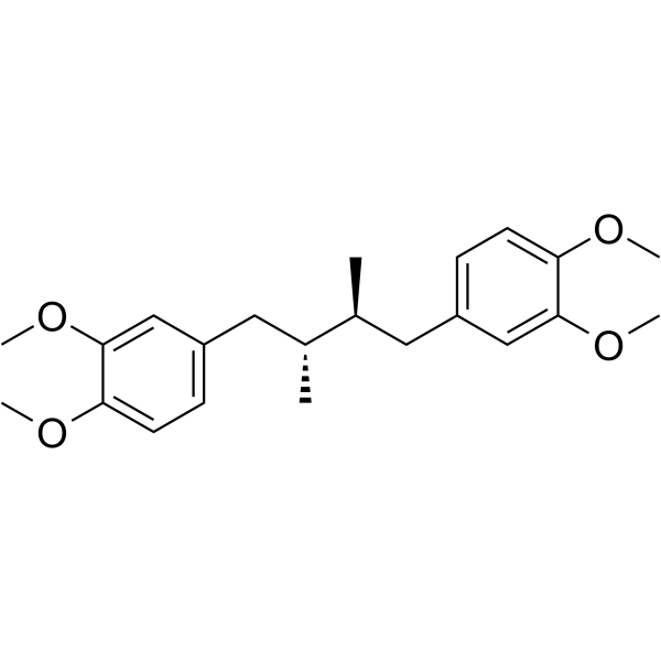
Terameprocol is an inhibitor targeting the Sp1 transcription factor, which can selectively inhibit the transcription of Sp1-dependent genes. Terameprocol exerts its effects by inhibiting Sp1-mediated gene transcription, such as reducing the expression of genes like CDC2, survivin and HMGB1, thereby arresting the cell cycle, inducing apoptosis, and suppressing the inflammatory response. Terameprocol exhibits anti-proliferative, pro-apoptotic, and anti-inflammatory activities and is currently mainly used in the research of diseases such as cancer and pulmonary arterial hypertension[1][2][3].
For research use only. We do not sell to patients.
- Purity : 99.50%
- CAS No.: 24150-24-1
- Formula: C22H30O4
- Molecular Weight:358.47
-
Storage:Powder -20°C, 3 years ; In solvent -80°C, 6 months , -20°C, 1 month

To place orders, for customer services and technical support, please contact: MedChemExpress USA
Tel: 609-228-6898 E-mail: [email protected] [email protected]


-
 Biological Activity
-
Chemical Information
-
Solvent & Solubility
- Protocols
- Purity & Documentation
- References
-
 Help & FAQs


-

HY-15763
ErastinFerroptosis Inducer -
HY-100218A
RSL3GPX4 Inhibitor -
HY-B0673
PirfenidoneAntifibrotic Agent -
HY-B0988
Deferoxamine mesylateIron Chelator -

HY-10071
Y-27632ROCK Inhibitor -
HY-A0122
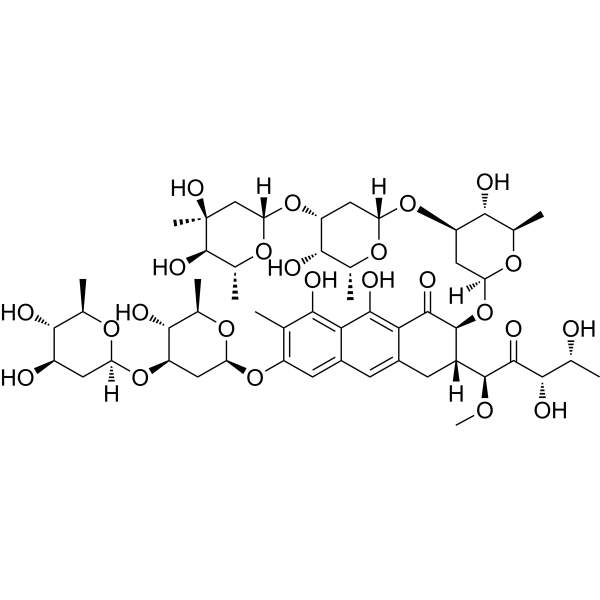
PlicamycinSp1 Inhibitor -
HY-109500
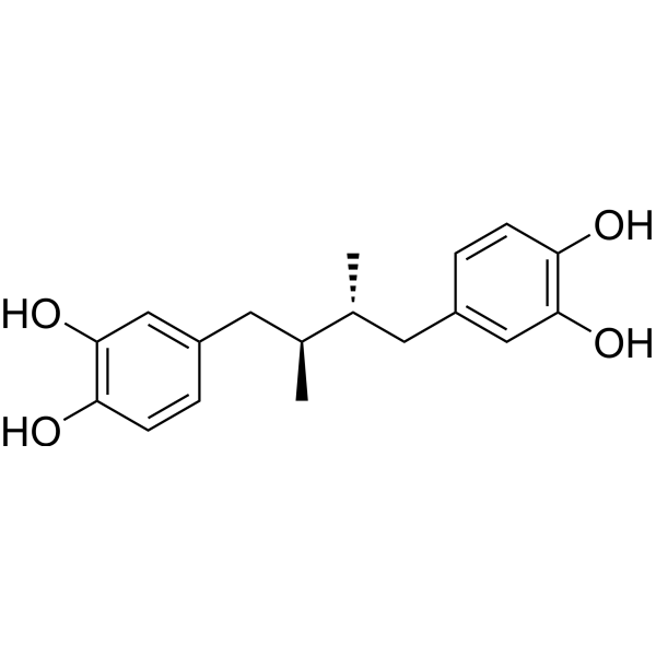
MasoprocolLipoxygenase Inhibitor -

HY-W100287
Murrayafoline AAnti-neuroinflammatory Agent -
HY-135960
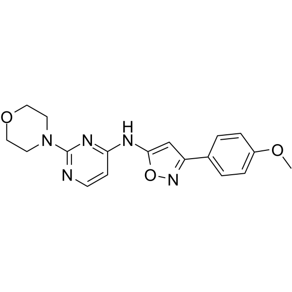
BO-264TACC3 Inhibitor -
HY-W012399
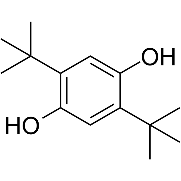
2,5-Di-tert-butylhydroquinoneSERCA Inhibitor -
HY-17425A
Valacyclovir hydrochlorideAntiviral Agent -
HY-13269
BMS-707035HIV-1 Integrase Strand Transfer Inhibitor


Biological Activity
Description
IC50 & Target
|
COX-2 |
Cellular Effect
|
Cell Line

|
Type | Value | Description | References |
|---|---|---|---|---|
| 786-0 | GI50 |
4.54 μM
Compound: Terameprocol, M4N, EM-1421
|
Cytotoxicity against human 786-0 cells after 72 hrs by MTT assay
Cytotoxicity against human 786-0 cells after 72 hrs by MTT assay
|
[PMID: 24080463] |
| A-375 | IC50 |
16.6 μM
Compound: 1a, M4N
|
Cytotoxicity against human A375 cells after 5 days by MTT assay
Cytotoxicity against human A375 cells after 5 days by MTT assay
|
[PMID: 19615898] |
| A-375 | IC50 |
2.5 μM
Compound: 1a, M4N
|
Cytotoxicity against human A375 cells
Cytotoxicity against human A375 cells
|
[PMID: 19615898] |
| A-375 | IC50 |
16.6 μM
Compound: 2; EM-1424, M4N
|
Antiproliferative activity against human A-375 cells assessed as reduction in cell growth
Antiproliferative activity against human A-375 cells assessed as reduction in cell growth
|
[PMID: 38092421] |
| BXPC-3 | GI50 |
17.1 μM
Compound: Terameprocol, M4N, EM-1421
|
Cytotoxicity against human BxPC3 cells after 72 hrs by MTT assay
Cytotoxicity against human BxPC3 cells after 72 hrs by MTT assay
|
[PMID: 24080463] |
| C8166 | CC50 |
28.8 μM
Compound: 21
|
Cytotoxicity against human C8166 cells by MTT assay
Cytotoxicity against human C8166 cells by MTT assay
|
[PMID: 19413342] |
| C8166 | EC50 |
14.8 μM
Compound: 21
|
Antiviral activity against HIV1 3B in human C8166 cells assessed as inhibition of virus-induced cytopathogenicity
Antiviral activity against HIV1 3B in human C8166 cells assessed as inhibition of virus-induced cytopathogenicity
|
[PMID: 19413342] |
| COS-1 | IC50 |
11.09 μM
Compound: 10
|
Inhibitory activity against HIV Tat-regulated Transactivation in COS cells.
Inhibitory activity against HIV Tat-regulated Transactivation in COS cells.
|
[PMID: 9685238] |
| HaCaT | IC50 |
85 μM
Compound: 2; EM-1424, M4N
|
Cytotoxicity against human HaCaT cells assessed as reduction in cell viability
Cytotoxicity against human HaCaT cells assessed as reduction in cell viability
|
[PMID: 38092421] |
| HeLa | GI50 |
>50 μM
Compound: Terameprocol, M4N, EM-1421
|
Cytotoxicity against human HeLa cells after 72 hrs by MTT assay
Cytotoxicity against human HeLa cells after 72 hrs by MTT assay
|
[PMID: 24080463] |
| Hep 3B2 | IC50 |
≤10 μM
Compound: 3, M4N
|
Cytotoxicity against human Hep3B cells after 3 days by MTT assay
Cytotoxicity against human Hep3B cells after 3 days by MTT assay
|
[PMID: 21123067] |
| HepG2 | IC50 |
44.5 μM
Compound: 1a, M4N
|
Cytotoxicity against human HepG2 cells after 5 days by MTT assay
Cytotoxicity against human HepG2 cells after 5 days by MTT assay
|
[PMID: 19615898] |
| HepG2 | IC50 |
44.5 μM
Compound: 2; EM-1424, M4N
|
Antiproliferative activity against human HepG2 cells assessed as reduction in cell viability incubated for 24 hrs by MTT assay
Antiproliferative activity against human HepG2 cells assessed as reduction in cell viability incubated for 24 hrs by MTT assay
|
[PMID: 38092421] |
| HT-29 | IC50 |
61.5 μM
Compound: 1a, M4N
|
Cytotoxicity against human HT-29 cells after 5 days by MTT assay
Cytotoxicity against human HT-29 cells after 5 days by MTT assay
|
[PMID: 19615898] |
| HT-29 | IC50 |
≤10 μM
Compound: 3, M4N
|
Cytotoxicity against human HT-29 cells after 3 days by MTT assay
Cytotoxicity against human HT-29 cells after 3 days by MTT assay
|
[PMID: 21123067] |
| HT-29 | IC50 |
61.5 μM
Compound: 2; EM-1424, M4N
|
Antiproliferative activity against human HT-29 cells assessed as cell growth inhibition
Antiproliferative activity against human HT-29 cells assessed as cell growth inhibition
|
[PMID: 38092421] |
| K562 | IC50 |
5.87 mg/L
Compound: 2, EM-1421, M4N, Terameprocol
|
Inhibition of growth metabolism of human K562 cells at 37 degC by TAM air microcalorimetry
Inhibition of growth metabolism of human K562 cells at 37 degC by TAM air microcalorimetry
|
[PMID: 23434529] |
| LNCaP | IC50 |
≤10 μM
Compound: 3, M4N
|
Cytotoxicity against human LNCAP cells after 3 days by MTT assay
Cytotoxicity against human LNCAP cells after 3 days by MTT assay
|
[PMID: 21123067] |
| MCF7 | IC50 |
42.4 μM
Compound: 1a, M4N
|
Cytotoxicity against human MCF7 cells after 5 days by MTT assay
Cytotoxicity against human MCF7 cells after 5 days by MTT assay
|
[PMID: 19615898] |
| MCF7 | IC50 |
≤10 μM
Compound: 3, M4N
|
Cytotoxicity against human MCF7 cells after 3 days by MTT assay
Cytotoxicity against human MCF7 cells after 3 days by MTT assay
|
[PMID: 21123067] |
| MCF7 | IC50 |
42.4 μM
Compound: 2; EM-1424, M4N
|
Antiproliferative activity against human MCF7 cells assessed as cell growth inhibition
Antiproliferative activity against human MCF7 cells assessed as cell growth inhibition
|
[PMID: 38092421] |
| NCI/ADR-RES | IC50 |
≤10 μM
Compound: 3, M4N
|
Cytotoxicity against human NCI/ADR-RES cells after 3 days by MTT assay
Cytotoxicity against human NCI/ADR-RES cells after 3 days by MTT assay
|
[PMID: 21123067] |
| T47D | GI50 |
5.36 μM
Compound: Terameprocol, M4N, EM-1421
|
Cytotoxicity against human T47D cells after 72 hrs by MTT assay
Cytotoxicity against human T47D cells after 72 hrs by MTT assay
|
[PMID: 24080463] |
| T98G | GI50 |
>50 μM
Compound: Terameprocol, M4N, EM-1421
|
Cytotoxicity against human T98G cells after 72 hrs by MTT assay
Cytotoxicity against human T98G cells after 72 hrs by MTT assay
|
[PMID: 24080463] |
| U-87MG ATCC | GI50 |
>50 μM
Compound: Terameprocol, M4N, EM-1421
|
Cytotoxicity against human U87 cells after 72 hrs by MTT assay
Cytotoxicity against human U87 cells after 72 hrs by MTT assay
|
[PMID: 24080463] |
| Vero | IC50 |
11.08 μM
Compound: M4N
|
Comparative Potency (IC50s) at HSV-2 passage 2 in vero cells
Comparative Potency (IC50s) at HSV-2 passage 2 in vero cells
|
[PMID: 9685239] |
| Vero | IC50 |
11.08 μM
Compound: M4N
|
Comparative Potency (IC50s) at HSV-2 passage 3 in vero cells
Comparative Potency (IC50s) at HSV-2 passage 3 in vero cells
|
[PMID: 9685239] |
| Vero | IC50 |
11.08 μM
Compound: M4N
|
Comparative Potency (IC50s) at HSV-2 passage 4 in vero cells
Comparative Potency (IC50s) at HSV-2 passage 4 in vero cells
|
[PMID: 9685239] |
| Vero | IC50 |
4.18 μM
Compound: M4N
|
Comparative Potency (IC50s) at HSV-2 passage 1 in vero cells
Comparative Potency (IC50s) at HSV-2 passage 1 in vero cells
|
[PMID: 9685239] |
| Vero | IC50 |
4.4 μM
Compound: M4N
|
Comparative Potency (IC50s) at HSV-1 passage 2 in vero cells
Comparative Potency (IC50s) at HSV-1 passage 2 in vero cells
|
[PMID: 9685239] |
| Vero | IC50 |
5.97 μM
Compound: M4N
|
Comparative Potency (IC50s) at HSV-1 passage 4 in vero cells
Comparative Potency (IC50s) at HSV-1 passage 4 in vero cells
|
[PMID: 9685239] |
| Vero | IC50 |
6.48 μM
Compound: M4N
|
Comparative Potency (IC50s) at HSV-1 passage 5 in vero cells
Comparative Potency (IC50s) at HSV-1 passage 5 in vero cells
|
[PMID: 9685239] |
| Vero | IC50 |
6.98 μM
Compound: M4N
|
Comparative Potency (IC50s) at HSV-1 passage 6 in vero cells
Comparative Potency (IC50s) at HSV-1 passage 6 in vero cells
|
[PMID: 9685239] |
| Vero | IC50 |
6.98 μM
Compound: M4N
|
Comparative Potency (IC50s) at HSV-1 passage 7 in vero cells
Comparative Potency (IC50s) at HSV-1 passage 7 in vero cells
|
[PMID: 9685239] |
| Vero | IC50 |
8.24 μM
Compound: M4N
|
Comparative Potency (IC50s) at HSV-1 passage 3 in vero cells
Comparative Potency (IC50s) at HSV-1 passage 3 in vero cells
|
[PMID: 9685239] |
| Vero | IC50 |
8.82 μM
Compound: M4N
|
Comparative Potency (IC50s) at HSV-1 passage 9 in vero cells
Comparative Potency (IC50s) at HSV-1 passage 9 in vero cells
|
[PMID: 9685239] |
| Vero | IC50 |
9.91 μM
Compound: M4N
|
Comparative Potency (IC50s) at HSV-1 passage 8 in vero cells
Comparative Potency (IC50s) at HSV-1 passage 8 in vero cells
|
[PMID: 9685239] |
In Vitro
Terameprocol (25 μM; 16 h) inhibited the production of prostaglandins (PGE2), some cytokines and chemokines induced by lipopolysaccharide (LPS) in RAW 264.7 cells, and also inhibited the expression and activity of COX-2, and inhibited the growth of RAW264.7 cells, but did not induce cell apoptosis[1]. Terameprocol (10 μM; 24 h, 48 h) downregulated survivin transcription and protein expression in HCC2429 and H460 non-small cell lung cancer cell experiments, while enhancing the sensitivity of cells to radiotherapy, but did not induce cell apoptosis and did not affect the cell cycle[2]. Terameprocol (0.1-20 μM; 24 h) inhibited cell proliferation in a dose-dependent manner in rat pulmonary artery smooth muscle cells (PASMCs) experiments, and induced cell apoptosis at 20 μM[3].
MedChemExpress (MCE) has not independently confirmed the accuracy of these methods. They are for reference only. Further protocols information, click here.
-
Cell Line:RAW 264.7 cells
-
Concentration:25 μM
-
Incubation Time:16 h
-
Result:Inhibited the LPS-induced production of prostaglandins, including PGE2, PGF2α, and 6-keto - PGF1α.
Suppressed the production of several cytokines and chemokines, such as TNF-α and MCP-1.
Reduced the expression of COX-2 mRNA and protein, inhibited the enzymatic activity of COX-2, and inhibited the growth of RAW 264.7 cells, but did not induce apoptosis.
-
Cell Line:HCC2429 and H460 cells
-
Concentration:10 μM
-
Incubation Time:24 h or 48 h
-
Result:Down-regulated the transcription and protein expression of survivin.
Enhanced the radiosensitivity of both HCC2429 and H460 cells.
Did not induce apoptosis in either cell line and had no significant effect on the cell cycle.
-
Cell Line:Rat pulmonary artery smooth muscle cells (PASMCs)
-
Concentration:0.1 μM, 1 μM, 10 μM, 20 μM
-
Incubation Time:24 h
-
Result:Inhibited the proliferation of PASMCs in a dose-dependent manner. At 20μM, induced apoptosis of PASMCs.
In Vivo
Terameprocol (166mg/kg; intraperitoneal injection; on days 7, 12, and 17; for 21 days) improves cardiac function, alleviates cardiac and pulmonary remodeling, inhibits the proliferation and induces the apoptosis of pulmonary artery smooth muscle cells in the monocrotaline (MCT)-induced pulmonary hypertension model of male Wistar rats (weighing 180-200g, age not mentioned)[3].
MedChemExpress (MCE) has not independently confirmed the accuracy of these methods. They are for reference only.
-
Animal Model:C57BL6/J mice (15-16g, 6-8 weeks old)[1]
-
Dosage:1 mg (in CPE vehicle)
-
Administration:Intraperitoneal injection, 1 time, 1 hour before LPS injection
-
Result:Offset the increase in serum TNF-α induced by LPS by 41% and significantly suppressed the LPS-induced accumulation of MCP-1 in serum.
Clinical Trial
| NCT Number | Sponsor | Condition | Start Date |
Phase
|
|---|---|---|---|---|
| NCT01329991 | Plexxikon| | 2011-05 | PHASE1 |


Chemical Information
-
CAS No. 24150-24-1
-
Appearance Solid
-
Molecular Weight 358.47
-
Formula C22H30O4
-
Color White to off-white
-
SMILES
COC1=CC(C[C@H](C)[C@H](C)CC2=CC=C(OC)C(OC)=C2)=CC=C1OC
-
Synonyms
EM-1421
-
Shipping
Room temperature in continental US; may vary elsewhere.
-
Storage
Powder -20°C 3 years In solvent -80°C 6 months -20°C 1 month


Solvent & Solubility
In Vitro:
DMSO : 100 mg/mL (278.96 mM; Need ultrasonic; Hygroscopic DMSO has a significant impact on the solubility of product, please use newly opened DMSO)
Please refer to the solubility information to select the appropriate solvent. Once prepared, please aliquot and store the solution to prevent product inactivation from repeated freeze-thaw cycles.
Storage method and period of stock solution: -80°C, 6 months; -20°C, 1 month. When stored at -80°C, please use it within 6 months. When stored at -20°C, please use it within 1 month.
Please refer to the solubility information to select the appropriate solvent. Once prepared, please aliquot and store the solution to prevent product inactivation from repeated freeze-thaw cycles.
Storage method and period of stock solution: -80°C, 6 months; -20°C, 1 month. When stored at -80°C, please use it within 6 months. When stored at -20°C, please use it within 1 month.
Concentration (start) × Volume (start) = Concentration (final) × Volume (final)

Protocols
-
Nuclear Protein Extraction (High-Salt/Hypotonic Fractionation)
The high-salt/hypotonic fractionation method for nuclear protein extraction is based on the differential solubility of cellular components. Cytoplasmic proteins are extracted first using a hypotonic buffer that causes cell swelling and membrane rupture, followed by centrifugation to separate the cytoplasmic supernatant from the nuclear pellet. The nuclear pellet is then subjected to high-salt extraction (e. g. , 0. 4 M (NH4)2SO4 or 1 M NaCl) to solubilize tightly bound nuclear matrix proteins, including transcription factors, histones, and structural proteins associated with chromatin and the nuclear scaffold. This approach allows for the isolation of both soluble cytoplasmic proteins and salt-resistant nuclear proteins while minimizing cross-contamination.
-
RNA extraction experimental
By lysing cells, releasing RNA, and removing impurities such as proteins and DNA, high-purity RNA products are finally obtained. The commonly used traditional method is the guanidine isothiocyanate/phenol/chloroform method (Trizol), which is suitable for a variety of animal materials including animal tissues, microorganisms, cultured cells, etc., and most plant materials.
-
Apoptosis
Apoptosis, also called programmed cell death, is generally characterized by distinct morphological characteristics.
-
TUNEL staining for apoptotic DNA fragmentation
TUNEL staining detects DNA strand breaks by using terminal deoxynucleotidyl transferase to add labeled nucleotides to exposed 3′-OH DNA termini, generating either microscopic staining in fixed cells or tissue sections, or fluorescence/cytometric signal in cell suspensions. TUNEL positivity reflects DNA fragmentation but should not be interpreted alone as definitive apoptosis, because TUNEL can also label necrotic, autolytic, mechanically damaged, or DNA-repair-associated DNA breaks.
-
Flow cytometric DNA-content cell-cycle staining
Flow cytometric DNA-content cell-cycle staining measures the fluorescence intensity of DNA-bound fluorochromes in single cells or nuclei to estimate DNA content distributions, allowing assignment of populations to G0/G1, S, and G2/M phases by DNA histogram deconvolution. Propidium iodide (PI) intercalates into DNA, and PI fluorescence is proportional to cellular DNA content when staining is performed under conditions that make DNA accessible and minimize non-DNA signal. Cells with G2/M DNA content are expected to show approximately twice the fluorescence intensity of G0/G1 cells, while S-phase cells occupy intermediate fluorescence values. PI-based DNA-content analysis can also detect cells with fractional DNA content, often reported as sub-G1, when DNA fragmentation and extraction during staining reduce retained DNA signal in apoptotic cells. DAPI is an alternative DNA fluorochrome for univariate DNA-content analysis, while bivariate approaches combining DNA content with proliferation
-
Annexin V plus membrane-impermeant dye apoptosis staining
Annexin V-based apoptosis assays rely on the detection of phosphatidylserine (PS) externalization from the inner leaflet of the plasma membrane to the outer leaflet, an early biochemical hallmark of apoptosis. Fluorescently labeled Annexin V binds PS in a calcium-dependent manner, enabling identification of early apoptotic cells by flow cytometry or fluorescence microscopy. When combined with a membrane-impermeant DNA-binding dye (e. g. , propidium iodide), this approach allows discrimination between viable (Annexin V−/dye−), early apoptotic (Annexin V+/dye−), and late apoptotic or necrotic (Annexin V+/dye+) cell populations by assessing membrane integrity and PS exposure.
-
BrdU Incorporation Assay
Bromodeoxyuridine (BrdU) incorporation assay is based on the principle that BrdU, a thymidine analog, is incorporated into newly synthesized DNA during the S phase of the cell cycle, thereby serving as a marker of DNA replication and cellular proliferation. Incorporated BrdU can be detected using anti-BrdU antibodies following DNA denaturation, enabling visualization or quantification of proliferating cells through immunochemical detection methods such as immunofluorescence or immunohistochemistry.
-
Apoptosis Solutions
Apoptosis is a regulated, generally non-lytic cell-death pathway that removes unwanted, damaged, infected, or abnormal cells through coordinated morphological changes, caspase activation, DNA fragmentation, and membrane remodeling. The intrinsic apoptosis pathway is controlled mainly by mitochondrial outer membrane permeabilization, BCL-2 family proteins, cytochrome c release, apoptosome formation, caspase-9 activation, and downstream executioner caspase-3/7 activation. The extrinsic apoptosis pathway is initiated by death receptors such as Fas, TNFR, and TRAIL receptors, which recruit adaptor proteins and activate caspase-8 before engaging executioner caspases or mitochondrial amplification through BID cleavage. Apoptosis is linked to many phenotypes, including cancer cell killing, tissue homeostasis, immune regulation, neurodegeneration, infection response, and treatment-induced cytotoxicity; unresolved questions include how apoptosis interacts with necroptosis, pyroptosis, ferroptos
-
Protocol for Cell Cycle
Cell-cycle analysis by flow cytometry measures DNA content in single cells to estimate the fraction of cells in G0/G1, S, and G2/M phases. Propidium iodide intercalates into DNA, and after RNA removal with RNase, fluorescence intensity reflects cellular DNA content: 2N cells are assigned to G0/G1, cells between 2N and 4N to S phase, and 4N cells to G2/M. DNA-content analysis alone cannot reliably separate G0 from G1 or G2 from M. Ki-67 can distinguish quiescent G0 cells from cycling cells, EdU or BrdU incorporation marks active DNA synthesis in S phase, and phospho-histone H3 staining identifies mitotic cells within the 4N population.
-
Research Protocol for Cardiovascular Diseases
Cardiovascular disease can be modeled as maladaptive cardiac remodeling, where ischemic injury or pressure overload activates inflammatory signaling, fibroblast activation, extracellular-matrix deposition, cardiomyocyte hypertrophy, vascular remodeling, and progressive ventricular dysfunction. The TGF-β/SMAD axis is a central profibrotic pathway after myocardial injury and pressure overload, while innate immune and cytokine pathways regulate leukocyte recruitment, scar formation, and adverse remodeling. Key unresolved questions include which inflammatory signals are reparative versus harmful, when fibrosis is protective versus maladaptive, and whether pathway inhibition improves function without weakening necessary infarct healing or compensatory remodeling.
-
Research Protocol for Inflammation-related Diseases
The NLRP3 inflammasome is a cytosolic innate immune signaling platform that integrates priming signals and danger-signal activation to promote caspase-1 activation, maturation of IL-1β and IL-18, and gasdermin D-mediated pyroptotic cell death. The core experimental logic is to determine whether inflammatory disease phenotypes are driven by increased NLRP3 expression, ASC-containing inflammasome assembly, caspase-1 cleavage, GSDMD cleavage, and extracellular release of IL-1β/IL-18 rather than by nonspecific cell injury alone. The pathway is strongly linked to inflammation-related disease phenotypes because monosodium urate crystals activate NALP3/NLRP3 inflammasome signaling in gout-like crystal inflammation, cholesterol crystals activate NLRP3 inflammasomes in atherogenesis models, and DSS-induced intestinal inflammation has been reported to involve NLRP3 inflammasome activity. However, experimental colitis studies also show context-dependent protective effects of NLRP3 inflammasome co
-
Cell Viability Determination by MTT Colorimetric Assay
The following protocol uses the MTT colorimetric assay as a classic literature-established method for assessing cell viability/metabolic activity in cultured mammalian cells. MTT[3-(4,5-dimethylthiazol-2-yl)-2,5-diphenyltetrazolium bromide] is reduced by metabolically active cells to a colored formazan product; the amount of formazan is quantified spectrophotometrically and provides an indirect measure of metabolically active viable cells. Importantly, MTT reduction reflects cellular oxidoreductase/metabolic activity rather than an absolute direct count of living cells, so changes in cellular metabolism can alter the signal independently of cell number.


Purity & Documentation
-
Data Sheet (277 KB)
-
SDS (459 KB)
- English - EN (459 KB)
- Français - FR (459 KB)
- Deutsch - DE (459 KB)
- Norwegian - NO (459 KB)
- Español - ES (459 KB)
- Swedish - SV (459 KB)
- Italian - IT (459 KB)
- Korean - KR (459 KB)
- Portuguese - PT (459 KB)
-
Handling Instructions (2659 KB)
References
[1]. Eads D, et al. Terameprocol, a methylated derivative of nordihydroguaiaretic acid, inhibits production of prostaglandins and several key inflammatory cytokines and chemokines. J Inflamm (Lond). 2009 Jan 8;6:2. [Content Brief]
[2]. Sun Y, et al. Terameprocol (tetra-O-methyl nordihydroguaiaretic acid), an inhibitor of Sp1-mediated survivin transcription, induces radiosensitization in non-small cell lung carcinoma. J Thorac Oncol. 2011 Jan;6(1):8-14. [Content Brief]
[3]. Nogueira-Ferreira R, et al. HMGB1 down-regulation mediates terameprocol vascular anti-proliferative effect in experimental pulmonary hypertension. J Cell Physiol. 2017 Nov;232(11):3128-3138. [Content Brief]


Complete Stock Solution Preparation Table
Please refer to the solubility information to select the appropriate solvent. Once prepared, please aliquot and store the solution to prevent product inactivation from repeated freeze-thaw cycles.
Storage method and period of stock solution: -80°C, 6 months; -20°C, 1 month. When stored at -80°C, please use it within 6 months. When stored at -20°C, please use it within 1 month.
| Optional Solvent | Concentration Solvent Mass | 1 mg | 5 mg | 10 mg | 25 mg |
|---|---|---|---|---|---|
| DMSO | 1 mM | 2.7896 mL | 13.9482 mL | 27.8963 mL | 69.7408 mL |
| 5 mM | 0.5579 mL | 2.7896 mL | 5.5793 mL | 13.9482 mL | |
| 10 mM | 0.2790 mL | 1.3948 mL | 2.7896 mL | 6.9741 mL | |
| 15 mM | 0.1860 mL | 0.9299 mL | 1.8598 mL | 4.6494 mL | |
| 20 mM | 0.1395 mL | 0.6974 mL | 1.3948 mL | 3.4870 mL | |
| 25 mM | 0.1116 mL | 0.5579 mL | 1.1159 mL | 2.7896 mL | |
| 30 mM | 0.0930 mL | 0.4649 mL | 0.9299 mL | 2.3247 mL | |
| 40 mM | 0.0697 mL | 0.3487 mL | 0.6974 mL | 1.7435 mL | |
| 50 mM | 0.0558 mL | 0.2790 mL | 0.5579 mL | 1.3948 mL | |
| 60 mM | 0.0465 mL | 0.2325 mL | 0.4649 mL | 1.1623 mL | |
| 80 mM | 0.0349 mL | 0.1744 mL | 0.3487 mL | 0.8718 mL | |
| 100 mM | 0.0279 mL | 0.1395 mL | 0.2790 mL | 0.6974 mL |


Terameprocol Related Classifications
HY-10447 Related Classifications
 Powered by Bioz