



Tosufloxacin
Based on 2 publication(s) in Google Scholar
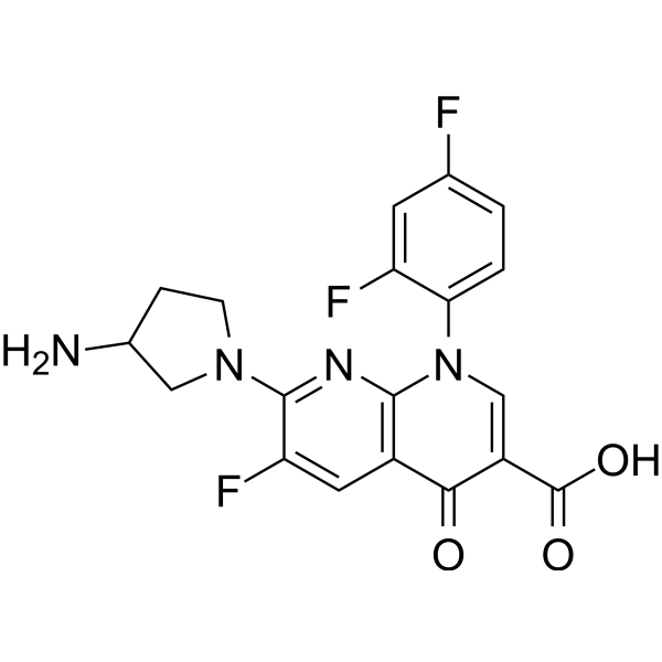
Tosufloxacin (A-61827) is an orally active fluoroquinolone antibiotic. Tosufloxacin shows a broad spectrum of antibacterial activity against gram-positive and gram-negative bacteria.
For research use only. We do not sell to patients.
- CAS No.: 100490-36-6
- Formula: C19H15F3N4O3
- Molecular Weight:404.34
-
Storage:
Please store the product under the recommended conditions in the Certificate of Analysis.

To place orders, for customer services and technical support, please contact: MedChemExpress USA
Tel: 609-228-6898 E-mail: [email protected] [email protected]


-
 Biological Activity
-
Chemical Information
- Protocols
- Purity & Documentation
- References
-
 Help & FAQs


Publications Citing Use of MedChemExpress (MCE) Tosufloxacin
MoreAll Antibiotic Isoforms
More

Biological Activity
Description
IC50 & Target
|
Quinolone |
Cellular Effect
|
Cell Line

|
Type | Value | Description | References |
|---|---|---|---|---|
| HeLa | IC50 |
32 μM
Compound: Tosufloxacin
|
Inhibition of human HeLa cell proliferation
Inhibition of human HeLa cell proliferation
|
[PMID: 19595598] |
| V79 | IC50 |
128 μg/mL
Compound: 6 (Tosufloxacin)
|
Mammalian cell cytotoxicity test in chinese hamster V79 cells (clonogenic cytotoxicity)
Mammalian cell cytotoxicity test in chinese hamster V79 cells (clonogenic cytotoxicity)
|
[PMID: 1469702] |
In Vitro
Tosufloxacin tosylate hydrate (T-3262) (0.05-3.13 μg/mL; 18 h) shows antibacterial activities against S. aureus, Staphylococcus epidermidis, streptococci, enterococci, Bacteroides fragilis, Clostridium difficile, and Clostridium perfringens[2].
MedChemExpress (MCE) has not independently confirmed the accuracy of these methods. They are for reference only.
-
Cell Line:S. aureus, Staphylococcus epidermidis, streptococci, enterococci, Bacteroides fragilis, Clostridium difficile, and Clostridium perfringens
-
Concentration:0.05-3.13 μg/mL
-
Incubation Time:18 hours
-
Result:Showed MIC90s (MICs for 90% of the isolates tested) ranging from 0.05 to 1.56 μg/mL for S. aureus, Staphylococcus epidermidis, streptococci, and enterococci.
Showed MIC90s of 1.56, 3.13, and 0.20 μg/mL for Bacteroides fragilis, Clostridium difficile, and Clostridium perfringens, respectively.
In Vivo
MedChemExpress (MCE) has not independently confirmed the accuracy of these methods. They are for reference only.
-
Animal Model:Male Slc:ICR mice infected with S. aureus[2]
-
Dosage:1.27-2.15 mg/kg
-
Administration:Oral gavage; 1.27-2.15 mg/kg; once
-
Result:Showed 50% effective dose (ED50) of 1.62 mg/kg (body weight) at 7 days after infection.
Showed MIC value of 0.0125 μg/mL.
-
Animal Model:Male Slc:ICR mice infected with E. coli[2]
-
Dosage:0.16-0.30 mg/kg
-
Administration:Oral gavage; 0.16-0.30 mg/kg; once
-
Result:Showed 50% effective dose (ED50) of 0.22 mg/kg (body weight) at 7 days after infection.
Showed MIC value of 0.0125 μg/mL.
-
Animal Model:Male Slc:ICR mice infected with P. aeruginosa[2]
-
Dosage:7.66-13.39 mg/kg
-
Administration:Oral gavage; 7.66-13.39 mg/kg; once
-
Result:Showed 50% effective dose (ED50) of 10.13 mg/kg (body weight) at 7 days after infection.
Showed MIC value of 0.78 μg/mL.


Chemical Information
-
CAS No. 100490-36-6
-
Molecular Weight 404.34
-
Formula C19H15F3N4O3
-
SMILES
O=C(C1=CN(C2=CC=C(F)C=C2F)C3=C(C=C(F)C(N4CC(N)CC4)=N3)C1=O)O
-
Synonyms
A-61827
-
Shipping
Room temperature in continental US; may vary elsewhere.
-
Storage
Please store the product under the recommended conditions in the Certificate of Analysis.


Publications (2)
-
Journal Impact Factor
-
Most Recent
-
PLoS Negl Trop Dis
Identification of anti-flaviviral drugs with mosquitocidal and anti-Zika virus activity in Aedes aegypti. [Abstract]2019 Aug 20;13(8):e0007681. PMID: 31430351 -
Curr Microbiol
Repurposing Sitafloxacin, Prulifloxacin, Tosufloxacin, and Sisomicin as Antimicrobials Against Biofilm and Persister Cells of Pseudomonas aeruginosa. [Abstract]2021 Dec 14;79(1):12. PMID: 34905092


Purity & Documentation
References
[1]. Chu DT, et al. Synthesis and biological properties of A-71497: a prodrug of tosufloxacin. Drugs Exp Clin Res. 1990;16(9):435-43. [Content Brief]
[2]. Fujimaki K, et al. In vitro and in vivo antibacterial activities of T-3262, a new fluoroquinolone. Antimicrob Agents Chemother. 1988 Jun;32(6):827-33. [Content Brief]

Calculators
Concentration (start) × Volume (start) = Concentration (final) × Volume (final)


Tosufloxacin Related Classifications
HY-B1802 Related Classifications
 Powered by Bioz