



Vps34-IN-4

Based on 1 Customer Validation
Vps34-IN-4 is an orally active and selective VPS34 inhibitor with an IC50 of 15 nM against human VPS34. Vps34-IN-4 binds to the ATP-binding pocket of VPS34. Vps34-IN-4 inhibits autophagy (autophagy) by blocking the degradation of autophagic substrates and increasing both lipidated and non-lipidated forms of LC3. Vps34-IN-4 induces time-dependent LC3-II accumulation in tumor tissues of tumor-bearing mice. Vps34-IN-4 can be used for research on cancers such as colon cancer.
For research use only. We do not sell to patients.
- Purity : 98.79%
- CAS No.: 1383716-46-8
- Formula: C21H25N7O
- Molecular Weight:391.47
-
Storage:Powder -20°C, 3 years ; In solvent -80°C, 6 months , -20°C, 1 month

To place orders, for customer services and technical support, please contact: MedChemExpress USA
Tel: 609-228-6898 E-mail: [email protected] [email protected]


-
 Biological Activity
-
Chemical Information
-
Solvent & Solubility
- Protocols
- Purity & Documentation
- References
-
 Help & FAQs


-
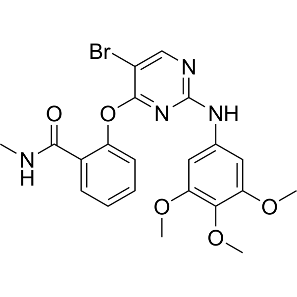
HY-16966
SBI-0206965ULK1/AMPK Inhibitor -
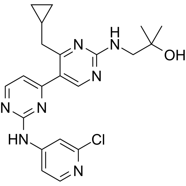
HY-12795
Vps34-IN-1Vps34 Inhibitor -
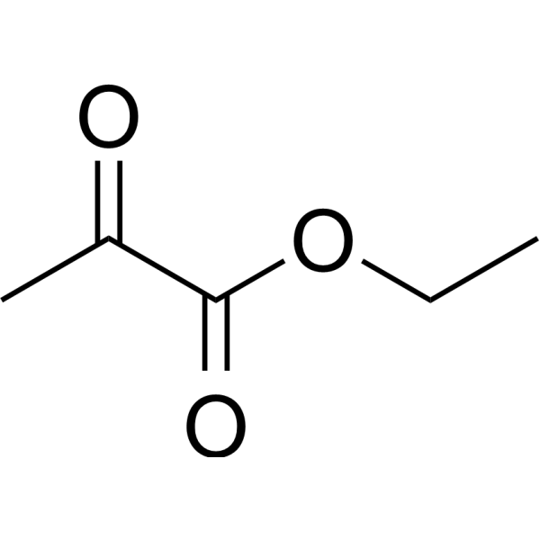
HY-Y1362
Ethyl pyruvateAnti-inflammatory Agent -
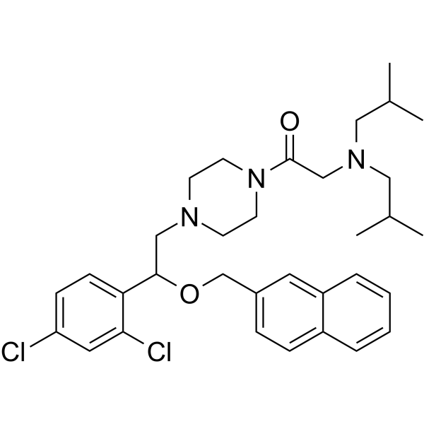
HY-101923
LYN-1604ULK1 Activator -
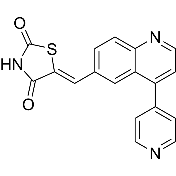
HY-12036
GSK1059615Apoptosis Inducer -
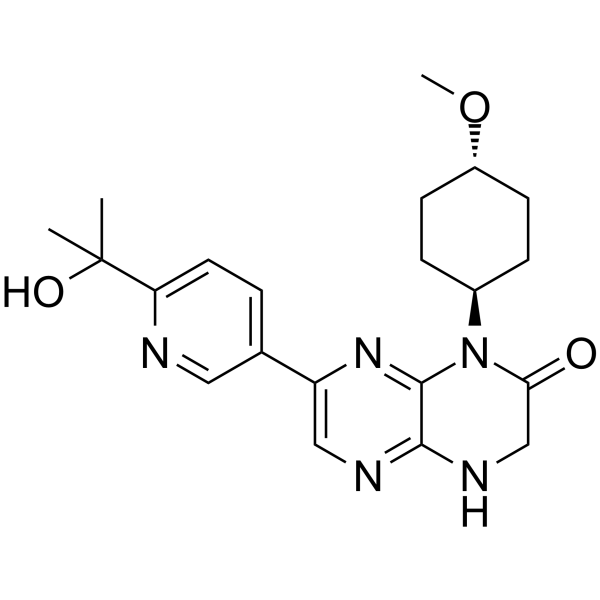
HY-16956
OnatasertibmTOR Inhibitor -
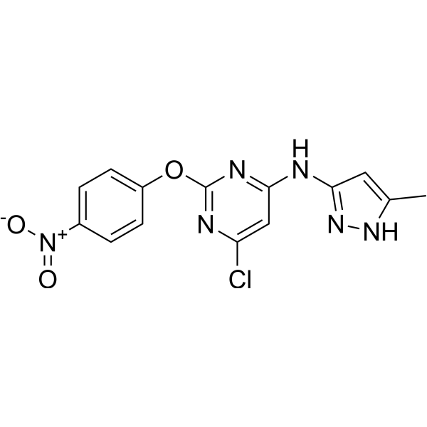
HY-101920
AutophinibVPS34 Inhibitor -
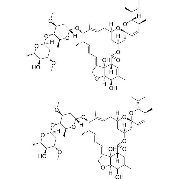
HY-15311
Avermectin B1Anti-infection Agent -
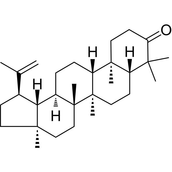
HY-N2590
LupenoneTriterpenoids Compound -

HY-Y1362R
Ethyl pyruvate (Standard)Anti-inflammatory Agent -

HY-101923R
LYN-1604 (Standard)Reference Standard -

HY-101920R
Autophinib (Standard)Reference Standard


Biological Activity
Description
IC50 & Target
[1]|
hVps34 15 nM (IC50) |
Cellular Effect
|
Cell Line

|
Type | Value | Description | References |
|---|---|---|---|---|
| U2OS | IC50 |
25 nM
|
Inhibition of VPS34-dependent phosphatidylinositol-3-phosphate production in cells measured via GFP-FYVE reporter cellular assay.
Inhibition of VPS34-dependent phosphatidylinositol-3-phosphate production in cells measured via GFP-FYVE reporter cellular assay.
|
26819669 |
In Vitro
Vps34-IN-4 (Compound 19) (1-10 μM; 24 h) potently inhibits autophagy in DLD1 cells and induces concentration-dependent accumulation of autophagic substrates and LC3[1].
Vps34-IN-4 potently inhibits VPS34 activity in U2OS cells with an IC50 of 25 nM in the GFP-FYVE reporter assay[1].
MedChemExpress (MCE) has not independently confirmed the accuracy of these methods. They are for reference only. Further protocols information, click here.
-
Cell Line:DLD1 human colon cancer cells
-
Concentration:1, 5, 10 μM
-
Incubation Time:24 h
-
Result:Caused a concentration-dependent increase in the protein levels of autophagy cargo receptors NBR1, NCOA4, SQSTM1/p62, NDP52, and ferritin heavy chain (FTH1).
Caused a concentration-dependent increase in both the lipidated (LC3-II) and nonlipidated (LC3-I) forms of LC3.
Showed no change in GAPDH levels across conditions, serving as a loading control.
Parmacokinetics
In Vivo
MedChemExpress (MCE) has not independently confirmed the accuracy of these methods. They are for reference only.
-
Animal Model:Nude mice (tumor-bearing; C57BL/6 mice used for PK only)[1]
-
Dosage:50 mg/kg
-
Administration:p.o.; twice daily; 7 days
-
Result:Resulted in time-dependent accumulation of LC3-II in tumor tissue, consistent with reduced autophagic capacity.
Showed no reduction in tumor volume over the 7-day treatment period.


Chemical Information
-
CAS No. 1383716-46-8
-
Appearance Solid
-
Molecular Weight 391.47
-
Formula C21H25N7O
-
Color White to off-white
-
SMILES
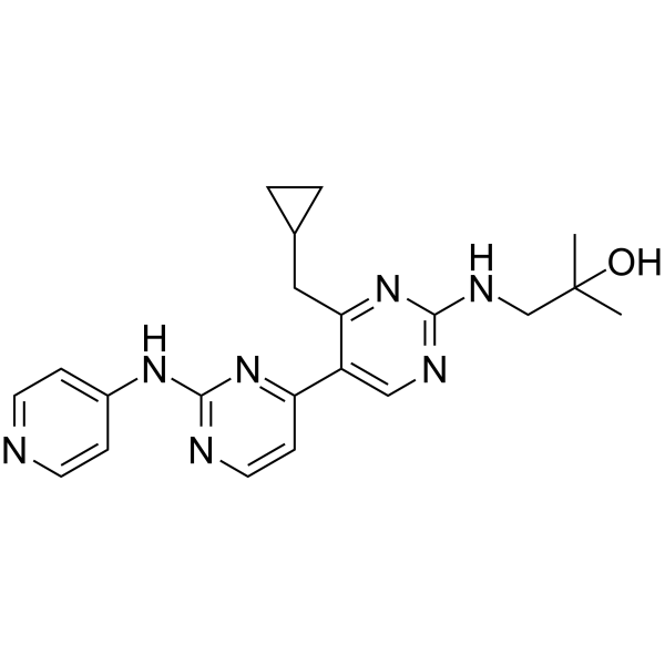
CC(O)(C)CNC1=NC=C(C2=NC(NC3=CC=NC=C3)=NC=C2)C(CC4CC4)=N1
-
Shipping
Room temperature in continental US; may vary elsewhere.
-
Storage
Powder -20°C 3 years In solvent -80°C 6 months -20°C 1 month


Solvent & Solubility
In Vitro:
DMSO : 78 mg/mL (199.25 mM; Need ultrasonic; Hygroscopic DMSO has a significant impact on the solubility of product, please use newly opened DMSO)
Please refer to the solubility information to select the appropriate solvent. Once prepared, please aliquot and store the solution to prevent product inactivation from repeated freeze-thaw cycles.
Storage method and period of stock solution: -80°C, 6 months; -20°C, 1 month. When stored at -80°C, please use it within 6 months. When stored at -20°C, please use it within 1 month.
Please refer to the solubility information to select the appropriate solvent. Once prepared, please aliquot and store the solution to prevent product inactivation from repeated freeze-thaw cycles.
Storage method and period of stock solution: -80°C, 6 months; -20°C, 1 month. When stored at -80°C, please use it within 6 months. When stored at -20°C, please use it within 1 month.
Concentration (start) × Volume (start) = Concentration (final) × Volume (final)

Protocols
-
Cell Cytotoxicity Assay
Cytotoxicity assays are usually based on the assessment of cell membrane damage, which can also be indirectly detected by measuring cell viability. Detection methods include MTT assay, CKK-8 assay, LDH assay and ATP assay, etc.
-
Autophagy
Autophagy is a process in which eukaryotic cells use lysosomes to degrade their own cytoplasmic proteins and damaged organelles under the regulation of autophagy related gene (Atg). Microtubule-associated proteins light chain 3 (LC3) is recognized as autophagy marker, which transfers from cytoplasmic LC3 (LC3-I) to membrane type (LC3-II). LC3-II/I ratio could be detected by Western Blot and fluorescence microscopy.
-
Lysosome and acidic-vesicle live-cell staining
Lysosome and acidic-vesicle live-cell staining detects acidic intracellular compartments by using membrane-permeant acidotropic probes that accumulate in low-pH vesicles, including lysosomes, late endosomes, autolysosomes, and acidic phagosomes. LysoTracker staining is commonly used as an intensity-based readout of acidic lysosomal compartment abundance or enlargement, while acridine orange produces green fluorescence in less concentrated compartments and red fluorescence after concentration-dependent accumulation in acidic vesicular organelles. Loss or reduction of acridine-orange red signal can be used as a readout of lysosomal membrane permeabilization or reduced acidic-vesicle integrity. This protocol is designed for live cultured cells and can be adapted for fluorescence microscopy, high-content imaging, plate-reader readout, or flow cytometry when the selected literature supports the readout. Because these dyes report acidotropic accumulation rather than lysosome identity alone,
-
Macroautophagy Solutions
Macroautophagy is a conserved lysosome-dependent degradation pathway in which cytoplasmic material is sequestered into double-membrane autophagosomes and delivered to lysosomes for degradation and recycling. The pathway supports cellular homeostasis during nutrient limitation, organelle stress, protein-aggregate accumulation, infection, differentiation, and tissue remodeling by coupling cargo sequestration, autophagosome maturation, lysosomal fusion, and degradation of cargo-derived macromolecules. The core molecular sequence includes initiation by nutrient- and stress-regulated autophagy machinery, autophagosome nucleation, LC3/ATG8-family conjugation to autophagosomal membranes, cargo selection through receptors such as SQSTM1/p62, autophagosome-lysosome fusion, and lysosomal degradation. LC3 was identified as a mammalian homolog of yeast Atg8 that localizes to autophagosomal membranes after processing, and p62/SQSTM1 was shown to connect ubiquitinated cargo with autophagic degradati


Purity & Documentation
-
Data Sheet (279 KB)
-
SDS (394 KB)
- English - EN (394 KB)
- Français - FR (394 KB)
- Deutsch - DE (394 KB)
- Norwegian - NO (394 KB)
- Español - ES (394 KB)
- Swedish - SV (394 KB)
- Italian - IT (394 KB)
- Korean - KR (394 KB)
- Portuguese - PT (394 KB)
-
Handling Instructions (2659 KB)
References


Complete Stock Solution Preparation Table
Please refer to the solubility information to select the appropriate solvent. Once prepared, please aliquot and store the solution to prevent product inactivation from repeated freeze-thaw cycles.
Storage method and period of stock solution: -80°C, 6 months; -20°C, 1 month. When stored at -80°C, please use it within 6 months. When stored at -20°C, please use it within 1 month.
| Optional Solvent | Concentration Solvent Mass | 1 mg | 5 mg | 10 mg | 25 mg |
|---|---|---|---|---|---|
| DMSO | 1 mM | 2.5545 mL | 12.7724 mL | 25.5447 mL | 63.8619 mL |
| 5 mM | 0.5109 mL | 2.5545 mL | 5.1089 mL | 12.7724 mL | |
| 10 mM | 0.2554 mL | 1.2772 mL | 2.5545 mL | 6.3862 mL | |
| 15 mM | 0.1703 mL | 0.8515 mL | 1.7030 mL | 4.2575 mL | |
| 20 mM | 0.1277 mL | 0.6386 mL | 1.2772 mL | 3.1931 mL | |
| 25 mM | 0.1022 mL | 0.5109 mL | 1.0218 mL | 2.5545 mL | |
| 30 mM | 0.0851 mL | 0.4257 mL | 0.8515 mL | 2.1287 mL | |
| 40 mM | 0.0639 mL | 0.3193 mL | 0.6386 mL | 1.5965 mL | |
| 50 mM | 0.0511 mL | 0.2554 mL | 0.5109 mL | 1.2772 mL | |
| 60 mM | 0.0426 mL | 0.2129 mL | 0.4257 mL | 1.0644 mL | |
| 80 mM | 0.0319 mL | 0.1597 mL | 0.3193 mL | 0.7983 mL | |
| 100 mM | 0.0255 mL | 0.1277 mL | 0.2554 mL | 0.6386 mL |


Vps34-IN-4 Related Classifications
HY-123058 Related Classifications
 Powered by Bioz