



XL177A
Based on 6 publication(s) in Google Scholar
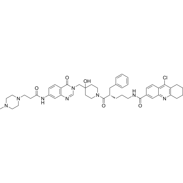
XL177A is a covalent USP7 inhibitor that blocks the deubiquitinase activity of USP7. XL177A destabilizes non-canonical PRC1 complexes or KDM6A and reduces chromatin deposition of H2AK119Ub, thereby relieving the repression of neuronal differentiation programs. Meanwhile, XL177A also regulates the ELOF1-UVSSA-USP7-nuclear β-catenin axis, decreasing the transcription levels of related proteins and the accumulation of nuclear β-catenin. XL177A exerts antiviral effects by reducing the expression levels of coronavirus receptors, and exhibits inhibitory activity against APC-mutated colorectal cancer cells, neuroblastoma, and coronaviruses including SARS-CoV-2 variants. XL177A is mainly used in studies related to colorectal cancer, neuroblastoma, and coronavirus infections.
For research use only. We do not sell to patients.
- Purity : 99.60%
- CAS No.: 2417089-74-6
- Formula: C48H57ClN8O5
- Molecular Weight:861.47
-
Storage:Powder -20°C, 3 years , 4°C, 2 years ; In solvent -80°C, 6 months , -20°C, 1 month

To place orders, for customer services and technical support, please contact: MedChemExpress USA
Tel: 609-228-6898 E-mail: [email protected] [email protected]


-
 Biological Activity
-
Chemical Information
-
Solvent & Solubility
- Protocols
- Purity & Documentation
- References
-
 Help & FAQs


-
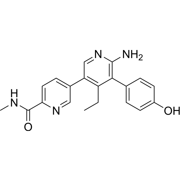
HY-107986
GNE-6776USP7 Inhibitor -
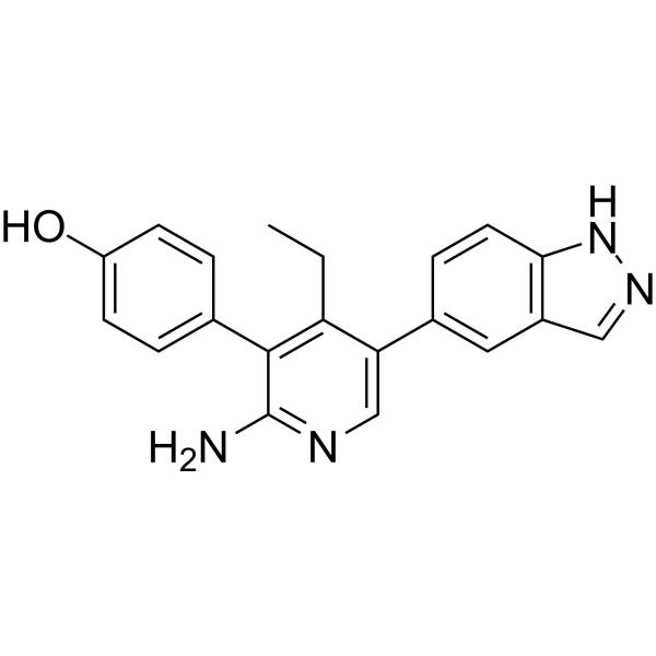
HY-112937
GNE-6640USP7 Inhibitor -
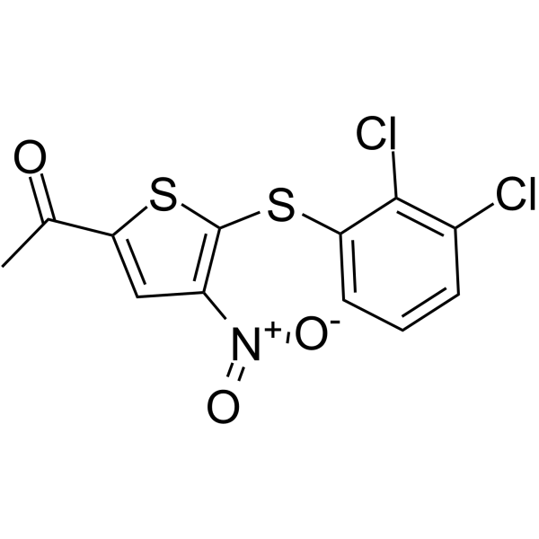
HY-15667
P005091USP7 Inhibitor -
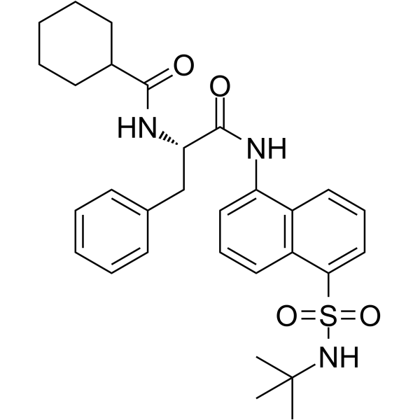
HY-112438
MF-094USP30 Inhibitor -
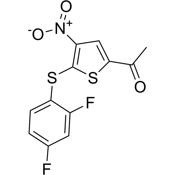
HY-13865
P 22077USP7/47 Inhibitor -
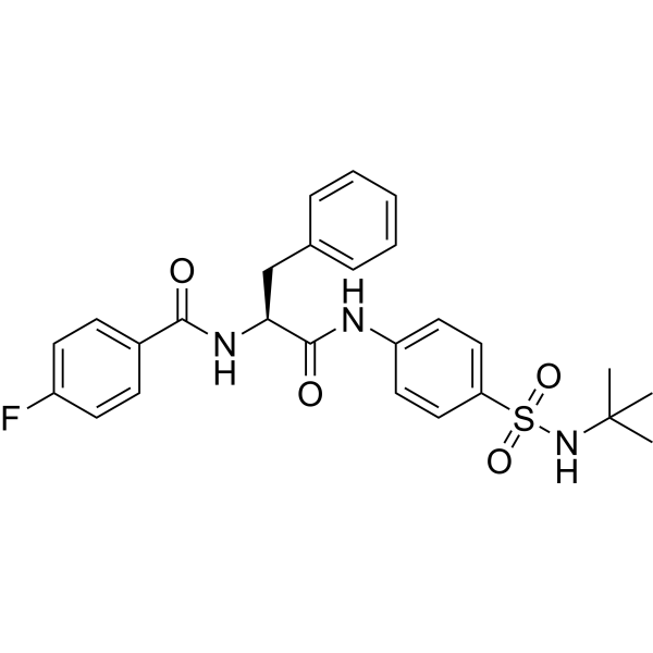
HY-141659
CMPD-39USP30 Inhibitor -
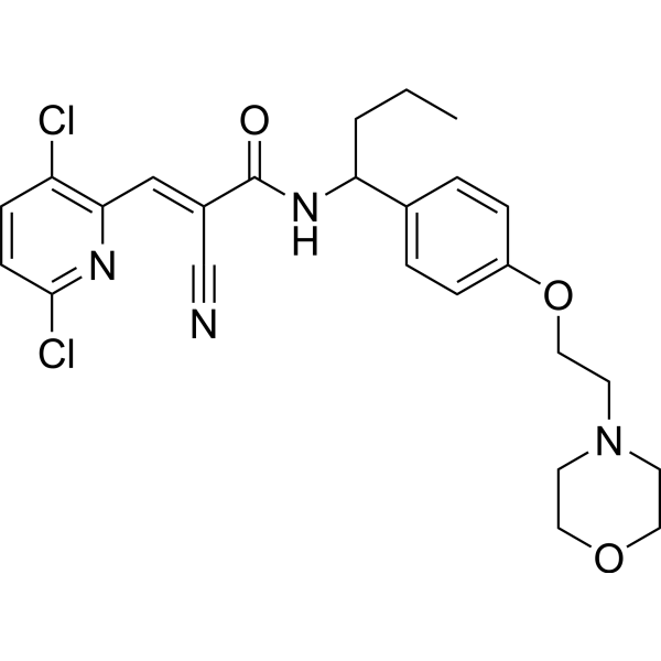
HY-111408
EOAI3402143DUB Inhibitor -
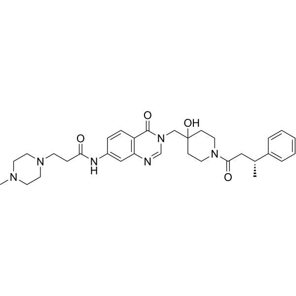
HY-122886
XL 188USP7 Inhibitor -
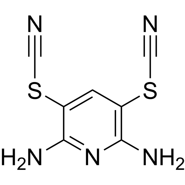
HY-13814
PR-619DUB Inhibitor -
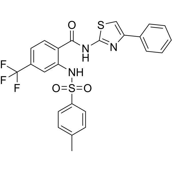
HY-100900
ML364USP2 Inhibitor -
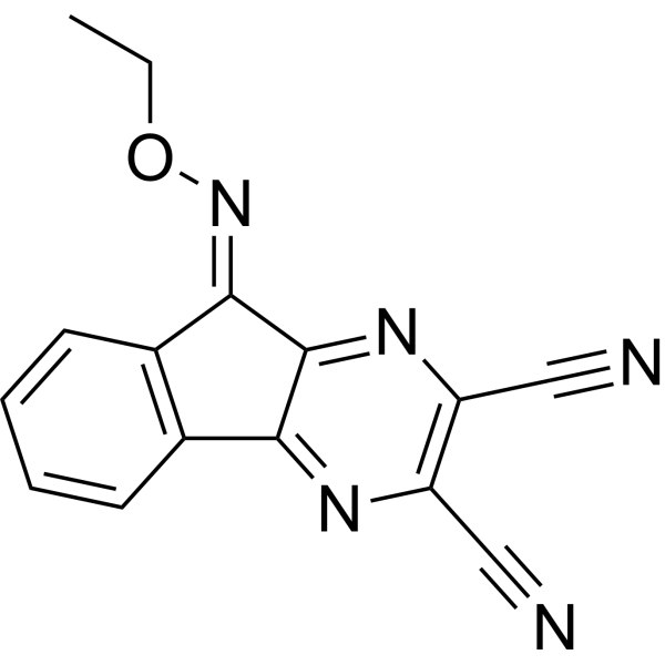
HY-50737A
DUB-IN-2Deubiquitinase Inhibitor -
HY-17543
ML-323USP1-UAF1 Inhibitor
Publications Citing Use of MedChemExpress (MCE) XL177A
More

Biological Activity
Description
In Vitro
XL177A GMP (1-10 μM; 7 d) enhances the cytotoxicity of carboplatin and the chemosensitivity of stem cell-like organoids derived from ovarian cancer patients. XL177A GMP also inhibits the TNFα/NF-κB signaling pathway, upregulates TNF-related apoptotic pathways, and induces cells to transition to an early stem cell-like state[1].
XL177A GMP (5 μM) potently inhibits the proliferation of SW480 colorectal cancer cells carrying APC mutations, while exerting minimal effects on CCD 841 CoN normal colonic epithelial cells with wild-type APC[2].
XL177A GMP (1 μM; 14 d) significantly reduces the population doubling number of KELLY, NB-SD and NGP neuroblastoma cell lines[3].
XL177A GMP (1 μM; 10 d) induces neuronal differentiation in NB-SD neuroblastoma cells, resulting in neurite formation and upregulated expression of the differentiation markers SEMA6B, CHGA and GAP43[3].
XL177A GMP (7.5-15 μM; 2 d) reduces the expression of ACE2 and DPP4 in Huh7.5 cells, and inhibits the entry of SARS-CoV-2-S and MERS-CoV-S pseudoviruses at concentrations of 7.5 and 15 μM after 2 days of pretreatment[4].
MedChemExpress (MCE) has not independently confirmed the accuracy of these methods. They are for reference only. Further protocols information, click here.
-
Cell Line:MCF7 cells
-
Concentration:0.001, 0.01, 0.1, 1, 10 μM
-
Incubation Time:18-24 hours
-
Result:The p53 and p21 protein levels remained high, but MDM2 protein levels matched DMSO control.


Chemical Information
-
CAS No. 2417089-74-6
-
Appearance Solid
-
Molecular Weight 861.47
-
Formula C48H57ClN8O5
-
Color White to light yellow
-
SMILES
O=C(C(C=CC1=C2Cl)=CC1=NC3=C2CCCC3)NCCC[C@@H](CC4=CC=CC=C4)C(N5CCC(CN6C=NC7=C(C=CC(NC(CCN8CCN(C)CC8)=O)=C7)C6=O)(O)CC5)=O
-
Shipping
Room temperature in continental US; may vary elsewhere.
-
Storage
Powder -20°C 3 years 4°C 2 years In solvent -80°C 6 months -20°C 1 month


Publications (6)
-
Journal Impact Factor
-
Most Recent
-
Nat Commun
Targeted degradation of USP7 in solid cancer cells reveals distinct effects of deubiquitinase degraders and inhibitors. [Abstract]2026 May 13;17(1):4331. PMID: 42129197 -
Redox Biol
USP20 governs tyrosine kinase inhibitors resistance through ferroptosis evasion by targeting GPX4 in cancers. [Abstract]2026 May:92:104086. PMID: 41844497 -
Adv Sci (Weinh)
Antagonistic Ubiquitin Switching by USP7 and RNF40 Orchestrates KDM6A Homeostasis to License Coronavirus Susceptibility. [Abstract]2026 Apr;13(23):e18058. PMID: 41691482 -
Cell Rep
2024 Oct 15;43(11):114872. PMID: 39412987 -
J Med Virol
2025 Jan;97(1):e70168. PMID: 39821265 -
Anim Cells Syst
Blocking SLC7A11 attenuates the proliferation of esophageal squamous cell carcinoma cells. [Abstract]2024 May 11;28(1):237-250. PMID: 38741950


Solvent & Solubility
In Vitro:
DMSO : 12.5 mg/mL (14.51 mM; ultrasonic and warming and heat to 60°C; Hygroscopic DMSO has a significant impact on the solubility of product, please use newly opened DMSO)
Please refer to the solubility information to select the appropriate solvent. Once prepared, please aliquot and store the solution to prevent product inactivation from repeated freeze-thaw cycles.
Storage method and period of stock solution: -80°C, 6 months; -20°C, 1 month. When stored at -80°C, please use it within 6 months. When stored at -20°C, please use it within 1 month.
Please refer to the solubility information to select the appropriate solvent. Once prepared, please aliquot and store the solution to prevent product inactivation from repeated freeze-thaw cycles.
Storage method and period of stock solution: -80°C, 6 months; -20°C, 1 month. When stored at -80°C, please use it within 6 months. When stored at -20°C, please use it within 1 month.
Concentration (start) × Volume (start) = Concentration (final) × Volume (final)
In Vivo:
Select the appropriate dissolution method based on your experimental animal and administration route.
- For the following dissolution methods, please ensure to first prepare a clear stock solution using an In Vitro approach and then sequentially add co-solvents:
- To ensure reliable experimental results, the clarified stock solution can be appropriately stored based on storage conditions. As for the working solution for In Vivo experiments, it is recommended to prepare freshly and use it on the same day.
- The percentages shown for the solvents indicate their volumetric ratio in the final prepared solution. If precipitation or phase separation occurs during preparation, heat and/or sonication can be used to aid dissolution.
Add each solvent one by one: 10% DMSO 40% PEG300 5% Tween-80 45% Saline
Solubility: ≥ 1.25 mg/mL (1.45 mM); Clear solution
This protocol yields a clear solution of ≥ 1.25 mg/mL (saturation unknown).
Taking 1 mL working solution as an example, add 100 μL DMSO stock solution (12.5 mg/mL) to 400 μL PEG300, and mix evenly; then add 50 μL Tween-80 and mix evenly; then add 450 μL Saline to adjust the volume to 1 mL.
Preparation of Saline: Dissolve 0.9 g sodium chloride in ddH₂O and dilute to 100 mL to obtain a clear Saline solution.
Add each solvent one by one: 10% DMSO 90% (20% SBE-β-CD in Saline)
Solubility: ≥ 1.25 mg/mL (1.45 mM); Clear solution
This protocol yields a clear solution of ≥ 1.25 mg/mL (saturation unknown).
Taking 1 mL working solution as an example, add 100 μL DMSO stock solution (12.5 mg/mL) to 900 μL 20% SBE-β-CD in Saline, and mix evenly.
Preparation of 20% SBE-β-CD in Saline (4°C, storage for one week): 2 g SBE-β-CD powder is dissolved in 10 mL Saline, completely dissolve until clear.
In Vivo Dissolution Calculator
Please enter the basic information of animal experiments:
-
-
-
-
Recommended: Prepare an additional quantity of animals to account for potential losses during experiments.
Please enter your animal formula composition:
-
%DMSO +
Recommended: Keep the proportion of DMSO in working solution below 2% if your animal is weak.
-
%+
-
+%Tween-80 + +
-
%Saline +
The co-solvents required include: DMSO, . All of co-solvents are available by MedChemExpress (MCE). , Tween 80. All of co-solvents are available by MedChemExpress (MCE).
Working solution concentration: 0.22 mg/mL
Method for preparing stock solution: mg drug dissolved in μL DMSO. Stock solution concentration: mg/mL.
1. Take μL DMSO stock solution;
2. Add μL .
μL , mix evenly;
3. Then add μL Tween 80, mix evenly;
4. Then add μL
Please ensure that the stock solution in the first step is dissolved to a clear state, and add co-solvents in sequence. You can use ultrasonic heating (ultrasonic cleaner, recommended frequency 20-40 kHz), vortexing, etc. to assist dissolution.

Protocols
-
PC12 NGF-Induced Neuronal Differentiation Culture
PC12 cells are a rat adrenal pheochromocytoma clonal line that responds to NGF by stopping proliferation and extending branching neurite-like processes; after longer NGF exposure, cells develop long processes and neuronal-like ultrastructural and functional features. NGF-induced differentiation is read out mainly by neurite outgrowth, reduced proliferation, microtubule assembly, and neuronal differentiation-associated proteins such as MAPs, tau, GAP-43, and synapsin-1.
-
iPSC/hPSC-Derived Neuron Differentiation Culture
iPSC/hPSC-derived neuron differentiation culture directs pluripotent cells toward neuroectoderm and then neuronal lineages by suppressing developmental signals that maintain non-neural fates; the classic monolayer dual-SMAD approach blocks BMP and Activin/TGF-β signaling with Noggin or dorsomorphin/LDN193189 plus SB431542, producing PAX6-positive neural progenitors that can be further matured into neurons. The readout is generated by morphology, neural progenitor markers, neuronal markers, subtype markers, and functional assays: PAX6/SOX1/NESTIN indicate neural progenitor induction, βIII-tubulin/TUJ1 and MAP2 indicate neuronal differentiation, cortical programs can be assessed by FOXG1, TBR1, CTIP2, SATB2, and synaptic maturation can be assessed by synaptic proteins, calcium activity, multielectrode arrays, or patch-clamp electrophysiology.
-
Research Protocol for Infectious Diseases
Infectious-disease experiments test how pathogens interact with host barriers, innate immune receptors, inflammatory signaling, pathogen replication, and tissue injury; pattern-recognition receptors such as TLRs, RIG-I-like receptors, NOD-like receptors, and inflammasomes detect microbial molecules and activate NF-κB, interferon, and cytokine responses. The central hypothesis is that infection severity reflects the balance between pathogen burden and host response: protective inflammation restricts pathogen growth, whereas excessive or mislocalized inflammation contributes to tissue damage and disease phenotype. Unresolved questions include which host pathways are protective versus pathogenic, why some infection models fail to translate to human disease, and which combined readouts best predict clinically relevant infection outcomes.
-
RNA extraction experimental
By lysing cells, releasing RNA, and removing impurities such as proteins and DNA, high-purity RNA products are finally obtained. The commonly used traditional method is the guanidine isothiocyanate/phenol/chloroform method (Trizol), which is suitable for a variety of animal materials including animal tissues, microorganisms, cultured cells, etc., and most plant materials.
-
SH-SY5Y Neuronal Differentiation Culture
SH-SY5Y neuronal differentiation culture uses sequential exposure to retinoic acid and neurotrophic factors to reduce proliferative neuroblastoma-like behavior and induce neuron-like morphology, including neurite extension, neuronal marker expression, and, in RA/BDNF protocols, greater synaptic-marker expression than undifferentiated culture. Retinoic acid is commonly used as the initiating differentiation cue, while BDNF in serum-reduced or serum-free medium supports later maturation and neurotrophic-factor-dependent neuron-like survival.
-
Human pluripotent stem cell neural induction and neuron differentiation
Human pluripotent stem cell neural induction can be achieved by blocking BMP and TGFβ/Activin/Nodal SMAD signaling, which suppresses non-neural differentiation and promotes early neuroectodermal identity; the expected readout is loss of pluripotency markers such as OCT4 and induction of neural markers such as PAX6, followed by neural progenitor and neuron marker acquisition during differentiation. This protocol uses dual-SMAD neural induction as the core induction method, followed by cortical neuron differentiation as a representative neuron differentiation model; published cortical protocols describe generation of cortical progenitors, temporally ordered cortical projection neurons, action-potential firing, synaptogenesis, and neural network formation over an approximately 80-day process.
-
SH-SY5Y neuronal-like differentiation
SH-SY5Y neuronal-like differentiation uses defined culture conditions to shift proliferative human neuroblastoma cells toward a neuron-like state, mainly assessed by reduced proliferation, neurite extension, neuronal-marker expression, and, in some protocols, increased dependence on neurotrophic support. Retinoic acid (RA) is commonly used for the first differentiation phase, and sequential RA followed by brain-derived neurotrophic factor (BDNF) in serum-free medium is a well-characterized approach for generating neuron-like SH-SY5Y cultures with extensive neurite outgrowth. The primary readouts are morphology-based neurite outgrowth and marker-based confirmation using proteins such as βIII-tubulin, MAP2, GAP43, synaptophysin, NeuN, NSE, TH, or related neuronal/synaptic markers, depending on the study endpoint.
-
Cell differentiation
Cell differentiation refers to the process in which cells of the same origin gradually produce cell groups with different morphological structure and functional characteristics.
-
PC12 NGF-induced neuronal-like differentiation
PC12 cells are a rat adrenal pheochromocytoma-derived clonal cell line that responds to nerve growth factor by stopping proliferation and extending neurites, producing a sympathetic neuron-like phenotype used to study neuronal differentiation and neurite outgrowth. NGF acts through TrkA-dependent signaling, and neurite outgrowth is associated with ERK/Akt signaling, microtubule organization, neuronal-marker expression, and increased electrophysiological neuronal features such as sodium-channel density. The main assay readout is morphological differentiation, usually measured as the percentage of neurite-bearing cells, neurite length, neurite number, or total neurite length per cell. Additional readouts include GAP-43, tyrosine hydroxylase, βIII-tubulin, neurofilament, synapsin I, synaptophysin, ERK phosphorylation, Akt phosphorylation, and sodium-channel current density.
-
Directly Induced Neuron Culture
Directly induced neuron culture converts somatic cells, most commonly fibroblasts, into induced neurons without passing through a pluripotent or neural progenitor stage; classic evidence shows that mouse fibroblasts can be converted by Ascl1, Brn2/Pou3f2, and Myt1l, human fibroblasts can be converted by defined neuronal transcription factors, and human fibroblasts can also be converted by miR-9/9-124 with neurogenic or subtype-specifying transcription factors. The readout is acquisition of neuronal identity and function, assessed by neuronal morphology, neuronal markers such as Tuj1/βIII-tubulin, MAP2, synapsin, and subtype markers when relevant, together with functional assays such as action-potential firing, synaptic activity, and electrophysiology.


Purity & Documentation
-
Data Sheet (273 KB)
-
SDS (254 KB)
- English - EN (254 KB)
- Français - FR (254 KB)
- Deutsch - DE (254 KB)
- Norwegian - NO (254 KB)
- Español - ES (254 KB)
- Swedish - SV (254 KB)
- Italian - IT (254 KB)
- Korean - KR (254 KB)
- Portuguese - PT (254 KB)
-
Handling Instructions (2659 KB)
References
[2]. Abdrabou AM, et al. nuPRISM: Microfluidic Genome-Wide Phenotypic Screening Platform for Cellular Nuclei. ACS Cent Sci. 2022;8(12):1618-1626. [Content Brief]
[3]. Cmarik EA, et al. Inhibition of USP7 destabilizes the noncanonical PRC1.1 complex and induces neuroblastoma differentiation. Mol Cancer Res. Published online March 19, 2026. [Content Brief]


Complete Stock Solution Preparation Table
Please refer to the solubility information to select the appropriate solvent. Once prepared, please aliquot and store the solution to prevent product inactivation from repeated freeze-thaw cycles.
Storage method and period of stock solution: -80°C, 6 months; -20°C, 1 month. When stored at -80°C, please use it within 6 months. When stored at -20°C, please use it within 1 month.
| Optional Solvent | Concentration Solvent Mass | 1 mg | 5 mg | 10 mg | 25 mg |
|---|---|---|---|---|---|
| DMSO | 1 mM | 1.1608 mL | 5.8040 mL | 11.6081 mL | 29.0202 mL |
| 5 mM | 0.2322 mL | 1.1608 mL | 2.3216 mL | 5.8040 mL | |
| 10 mM | 0.1161 mL | 0.5804 mL | 1.1608 mL | 2.9020 mL |


XL177A Related Classifications
HY-138794 Related Classifications
 Powered by Bioz