



Xylobiose

Based on 1 Customer Validation
Xylobiose (1,4-β-D-Xylobiose; 1,4-D-Xylobiose) is an orally active Claudin 2/CLDN2 inhibitor and HSP27 inducer. Xylobiose works by regulating intestinal barrier function and glucose and lipid metabolism-related signaling pathways. Xylobiose inhibits CLDN2 expression to reduce intestinal permeability, induces HSP27 to enhance cell protection, and regulates the miR-122a/miR-33a axis to inhibit liver lipid synthesis and improve insulin resistance. Xylobiose can strengthen intestinal barrier integrity, reduce blood sugar and blood lipid levels, and reduce oxidative stress and inflammatory response. Xylobiose can be used in the study of type 2 diabetes and metabolic syndrome.
For research use only. We do not sell to patients.
- Purity : 98.0%
- CAS No.: 6860-47-5
- Formula: C10H18O9
- Molecular Weight:282.24
-
Storage:Powder -20°C, 3 years , 4°C, 2 years ; In solvent -80°C, 6 months , -20°C, 1 month

To place orders, for customer services and technical support, please contact: MedChemExpress USA
Tel: 609-228-6898 E-mail: [email protected] [email protected]


-
 Biological Activity
-
Chemical Information
-
Solvent & Solubility
- Protocols
- Purity & Documentation
- References
-
 Help & FAQs


-
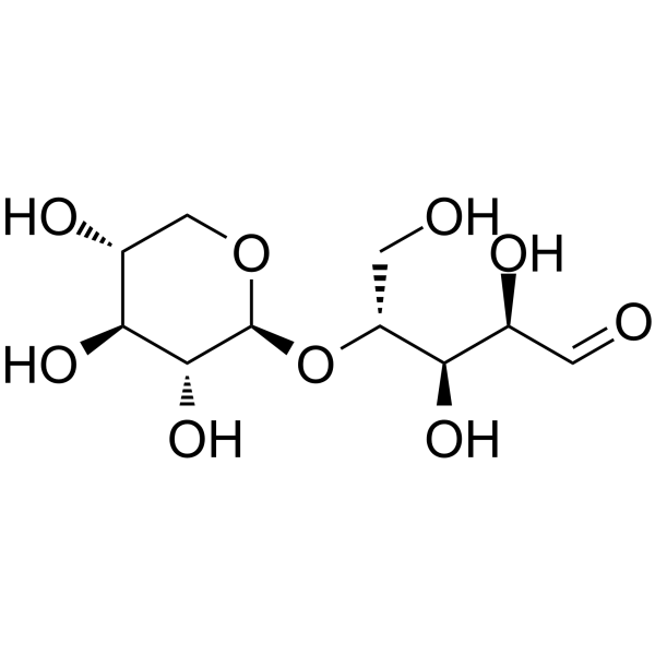
HY-N2469
XylotrioseBifidogenic Factor -
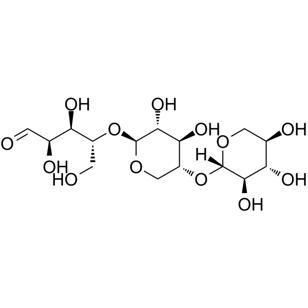
HY-N6840
XylotetraoseTaxy11 Substrate -
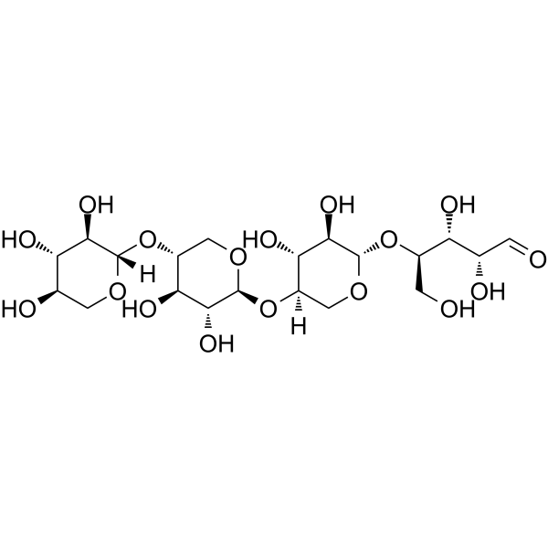
HY-N2325
D-(+)-CellobioseEndogenous Metabolite -
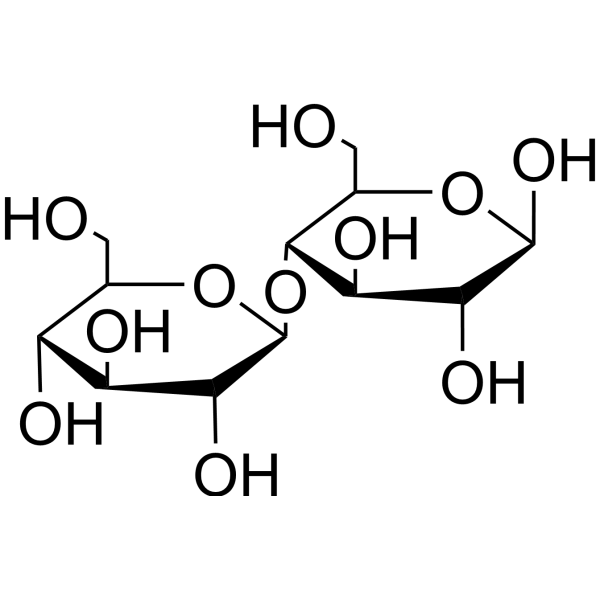
HY-N6839
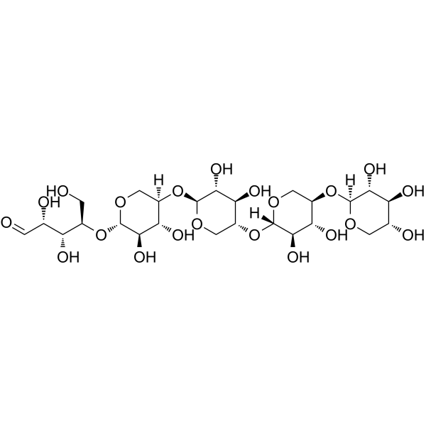
1,4-b-D-Xylopentaoseα-L-arabinofuranosidase Substrate -
HY-N6831
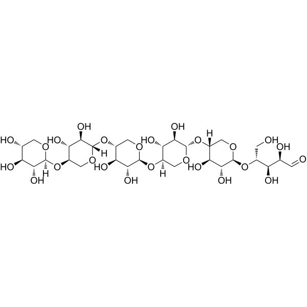
XylohexaoseXylooligosaccharide -
HY-N3018
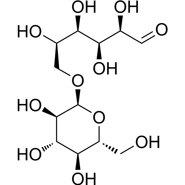
IsomaltoseGlucose Disaccharide -
HY-N1132
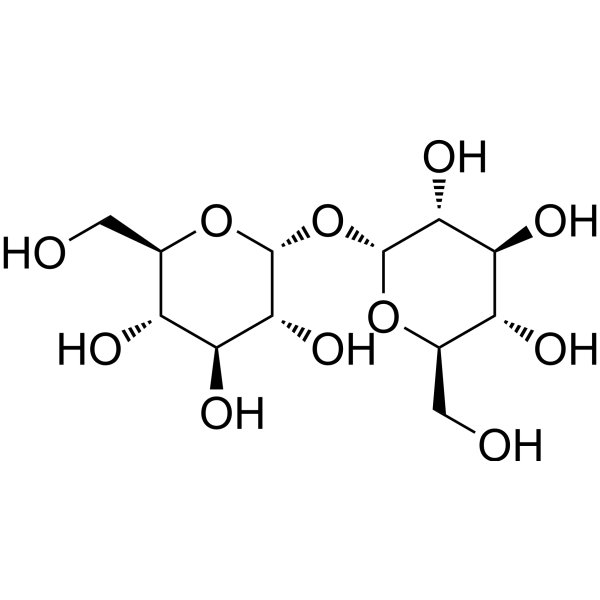
D-(+)-TrehaloseDisaccharide -
HY-N1495
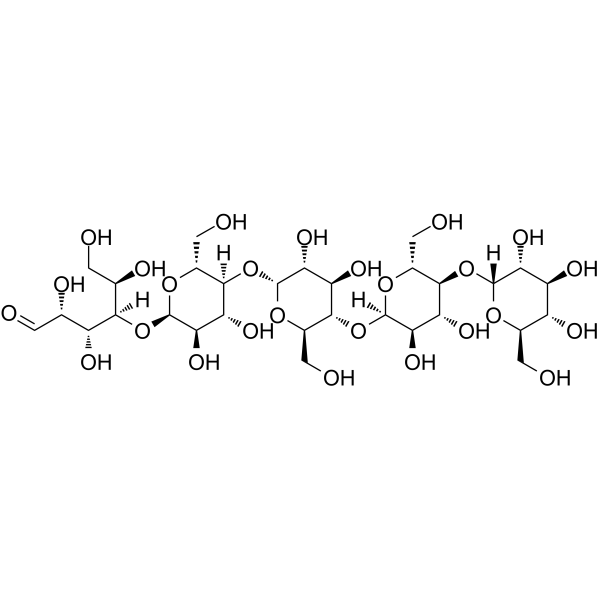
Maltopentaoseα-amylases Substrate -
HY-N0379
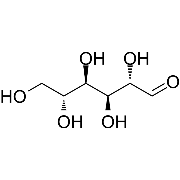
D-MannoseEndogenous Metabolite -

HY-N2468R
Xylobiose (Standard)CLDN2 Inhibitor -
HY-B0089
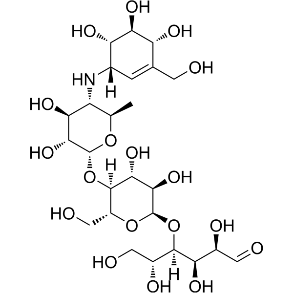
AcarboseAlpha-Glucosidase Inhibitor -
HY-N0059
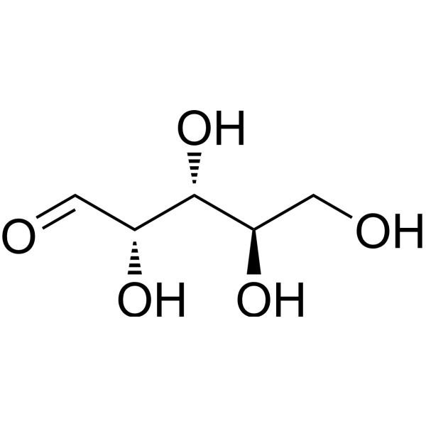
D-ArabinoseAnti-parasite Agent


Biological Activity
Description
IC50 & Target
|
Claudin-2 |
HSP27 |
In Vitro
Xylobiose (5-20 mg/mL; 3-24 h) down-regulates the gene and protein expression of Claudin 2 in human intestinal Caco-2 cells, up-regulates the gene and protein expression of heat shock proteins HSP27 and HSP70, and enhances the transepithelial electrical resistance (TER) of the cell monolayer, which may strengthen the intestinal barrier function by regulating signaling pathways such as PI3K[1].
MedChemExpress (MCE) has not independently confirmed the accuracy of these methods. They are for reference only. Further protocols information, click here.
-
Cell Line:Human intestinal Caco-2 cells
-
Concentration:5, 10, 20 mg/mL
-
Incubation Time:3, 6, 12, 24 h
-
Result:Downregulated CLDN2 expression, which is involved in paracellular permeability, and upregulated HSP27 and HSP70 expression, which are cytoprotective proteins. These changes indicated an improvement in the cell's functional state rather than a direct impact on cell viability.
In Vivo
MedChemExpress (MCE) has not independently confirmed the accuracy of these methods. They are for reference only.
-
Animal Model:
-
Dosage:
-
Administration:
-
Result:Animal Model: C57BL/KsJ-db/db male mice (17-24 g, 5-week-old), type 2 diabetes model induced by genetic factors[2] - Dosage (solvent): 5% xylobiose replacing sucrose in the diet (dissolved in the diet) - Administration: Dietary intake, daily, for 6 weeks - Result: Did not affect the body weight change compared with the diabetic control group, while significantly reduced food and water intake.


Chemical Information
-
CAS No. 6860-47-5
-
Appearance Solid
-
Molecular Weight 282.24
-
Formula C10H18O9
-
Color White to off-white
-
SMILES
O=C[C@H](O)[C@@H](O)[C@@H](CO)O[C@H]1[C@@H]([C@H]([C@H](O)CO1)O)O
-
Synonyms
1,4-β-D-Xylobiose; 1,4-D-Xylobiose
-
Structure Classification
-
Initial Source
-
Shipping
Room temperature in continental US; may vary elsewhere.
-
Storage
Powder -20°C 3 years 4°C 2 years In solvent -80°C 6 months -20°C 1 month


Solvent & Solubility
In Vitro:
H2O : ≥ 125 mg/mL (442.89 mM)
* "≥" means soluble, but saturation unknown.
Please refer to the solubility information to select the appropriate solvent. Once prepared, please aliquot and store the solution to prevent product inactivation from repeated freeze-thaw cycles.
Storage method and period of stock solution: -80°C, 6 months; -20°C, 1 month. When stored at -80°C, please use it within 6 months. When stored at -20°C, please use it within 1 month.
* Note: If you choose water as the stock solution, please dilute it to the working solution, then filter and sterilize it with a 0.22 μm filter before use.
Please refer to the solubility information to select the appropriate solvent. Once prepared, please aliquot and store the solution to prevent product inactivation from repeated freeze-thaw cycles.
Storage method and period of stock solution: -80°C, 6 months; -20°C, 1 month. When stored at -80°C, please use it within 6 months. When stored at -20°C, please use it within 1 month.
* Note: If you choose water as the stock solution, please dilute it to the working solution, then filter and sterilize it with a 0.22 μm filter before use.
Concentration (start) × Volume (start) = Concentration (final) × Volume (final)
In Vivo:
For the following dissolution methods, please prepare the working solution directly:
It is recommended to prepare fresh solutions and use them promptly within a short period of time.
The percentages shown for the solvents indicate their volumetric ratio in the final prepared solution. If precipitation or phase separation occurs during preparation, heat and/or sonication can be used to aid dissolution.
Add each solvent one by one: PBS
Solubility: 100 mg/mL (354.31 mM); Clear solution; Need ultrasonic

Protocols
-
RNA extraction experimental
By lysing cells, releasing RNA, and removing impurities such as proteins and DNA, high-purity RNA products are finally obtained. The commonly used traditional method is the guanidine isothiocyanate/phenol/chloroform method (Trizol), which is suitable for a variety of animal materials including animal tissues, microorganisms, cultured cells, etc., and most plant materials.
-
ROS/oxidative-stress fluorescent staining
ROS/oxidative-stress fluorescent staining uses cell-permeant fluorogenic probes that become fluorescent after oxidation inside cells or tissues; commonly used examples include DCFH-DA/DCFDA for broad cellular oxidant detection, DHE for superoxide-related signal detection, MitoSOX for mitochondrial superoxide-related signal detection, and CellROX probes for oxidative-stress-associated fluorescence readouts. The assay detects probe oxidation rather than a single ROS species unless the probe and analysis method have been chemically validated for that species. DCFH-DA enters cells, is deacetylated by intracellular esterases to DCFH, and produces fluorescent DCF after oxidation, so the readout is used as an operational measure of total cellular oxidative stress rather than a species-specific ROS measurement. DHE and MitoSOX can report superoxide-related oxidation, but red fluorescence alone can include non-specific ethidium-like oxidation products; HPLC or optimized spectral approaches are
-
Research Protocol for Endocrine Diseases
Endocrine diseases often arise from disrupted hormone production, hormone signaling, or target-tissue responsiveness; for diabetes-focused endocrine disease models, insulin signaling regulates glucose uptake, hepatic glucose output, lipid metabolism, and β-cell compensation. Type 2 diabetes develops through interacting defects in insulin resistance, β-cell dysfunction, adipose inflammation, hepatic glucose overproduction, altered incretin signaling, and ectopic lipid metabolism. A major unresolved question is whether endocrine dysfunction is driven primarily by target-tissue insulin resistance, intrinsic β-cell failure, immune/inflammatory stress, or combined multi-organ failure that differs by disease stage.
-
Transepithelial/transendothelial electrical resistance assay
TEER measures electrical resistance across epithelial or endothelial monolayers cultured on permeable supports, and the readout reflects ionic conductance through the cell barrier, especially the paracellular pathway regulated by junctional integrity. TEER can be measured without destroying the monolayer and is commonly used before or during transport, permeability, barrier-disruption, and barrier-maturation experiments. TEER values are influenced by biological maturation and technical conditions; reported factors include temperature, medium formulation, passage number, electrode geometry, membrane properties, and junctional length during early monolayer maturation. Therefore, TEER should be interpreted with blank-insert subtraction, area normalization, repeated readings, and, when possible, orthogonal barrier readouts such as FITC-dextran flux or tight-junction staining.
-
Research Protocol for Inflammation-related Diseases
The NLRP3 inflammasome is a cytosolic innate immune signaling platform that integrates priming signals and danger-signal activation to promote caspase-1 activation, maturation of IL-1β and IL-18, and gasdermin D-mediated pyroptotic cell death. The core experimental logic is to determine whether inflammatory disease phenotypes are driven by increased NLRP3 expression, ASC-containing inflammasome assembly, caspase-1 cleavage, GSDMD cleavage, and extracellular release of IL-1β/IL-18 rather than by nonspecific cell injury alone. The pathway is strongly linked to inflammation-related disease phenotypes because monosodium urate crystals activate NALP3/NLRP3 inflammasome signaling in gout-like crystal inflammation, cholesterol crystals activate NLRP3 inflammasomes in atherogenesis models, and DSS-induced intestinal inflammation has been reported to involve NLRP3 inflammasome activity. However, experimental colitis studies also show context-dependent protective effects of NLRP3 inflammasome co
-
Research Protocol for Metabolic Diseases
AMP-activated protein kinase, AMPK, is a conserved cellular energy sensor that responds to reduced cellular energy status and coordinates metabolism by increasing ATP-generating catabolic pathways while suppressing ATP-consuming anabolic processes. In metabolic disease research, the AMPK pathway is experimentally relevant because it regulates hepatic lipid synthesis, fatty acid oxidation, glucose production, skeletal-muscle glucose disposal, mTORC1-linked biosynthesis, autophagy, mitochondrial homeostasis, and whole-body energy balance. The central pathway logic is that energy stress, metformin, exercise-like stimulation, or direct AMPK activators increase AMPKα Thr172 phosphorylation and downstream substrate phosphorylation, including ACC and RAPTOR. Phosphorylation of ACC suppresses lipogenesis and supports fatty acid oxidation, whereas phosphorylation of RAPTOR suppresses mTORC1 signaling and links cellular energy status to growth and protein synthesis control. The pathway is linked


Purity & Documentation
-
Data Sheet (286 KB)
-
SDS (393 KB)
- English - EN (393 KB)
- Français - FR (393 KB)
- Deutsch - DE (393 KB)
- Norwegian - NO (393 KB)
- Español - ES (393 KB)
- Swedish - SV (393 KB)
- Italian - IT (393 KB)
- Korean - KR (393 KB)
- Portuguese - PT (393 KB)
-
Handling Instructions (2659 KB)
References
[1]. Rini DM, et al. Xylobiose treatment strengthens intestinal barrier function by regulating claudin 2 and heat shock protein 27 expression in human Caco-2 cells. J Sci Food Agric. 2024 Mar 15;104(4):2518-2525. [Content Brief]
[2]. Lim E, et al. Xylobiose, an Alternative Sweetener, Ameliorates Diabetes-Related Metabolic Changes by Regulating Hepatic Lipogenesis and miR-122a/33a in db/db Mice. Nutrients. 2016 Dec 5;8(12):791 [Content Brief]


Complete Stock Solution Preparation Table
Please refer to the solubility information to select the appropriate solvent. Once prepared, please aliquot and store the solution to prevent product inactivation from repeated freeze-thaw cycles.
Storage method and period of stock solution: -80°C, 6 months; -20°C, 1 month. When stored at -80°C, please use it within 6 months. When stored at -20°C, please use it within 1 month.
| Optional Solvent | Concentration Solvent Mass | 1 mg | 5 mg | 10 mg | 25 mg |
|---|---|---|---|---|---|
| H2O | 1 mM | 3.5431 mL | 17.7154 mL | 35.4308 mL | 88.5771 mL |
| 5 mM | 0.7086 mL | 3.5431 mL | 7.0862 mL | 17.7154 mL | |
| 10 mM | 0.3543 mL | 1.7715 mL | 3.5431 mL | 8.8577 mL | |
| 15 mM | 0.2362 mL | 1.1810 mL | 2.3621 mL | 5.9051 mL | |
| 20 mM | 0.1772 mL | 0.8858 mL | 1.7715 mL | 4.4289 mL | |
| 25 mM | 0.1417 mL | 0.7086 mL | 1.4172 mL | 3.5431 mL | |
| 30 mM | 0.1181 mL | 0.5905 mL | 1.1810 mL | 2.9526 mL | |
| 40 mM | 0.0886 mL | 0.4429 mL | 0.8858 mL | 2.2144 mL | |
| 50 mM | 0.0709 mL | 0.3543 mL | 0.7086 mL | 1.7715 mL | |
| 60 mM | 0.0591 mL | 0.2953 mL | 0.5905 mL | 1.4763 mL | |
| 80 mM | 0.0443 mL | 0.2214 mL | 0.4429 mL | 1.1072 mL | |
| 100 mM | 0.0354 mL | 0.1772 mL | 0.3543 mL | 0.8858 mL |
* Note: If you choose water as the stock solution, please dilute it to the working solution, then filter and sterilize it with a 0.22 μm filter before use.


Xylobiose Related Classifications
HY-N2468 Related Classifications
 Powered by Bioz