



ζ-Stat
Based on 4 publication(s) in Google Scholar
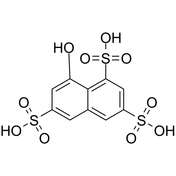
ζ-Stat (NSC37044) is a specific and atypical PKC-ζ inhibitor, with an IC50 of 5 μM. ζ-Stat can reduce melanoma cell lines proliferation and induce apoptosis, and has antitumor activity in vitro.
Nur für Forschungszwecke. Wir verkaufen nicht an Patienten.
- Reinheit : 98.98%
- CAS. Nr.: 3316-02-7
- Formel: C10H8O10S3
- Molecular Weight:384.36
-
Speicherung:Powder -20°C, 3 years , 4°C, 2 years ; In solvent -80°C, 2 years , -20°C, 1 year

To place orders, for customer services and technical support, please contact: MedChemExpress USA
Tel: 609-228-6898 E-mail: [email protected] [email protected]


-
 Biologische Aktivität
-
Chemical Information
-
Lösungsmittel & Löslichkeit
- Protokoll
- Reinheit & Dokumentation
- Verweise
-
 Help & FAQs


-
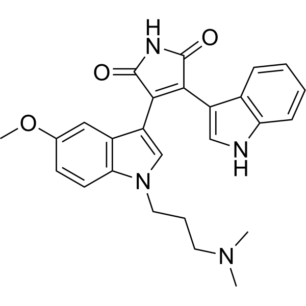
HY-13689
Go 6983PKC Inhibitor -
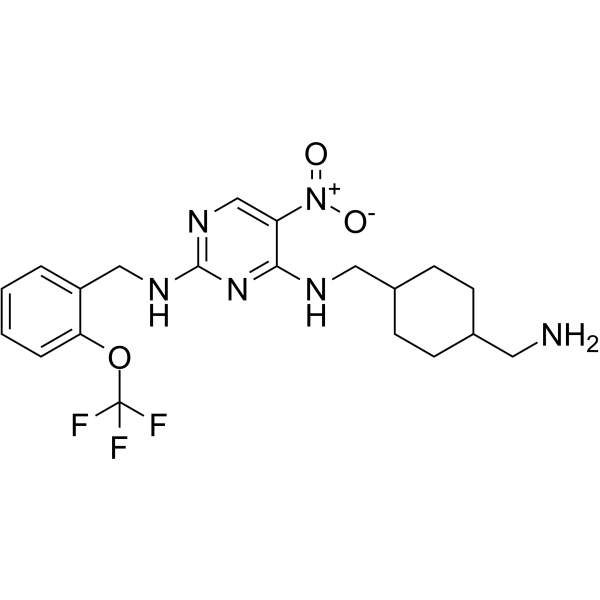
HY-112681
PKC-theta inhibitor 2PKC-θ Inhibitor -
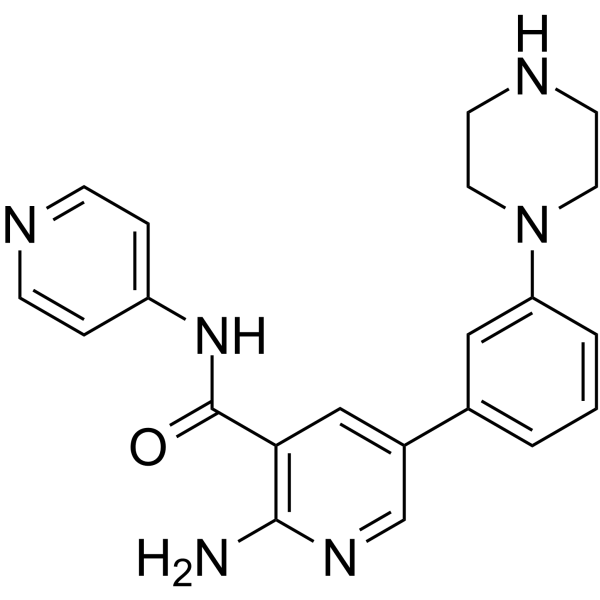
HY-126146
PKC-iota inhibitor 1PKC Inhibitor -
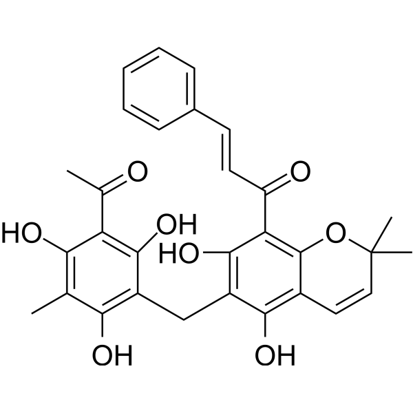
HY-18980
RottlerinPKC Inhibitor -
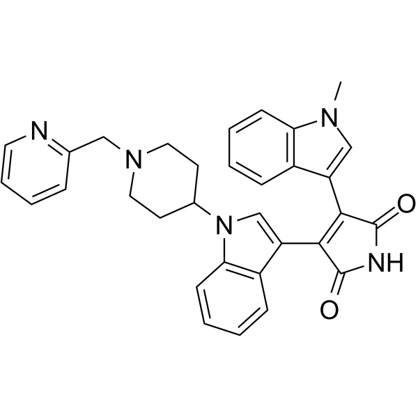
HY-10342
EnzastaurinPKCβ Inhibitor -
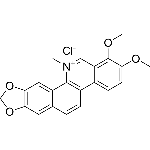
HY-12048
Chelerythrine chlorideBcl-2 Family Inhibitor -
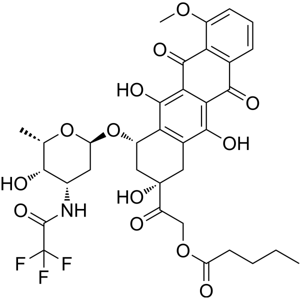
HY-13772
ValrubicinAntibiotic -
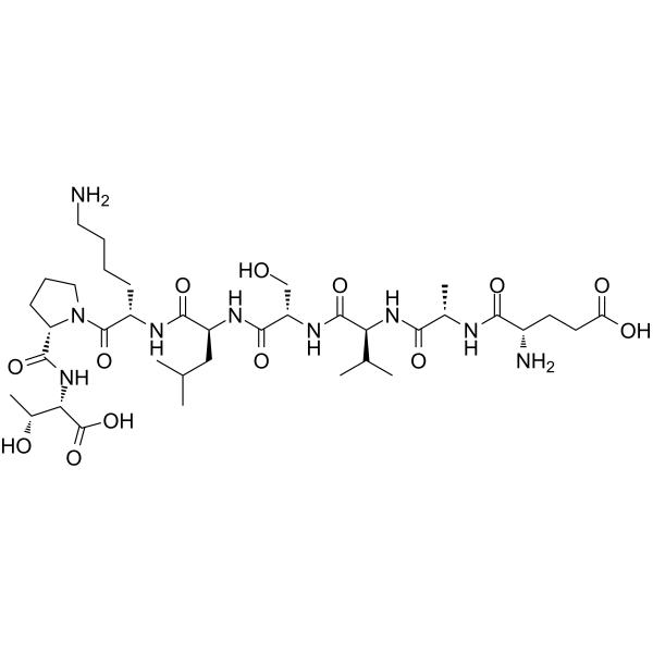
HY-P0154
Epsilon-V1-2PKCε Inhibitor -
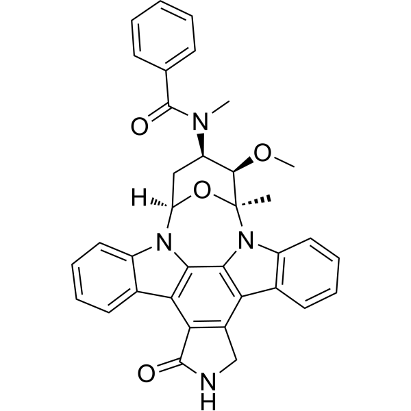
HY-10230
MidostaurinMulti-targeted Protein Kinase Inhibitor -
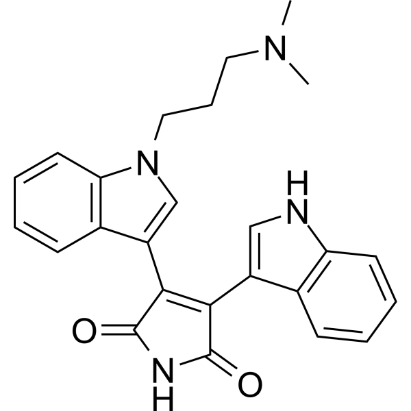
HY-13867
Bisindolylmaleimide IPKC Inhibitor -
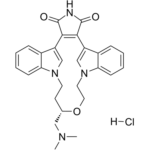
HY-10195B
Ruboxistaurin hydrochloridePKCβ Inhibitor -
HY-117800
BJE6-106PKCδ Inhibitor
Publications Citing Use of MedChemExpress (MCE) ζ-Stat
More

Biologische Aktivität
Beschreibung
IC50 & Target
[1]|
aPKC-ζ 5 μM (IC50) |
In Vitro
ζ-Stat (0.1-20 μM) shows only 13% inhibition on PKC-ι at 20 μM, but shows a significant inhibition on PKC-ζ as 51% at 5 μM level[1].
ζ-Stat (0.1-10 μM; 3 d) significantly decreases cell proliferation of SK-MEL-2 and MeWo upon increasing the concentrations[1].
ζ-Stat (7 or 10 μM; 24-72 h) and 5-FU in combination is able to decrease the viability of LoVo CRC cells by more than 75%[2].
ζ-Stat (5 μM; 3 d) shows a significant diminution of phosphorylated, total PKC-ζ, Bcl-2 and PARP levels, and increases Caspase-3 and cleaved-PARP levels in SK-MEL-2 and MeWo cells[1].
ζ-Stat (5 μM; 1-10 h) does not show significant cytotoxicity on MEL-F-NEO, SK-MEL-2 and MeWo cells[1].
MedChemExpress (MCE) has not independently confirmed the accuracy of these methods. They are for reference only.
-
Cell Line:MEL-F-NEO, SK-MEL-2 and MeWo cells
-
Concentration:0.1, 0.5, 1, 2.5, 5, 7.5, 10 µM
-
Incubation Time:3 days
-
Result:Decreased proliferation by 47.7% for 5 µM in SK-MEL-2 cells and by 50.6% for 5 µM in MeWo cells.
Showed significant inhibitions on MEL-F-NEO cells 19.3% (P ≤ 0.05) at 10 μM.
-
Cell Line:SK-MEL-2 and MeWo cells
-
Concentration:5 µM
-
Incubation Time:3 days
-
Result:Decreased phosphorylated and total PKC-ζ levels.


Chemical Information
-
CAS. Nr. 3316-02-7
-
Appearance Solid
-
Molecular Weight 384.36
-
Formel C10H8O10S3
-
Color Light brown to brown
-
SMILES
O=S(C1=C2C(O)=CC(S(=O)(O)=O)=CC2=CC(S(=O)(O)=O)=C1)(O)=O
-
Synonyms
NSC37044
-
Versand
Room temperature in continental US; may vary elsewhere.
-
Speicherung
Powder -20°C 3 years 4°C 2 years In solvent -80°C 2 years -20°C 1 year


Publications (4)
-
Journal Impact Factor
-
Most Recent
-
-
Br J Pharmacol
Oroxylin A attenuates psoriasiform skin inflammation by direct targeting p62 (sequestosome 1) via suppressing M1 macrophage polarization. [Abstract]2024 Dec;181(24):5110-5132. PMID: 39313956 -
Biochem Pharmacol
PKCζ regulates autophagy in pancreatic ductal adenocarcinoma via BAG3: a novel therapeutic vulnerability. [Abstract]2025 Jul 16:241:117176. PMID: 40675228 -


Lösungsmittel & Löslichkeit
In Vitro:
H2O : 62.5 mg/mL (162.61 mM; Need ultrasonic)
Please refer to the solubility information to select the appropriate solvent. Once prepared, please aliquot and store the solution to prevent product inactivation from repeated freeze-thaw cycles.
Storage method and period of stock solution: -80°C, 2 years; -20°C, 1 year. When stored at -80°C, please use it within 2 years. When stored at -20°C, please use it within 1 year.
* Note: If you choose water as the stock solution, please dilute it to the working solution, then filter and sterilize it with a 0.22 μm filter before use.
Please refer to the solubility information to select the appropriate solvent. Once prepared, please aliquot and store the solution to prevent product inactivation from repeated freeze-thaw cycles.
Storage method and period of stock solution: -80°C, 2 years; -20°C, 1 year. When stored at -80°C, please use it within 2 years. When stored at -20°C, please use it within 1 year.
* Note: If you choose water as the stock solution, please dilute it to the working solution, then filter and sterilize it with a 0.22 μm filter before use.
Konzentration (Stammlösung) × Volumen (Stammlösung) = Konzentration (Ziellösung) × Volumen (Ziellösung)
In Vivo:
For the following dissolution methods, please prepare the working solution directly:
It is recommended to prepare fresh solutions and use them promptly within a short period of time.
The percentages shown for the solvents indicate their volumetric ratio in the final prepared solution. If precipitation or phase separation occurs during preparation, heat and/or sonication can be used to aid dissolution.
Add each solvent one by one: PBS
Solubility: 100 mg/mL (260.17 mM); Clear solution; Need ultrasonic


Reinheit & Dokumentation
-
Data Sheet (281 KB)
-
SDS (252 KB)
- English - EN (252 KB)
- Français - FR (252 KB)
- Deutsch - DE (252 KB)
- Norwegian - NO (252 KB)
- Español - ES (252 KB)
- Swedish - SV (252 KB)
- Italian - IT (252 KB)
- Korean - KR (252 KB)
- Portuguese - PT (252 KB)
-
Handling Instructions (2659 KB)
Verweise
[1]. Ratnayake WS, et, al. Oncogenic PKC-ι activates Vimentin during epithelial-mesenchymal transition in melanoma; a study based on PKC-ι and PKC-ζ specific inhibitors. Cell Adh Migr. 2018; 12(5):447-463. [Content Brief]
[2]. Islam SMA, et, al. Atypical Protein Kinase-C inhibitors exhibit a synergistic effect in facilitating DNA damaging effect of 5-fluorouracil in colorectal cancer cells. Biomed Pharmacother. 2020 Jan; 121:109665. [Content Brief]


Complete Stock Solution Preparation Table
Please refer to the solubility information to select the appropriate solvent. Once prepared, please aliquot and store the solution to prevent product inactivation from repeated freeze-thaw cycles.
Storage method and period of stock solution: -80°C, 2 years; -20°C, 1 year. When stored at -80°C, please use it within 2 years. When stored at -20°C, please use it within 1 year.
| Optional Solvent | Concentration Solvent Mass | 1 mg | 5 mg | 10 mg | 25 mg |
|---|---|---|---|---|---|
| H2O | 1 mM | 2.6017 mL | 13.0086 mL | 26.0173 mL | 65.0432 mL |
| 5 mM | 0.5203 mL | 2.6017 mL | 5.2035 mL | 13.0086 mL | |
| 10 mM | 0.2602 mL | 1.3009 mL | 2.6017 mL | 6.5043 mL | |
| 15 mM | 0.1734 mL | 0.8672 mL | 1.7345 mL | 4.3362 mL | |
| 20 mM | 0.1301 mL | 0.6504 mL | 1.3009 mL | 3.2522 mL | |
| 25 mM | 0.1041 mL | 0.5203 mL | 1.0407 mL | 2.6017 mL | |
| 30 mM | 0.0867 mL | 0.4336 mL | 0.8672 mL | 2.1681 mL | |
| 40 mM | 0.0650 mL | 0.3252 mL | 0.6504 mL | 1.6261 mL | |
| 50 mM | 0.0520 mL | 0.2602 mL | 0.5203 mL | 1.3009 mL | |
| 60 mM | 0.0434 mL | 0.2168 mL | 0.4336 mL | 1.0841 mL | |
| 80 mM | 0.0325 mL | 0.1626 mL | 0.3252 mL | 0.8130 mL | |
| 100 mM | 0.0260 mL | 0.1301 mL | 0.2602 mL | 0.6504 mL |
* Note: If you choose water as the stock solution, please dilute it to the working solution, then filter and sterilize it with a 0.22 μm filter before use.


ζ-Stat Related Classifications
HY-123979 Related Classifications
 Powered by Bioz