



(5Z,2E)-CU-3
Based on 1 publication(s) in Google Scholar
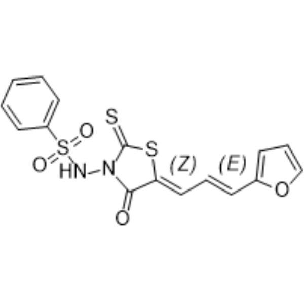
(5Z,2E)-CU-3 is an isomer of CU-3 (HY-121638). CU-3 is a DGKα inhibitor with an IC50 of 0.6 μM. CU-3 competitively reduces DGKα’s affinity for ATP via binding to the enzyme’s catalytic region. CU-3 induces apoptosis in cancer cells. CU-3 promotes T-cell activation and enhances IL-2 production. CU-3 can be used for the research of hepatocellular carcinoma and cervical cancer.
商品は「研究用試薬」です。人や動物の医療用・臨床診断用・食品用の製品ではありません。
研究用途以外に使用した場合、当社は一切の責任を負いかねます。
- 純度 : 95.03%
- CAS 番号: 1815598-71-0
- 分子式: C16H12N2O4S3
- 分子量:392.47
-
保管条件:Powder -20°C, 3 years , 4°C, 2 years ; In solvent -80°C, 2 years , -20°C, 1 year

To place orders, for customer services and technical support, please contact: MedChemExpress USA
Tel: 609-228-6898 E-mail: [email protected] [email protected]


-
 生物活性
生物活性
-
化学情報
-
溶剤 & 溶解度
- プロトコル
- 純度とドキュメンテーション
- 参考文献
-
 Help & FAQs


MedChemExpress(MCE)の使用を引用している文献 (5Z,2E)-CU-3
MoreCaspase アイソフォーム固有の製品をすべて表示
More

生物活性
製品説明
IC50 & Target
[1]|
IL-2 |
体外実験
CU-3 potently and selectively inhibits purified pig DGK α activity with an IC50 of 0.6 μM by targeting the catalytic domain and competitively interfering with ATP binding[1].
CU-3 (2 μM; 24 h) inhibits intracellular DGK α activity in COS-7 cells, reducing phosphatidic acid production induced by overexpressed constitutively active DGK α[1].
CU-3 (5 μM; 24 h) induces apoptosis in HepG2 and HeLa cancer cells, as measured by increased caspase-3/7 activity, without causing nonspecific toxicity to noncancerous COS-7 cells[1].
CU-3 (5 μM; 5 min) enhances Con A-induced IL-2 mRNA expression in Jurkat T cells by inhibiting DGK α, promoting T-cell activation[1].
MedChemExpress (MCE) has not independently confirmed the accuracy of these methods. They are for reference only.


化学情報
-
CAS 番号 1815598-71-0
-
性状 Solid
-
分子量 392.47
-
分子式 C16H12N2O4S3
-
Color Yellow to brown
-
SMILES
O=S(C1=CC=CC=C1)(NN(C/2=O)C(SC2=C/C=C/C3=CC=CO3)=S)=O
-
輸送条件
Room temperature in continental US; may vary elsewhere.
-
保管条件
Powder -20°C 3 years 4°C 2 years In solvent -80°C 2 years -20°C 1 year


Publications (1)
-
Journal Impact Factor
-
Most Recent


溶剤 & 溶解度
体外:
DMSO : 20 mg/mL (50.96 mM; ultrasonic and warming and heat to 60°C; Hygroscopic DMSO has a significant impact on the solubility of product, please use newly opened DMSO)
Please refer to the solubility information to select the appropriate solvent. Once prepared, please aliquot and store the solution to prevent product inactivation from repeated freeze-thaw cycles.
Storage method and period of stock solution: -80°C, 2 years; -20°C, 1 year. When stored at -80°C, please use it within 2 years. When stored at -20°C, please use it within 1 year.
Please refer to the solubility information to select the appropriate solvent. Once prepared, please aliquot and store the solution to prevent product inactivation from repeated freeze-thaw cycles.
Storage method and period of stock solution: -80°C, 2 years; -20°C, 1 year. When stored at -80°C, please use it within 2 years. When stored at -20°C, please use it within 1 year.
濃度 (開始) × 体積 (開始) = 濃度 (終了) × 体積 (終了)
体内:
Select the appropriate dissolution method based on your experimental animal and administration route.
- For the following dissolution methods, please ensure to first prepare a clear stock solution using an In Vitro approach and then sequentially add co-solvents:
- To ensure reliable experimental results, the clarified stock solution can be appropriately stored based on storage conditions. As for the working solution for In Vivo experiments, it is recommended to prepare freshly and use it on the same day.
- The percentages shown for the solvents indicate their volumetric ratio in the final prepared solution. If precipitation or phase separation occurs during preparation, heat and/or sonication can be used to aid dissolution.
Add each solvent one by one: 10% DMSO 90% (20% SBE-β-CD in Saline)
Solubility: 2 mg/mL (5.10 mM); Suspended solution; Need ultrasonic
This protocol yields a suspended solution of 2 mg/mL. Suspended solution can be used for oral and intraperitoneal injection.
Taking 1 mL working solution as an example, add 100 μL DMSO stock solution (20.0 mg/mL) to 900 μL 20% SBE-β-CD in Saline, and mix evenly.
Preparation of 20% SBE-β-CD in Saline (4°C, storage for one week): 2 g SBE-β-CD powder is dissolved in 10 mL Saline, completely dissolve until clear.
In Vivo Dissolution Calculator
Please enter the basic information of animal experiments:
-
-
-
-
Recommended: Prepare an additional quantity of animals to account for potential losses during experiments.
Please enter your animal formula composition:
-
%DMSO +
Recommended: Keep the proportion of DMSO in working solution below 2% if your animal is weak.
-
%+
-
+%Tween-80 + +
-
%Saline +
The co-solvents required include: DMSO, . All of co-solvents are available by MedChemExpress (MCE). , Tween 80. All of co-solvents are available by MedChemExpress (MCE).
Working solution concentration: 0.22 mg/mL
Method for preparing stock solution: mg drug dissolved in μL DMSO. Stock solution concentration: mg/mL.
1. Take μL DMSO stock solution;
2. Add μL .
μL , mix evenly;
3. Then add μL Tween 80, mix evenly;
4. Then add μL
Please ensure that the stock solution in the first step is dissolved to a clear state, and add co-solvents in sequence. You can use ultrasonic heating (ultrasonic cleaner, recommended frequency 20-40 kHz), vortexing, etc. to assist dissolution.


純度とドキュメンテーション
-
データシート (277 KB)
-
SDS (252 KB)
- English - EN (252 KB)
- Français - FR (252 KB)
- Deutsch - DE (252 KB)
- Norwegian - NO (252 KB)
- Español - ES (252 KB)
- Swedish - SV (252 KB)
- Italian - IT (252 KB)
- Korean - KR (252 KB)
- Portuguese - PT (252 KB)
-
取扱説明書 (2659 KB)
参考文献


Complete Stock Solution Preparation Table
Please refer to the solubility information to select the appropriate solvent. Once prepared, please aliquot and store the solution to prevent product inactivation from repeated freeze-thaw cycles.
Storage method and period of stock solution: -80°C, 2 years; -20°C, 1 year. When stored at -80°C, please use it within 2 years. When stored at -20°C, please use it within 1 year.
| Optional Solvent | Concentration Solvent Mass | 1 mg | 5 mg | 10 mg | 25 mg |
|---|---|---|---|---|---|
| DMSO | 1 mM | 2.5480 mL | 12.7398 mL | 25.4797 mL | 63.6991 mL |
| 5 mM | 0.5096 mL | 2.5480 mL | 5.0959 mL | 12.7398 mL | |
| 10 mM | 0.2548 mL | 1.2740 mL | 2.5480 mL | 6.3699 mL | |
| 15 mM | 0.1699 mL | 0.8493 mL | 1.6986 mL | 4.2466 mL | |
| 20 mM | 0.1274 mL | 0.6370 mL | 1.2740 mL | 3.1850 mL | |
| 25 mM | 0.1019 mL | 0.5096 mL | 1.0192 mL | 2.5480 mL | |
| 30 mM | 0.0849 mL | 0.4247 mL | 0.8493 mL | 2.1233 mL | |
| 40 mM | 0.0637 mL | 0.3185 mL | 0.6370 mL | 1.5925 mL | |
| 50 mM | 0.0510 mL | 0.2548 mL | 0.5096 mL | 1.2740 mL |


(5Z,2E)-CU-3 関連分類
HY-121638A 関連分類
 Powered by Bioz