



Hederagenin
Based on 8 publication(s) in Google Scholar
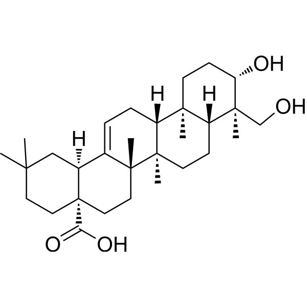
Hederagenin is a triterpenoid saponin with orally active and antitumor activity. Hederagenin can inhibit the expression of iNOS, COX-2, and NF-κB in cells induced by LPS stimulation. Hederagenin also increases ROS production in cancer cells, disrupts mitochondrial membrane potential, and induces apoptosis. Hederagenin also sensitizes cancer cells to Cisplatin (HY-17394) and Paclitaxel (HY-B0015), enhancing induced apoptosis. Hederagenin can also bind to SKP2, with KD = 67.9 μM. Hederagenin also has preventive potential against alcoholic liver injury.
For research use only. We do not sell to patients.
- Purity : 99.95%
- CAS No.: 465-99-6
- Formula: C30H48O4
- Molecular Weight:472.70
-
Storage:Powder -20°C, 3 years , 4°C, 2 years ; In solvent -80°C, 2 years , -20°C, 1 year

To place orders, for customer services and technical support, please contact: MedChemExpress USA
Tel: 609-228-6898 E-mail: [email protected] [email protected]


-
 Biological Activity
-
Chemical Information
-
Solvent & Solubility
- Protocols
- Purity & Documentation
- References
-
 Help & FAQs


-
HY-N0171A
Beta-Sitosterol (purity>98%)Dietary Plant Sterols -
HY-N0183
FormononetinFGFR2 Inhibitor -
HY-N0156
Oleanolic acidAutophagy Inducer -
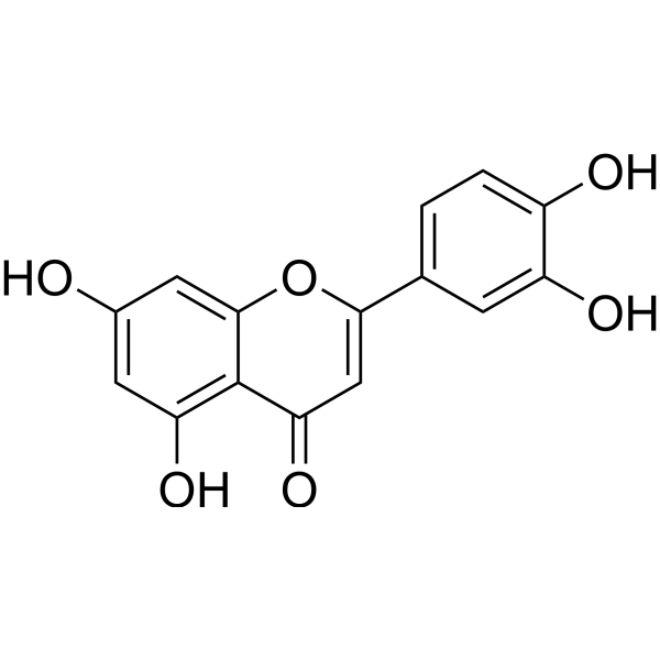
HY-N0162
LuteolinNrf2 Inhibitor -
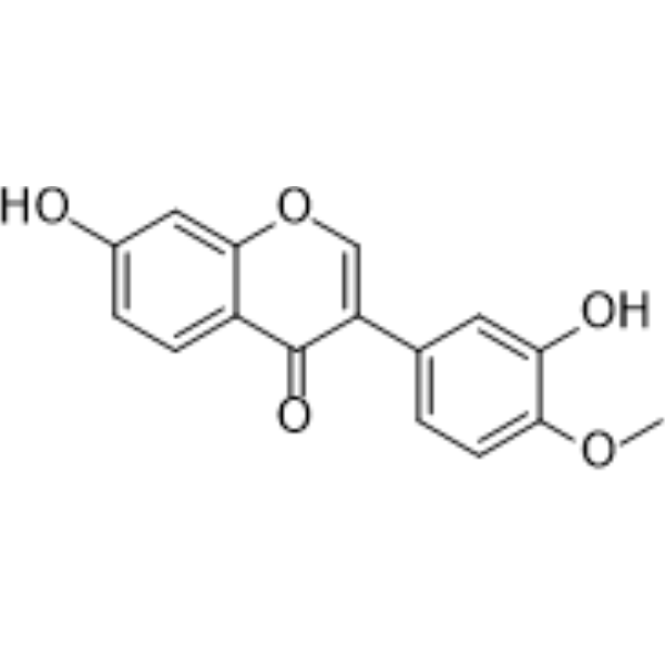
HY-N0519
CalycosinPhytoestrogen -
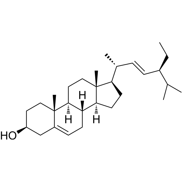
HY-N0131
StigmasterolPlant Sterol -
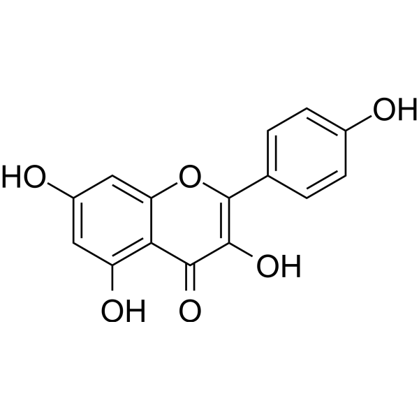
HY-14590
KaempferolEstrogen Receptor α Inhibitor -
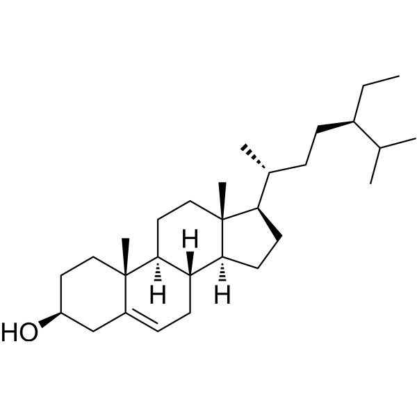
HY-N0171
Beta-Sitosterol (purity>80%)Anticancer Nutraceutical -
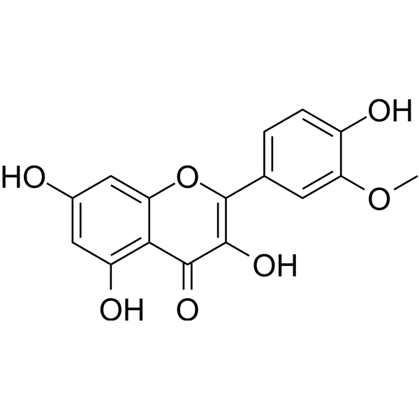
HY-N0776
IsorhamnetinEndogenous Metabolite -
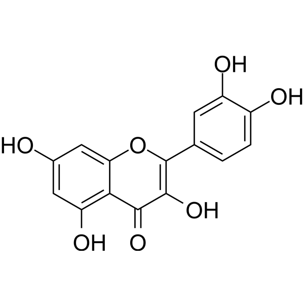
HY-18085
QuercetinFlavonoid Antioxidant -

HY-15141
StaurosporinePKC/PKA Inhibitor -
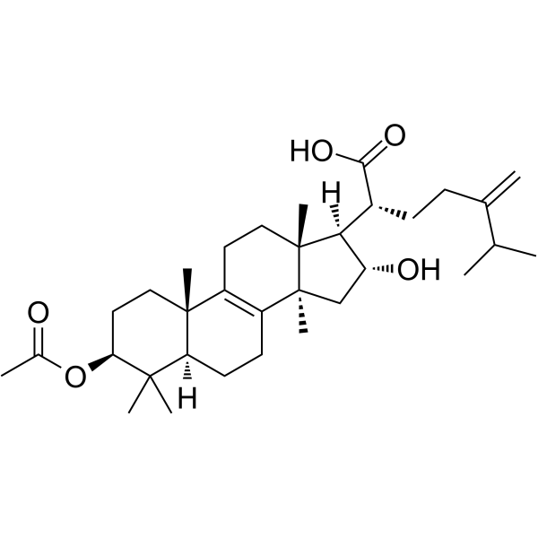
HY-N0371
Pachymic acidTriterpenoid Compound
Publications Citing Use of MedChemExpress (MCE) Hederagenin
More- J Agric Food Chem. 2026 May 13;74(18):14376-14392. [Abstract]
- CNS Neurosci Ther. 2026 Mar;32(3):e70814. [Abstract]
- Bone Joint Res. 2025 Jun 13;14(6):516-526. [Abstract]
- J Proteome Res. 2025 Mar 26. [Abstract]
- Biochem Biophys Res Commun. 2024 Jul 23:718:150085. [Abstract]
- Biol Pharm Bull. 2025;48(10):1514-1525. [Abstract]
- Ann Med Surg (Lond). 2024 Apr 23;86(6):3337-3348. [Abstract]
- Bioengineered. 2022 Apr;13(4):8689-8698. [Abstract]
 Customer Validation & Images

-
Cell Proliferation/Viability Assay
-
Apoptosis Analysis
-
WB
-
RT-PCR
-
Cell Imaging/Staining


Biological Activity
Description
IC50 & Target
[7]|
Skp2 |
Cellular Effect
|
Cell Line

|
Type | Value | Description | References |
|---|---|---|---|---|
| 518A2 | EC50 |
34.9 μM
Compound: He
|
Cytotoxicity against human 518A2 cells assessed as cell survival after 96 hrs by sulforhodamine B assay
Cytotoxicity against human 518A2 cells assessed as cell survival after 96 hrs by sulforhodamine B assay
|
[PMID: 26476750] |
| 518A2 | EC50 |
>30 μM
Compound: He; Hederagenin
|
Cytotoxicity against human 518A2 cells after 96 hrs by SRB assay
Cytotoxicity against human 518A2 cells after 96 hrs by SRB assay
|
[PMID: 27017553] |
| 8505C | EC50 |
38 μM
Compound: He
|
Cytotoxicity against human 8505C cells assessed as cell survival after 96 hrs by sulforhodamine B assay
Cytotoxicity against human 8505C cells assessed as cell survival after 96 hrs by sulforhodamine B assay
|
[PMID: 26476750] |
| 8505C | EC50 |
>30 μM
Compound: He; Hederagenin
|
Cytotoxicity against human 8505C cells after 96 hrs by SRB assay
Cytotoxicity against human 8505C cells after 96 hrs by SRB assay
|
[PMID: 27017553] |
| A2780 | EC50 |
19.9 μM
Compound: He
|
Cytotoxicity against human A2780 cells assessed as cell survival after 96 hrs by sulforhodamine B assay
Cytotoxicity against human A2780 cells assessed as cell survival after 96 hrs by sulforhodamine B assay
|
[PMID: 26476750] |
| A2780 | EC50 |
>30 μM
Compound: He; Hederagenin
|
Cytotoxicity against human A2780 cells after 96 hrs by SRB assay
Cytotoxicity against human A2780 cells after 96 hrs by SRB assay
|
[PMID: 27017553] |
| A2780 | EC50 |
>60 μM
Compound: He
|
Cytotoxicity against human A2780 cells assessed as reduction in cell viability incubated fro 96 hrs by SRB assay
Cytotoxicity against human A2780 cells assessed as reduction in cell viability incubated fro 96 hrs by SRB assay
|
[PMID: 29024910] |
| A2780 | EC50 |
>30 μM
Compound: He
|
Cytotoxicity against human A2780 cells assessed as reduction in cell viability after 96 hrs by SRB assay
Cytotoxicity against human A2780 cells assessed as reduction in cell viability after 96 hrs by SRB assay
|
[PMID: 30840925] |
| A-375 | EC50 |
>60 μM
Compound: He
|
Cytotoxicity against human A375 cells assessed as reduction in cell viability incubated fro 96 hrs by SRB assay
Cytotoxicity against human A375 cells assessed as reduction in cell viability incubated fro 96 hrs by SRB assay
|
[PMID: 29024910] |
| A549 | IC50 |
>200 μM
Compound: 3
|
Cell membrane permeabilization in human A549 cells assessed as drug level causing decrease in calcein fluorescence
Cell membrane permeabilization in human A549 cells assessed as drug level causing decrease in calcein fluorescence
|
[PMID: 19200744] |
| A549 | IC50 |
39 μM
Compound: 3
|
Cytotoxicity against human A549 cells after 48 hrs by resazurin reduction test
Cytotoxicity against human A549 cells after 48 hrs by resazurin reduction test
|
[PMID: 19200744] |
| A549 | IC50 |
38.64 μM
Compound: HE
|
Cytotoxicity against human A549 cells assessed as cell viability after 72 hrs by MTT assay
Cytotoxicity against human A549 cells assessed as cell viability after 72 hrs by MTT assay
|
[PMID: 23992864] |
| A549 | EC50 |
29 μM
Compound: He
|
Cytotoxicity against human A549 cells assessed as cell survival after 96 hrs by sulforhodamine B assay
Cytotoxicity against human A549 cells assessed as cell survival after 96 hrs by sulforhodamine B assay
|
[PMID: 26476750] |
| A549 | EC50 |
>30 μM
Compound: He; Hederagenin
|
Cytotoxicity against human A549 cells after 96 hrs by SRB assay
Cytotoxicity against human A549 cells after 96 hrs by SRB assay
|
[PMID: 27017553] |
| A549 | IC50 |
10.249 μM
Compound: Hederagenin
|
Cytotoxicity against human A549 cells assessed as reduction in cell viability after 48 hrs by MTT assay
Cytotoxicity against human A549 cells assessed as reduction in cell viability after 48 hrs by MTT assay
|
[PMID: 28237662] |
| A549 | IC50 |
>100 μM
Compound: alpha-hederagenin
|
Antiproliferative activity against human A549 cells after 72 hrs by MTT assay
Antiproliferative activity against human A549 cells after 72 hrs by MTT assay
|
[PMID: 29040953] |
| A549 | IC50 |
>10 μM
Compound: Hederagenin
|
Cytotoxicity against human A549 cells after 72 hrs by sulforhodamine B assay
Cytotoxicity against human A549 cells after 72 hrs by sulforhodamine B assay
|
[PMID: 29131631] |
| A549 | IC50 |
>10 μM
Compound: Hederagenin
|
Antiproliferative activity against human A549 cells harbouring wild type EGFR incubated for 72 hrs by SRB assay
Antiproliferative activity against human A549 cells harbouring wild type EGFR incubated for 72 hrs by SRB assay
|
[PMID: 31718182] |
| A549 | IC50 |
>50 μM
Compound: He
|
Cytotoxicity against human A549 cells assessed as reduction in cell viability measured after 24 hrs by MTT assay
Cytotoxicity against human A549 cells assessed as reduction in cell viability measured after 24 hrs by MTT assay
|
[PMID: 37859723] |
| A549 | IC50 |
26.23 μM
Compound: He
|
Cytotoxicity against human A549 cells assessed as reduction in cell growth incubated for 48 hrs by Alamar blue assay
Cytotoxicity against human A549 cells assessed as reduction in cell growth incubated for 48 hrs by Alamar blue assay
|
[PMID: 37859723] |
| AGS | IC50 |
36.14 μM
Compound: alpha-hederagenin
|
Antiproliferative activity against human AGS cells after 72 hrs by MTT assay
Antiproliferative activity against human AGS cells after 72 hrs by MTT assay
|
[PMID: 29040953] |
| BC | IC50 |
>5 μg/mL
Compound: Hederagenin
|
Cytotoxicity against human BC cells by colorimetric method
Cytotoxicity against human BC cells by colorimetric method
|
[PMID: 16038539] |
| BGC-823 | IC50 |
52.07 μM
Compound: alpha-hederagenin
|
Antiproliferative activity against human BGC823 cells after 72 hrs by MTT assay
Antiproliferative activity against human BGC823 cells after 72 hrs by MTT assay
|
[PMID: 29040953] |
| BT-20 | IC50 |
11.8 μM
Compound: He
|
Cytotoxicity against human BT-20 cells assessed as reduction in cell viability incubated for 48 hrs by Alamar blue assay
Cytotoxicity against human BT-20 cells assessed as reduction in cell viability incubated for 48 hrs by Alamar blue assay
|
[PMID: 37859723] |
| DLD-1 | IC50 |
>100 μM
Compound: 3
|
Cytotoxicity against human DLD1 cells after 48 hrs by resazurin reduction test
Cytotoxicity against human DLD1 cells after 48 hrs by resazurin reduction test
|
[PMID: 19200744] |
| DLD-1 | IC50 |
>200 μM
Compound: 3
|
Cell membrane permeabilization in human DLD1 cells assessed as drug level causing decrease in calcein fluorescence
Cell membrane permeabilization in human DLD1 cells assessed as drug level causing decrease in calcein fluorescence
|
[PMID: 19200744] |
| FaDu | EC50 |
>60 μM
Compound: He
|
Cytotoxicity against human FADU cells assessed as reduction in cell viability incubated fro 96 hrs by SRB assay
Cytotoxicity against human FADU cells assessed as reduction in cell viability incubated fro 96 hrs by SRB assay
|
[PMID: 29024910] |
| FaDu | EC50 |
>30 μM
Compound: He
|
Cytotoxicity against human FADU cells assessed as reduction in cell viability after 96 hrs by SRB assay
Cytotoxicity against human FADU cells assessed as reduction in cell viability after 96 hrs by SRB assay
|
[PMID: 30840925] |
| HeLa | IC50 |
42.27 μM
Compound: HE
|
Cytotoxicity against human HeLa cells assessed as cell viability after 72 hrs by MTT assay
Cytotoxicity against human HeLa cells assessed as cell viability after 72 hrs by MTT assay
|
[PMID: 23992864] |
| HeLa | IC50 |
53.96 μM
Compound: alpha-hederagenin
|
Antiproliferative activity against human HeLa cells after 72 hrs by MTT assay
Antiproliferative activity against human HeLa cells after 72 hrs by MTT assay
|
[PMID: 29040953] |
| HeLa | IC50 |
17.42 μg/mL
Compound: He
|
Antiproliferative activity against human HeLa cells assessed as reduction in cell viability measured after 72 hrs by MTT assay
Antiproliferative activity against human HeLa cells assessed as reduction in cell viability measured after 72 hrs by MTT assay
|
[PMID: 37859723] |
| HepG2 | IC50 |
>20 μM
Compound: 15
|
Inhibition of TNF-alpha-induced NFkappaB activation in human HepG2 cells after 1 hr by luciferase reporter assay
Inhibition of TNF-alpha-induced NFkappaB activation in human HepG2 cells after 1 hr by luciferase reporter assay
|
[PMID: 21870831] |
| HepG2 | IC50 |
28.05 μM
Compound: HE
|
Cytotoxicity against human HepG2 cells assessed as cell viability after 72 hrs by MTT assay
Cytotoxicity against human HepG2 cells assessed as cell viability after 72 hrs by MTT assay
|
[PMID: 23992864] |
| HepG2 | CC50 |
>1000 μM
Compound: He
|
Cytotoxicity in human HepG2 cells assessed as reduction in cell viability incubated for 48 hrs by MTT assay
Cytotoxicity in human HepG2 cells assessed as reduction in cell viability incubated for 48 hrs by MTT assay
|
[PMID: 29024910] |
| HepG2 | IC50 |
35.87 μM
Compound: He
|
Antiproliferative activity against human HepG2 cells assessed as reduction in cell viability after 48 hrs by MTT assay
Antiproliferative activity against human HepG2 cells assessed as reduction in cell viability after 48 hrs by MTT assay
|
[PMID: 37859723] |
| HL-60 | IC50 |
27.52 μM
Compound: HE
|
Cytotoxicity against human HL60 cells assessed as cell viability after 72 hrs by MTT assay
Cytotoxicity against human HL60 cells assessed as cell viability after 72 hrs by MTT assay
|
[PMID: 23992864] |
| HT-29 | EC50 |
50 μM
Compound: He
|
Cytotoxicity against human HT-29 cells assessed as cell survival after 96 hrs by sulforhodamine B assay
Cytotoxicity against human HT-29 cells assessed as cell survival after 96 hrs by sulforhodamine B assay
|
[PMID: 26476750] |
| HT-29 | EC50 |
>30 μM
Compound: He; Hederagenin
|
Cytotoxicity against human HT-29 cells after 96 hrs by SRB assay
Cytotoxicity against human HT-29 cells after 96 hrs by SRB assay
|
[PMID: 27017553] |
| HT-29 | EC50 |
>60 μM
Compound: He
|
Cytotoxicity against human HT-29 cells assessed as reduction in cell viability incubated fro 96 hrs by SRB assay
Cytotoxicity against human HT-29 cells assessed as reduction in cell viability incubated fro 96 hrs by SRB assay
|
[PMID: 29024910] |
| HT-29 | EC50 |
>30 μM
Compound: He
|
Cytotoxicity against human HT-29 cells assessed as reduction in cell viability after 96 hrs by SRB assay
Cytotoxicity against human HT-29 cells assessed as reduction in cell viability after 96 hrs by SRB assay
|
[PMID: 30840925] |
| KB | IC50 |
>5 μg/mL
Compound: Hederagenin
|
Cytotoxicity against human KB cells by colorimetric method
Cytotoxicity against human KB cells by colorimetric method
|
[PMID: 16038539] |
| KB | IC50 |
14.631 μM
Compound: Hederagenin
|
Cytotoxicity against human KB cells assessed as reduction in cell viability after 48 hrs by MTT assay
Cytotoxicity against human KB cells assessed as reduction in cell viability after 48 hrs by MTT assay
|
[PMID: 28237662] |
| KB | IC50 |
54.43 μM
Compound: alpha-hederagenin
|
Antiproliferative activity against human KB cells after 72 hrs by MTT assay
Antiproliferative activity against human KB cells after 72 hrs by MTT assay
|
[PMID: 29040953] |
| KB | IC50 |
>10 μM
Compound: Hederagenin
|
Cytotoxicity against human KB cells after 72 hrs by sulforhodamine B assay
Cytotoxicity against human KB cells after 72 hrs by sulforhodamine B assay
|
[PMID: 29131631] |
| KB | IC50 |
>10 μM
Compound: Hederagenin
|
Antiproliferative activity against human KB cells incubated for 72 hrs by SRB assay
Antiproliferative activity against human KB cells incubated for 72 hrs by SRB assay
|
[PMID: 31718182] |
| LoVo | IC50 |
1.17 μM
Compound: He
|
Antiproliferative activity against human LoVo cells assessed as reduction in cell viability incubated for 48 hrs by MTT assay
Antiproliferative activity against human LoVo cells assessed as reduction in cell viability incubated for 48 hrs by MTT assay
|
[PMID: 37859723] |
| LoVo | IC50 |
1.39 μM
Compound: He
|
Antiproliferative activity against human LoVo cells assessed as reduction in cell viability incubated for 24 hrs by MTT assay
Antiproliferative activity against human LoVo cells assessed as reduction in cell viability incubated for 24 hrs by MTT assay
|
[PMID: 37859723] |
| M14 | IC50 |
58 μM
Compound: 6
|
Cytotoxicity against human M14 cells after 48 hrs by MTT assay
Cytotoxicity against human M14 cells after 48 hrs by MTT assay
|
[PMID: 21954959] |
| MCF7 | EC50 |
25.7 μM
Compound: He
|
Cytotoxicity against human MCF7 cells assessed as cell survival after 96 hrs by sulforhodamine B assay
Cytotoxicity against human MCF7 cells assessed as cell survival after 96 hrs by sulforhodamine B assay
|
[PMID: 26476750] |
| MCF7 | EC50 |
>30 μM
Compound: He; Hederagenin
|
Cytotoxicity against human MCF7 cells after 96 hrs by SRB assay
Cytotoxicity against human MCF7 cells after 96 hrs by SRB assay
|
[PMID: 27017553] |
| MCF7 | IC50 |
39.389 μM
Compound: Hederagenin
|
Cytotoxicity against human MCF7 cells assessed as reduction in cell viability after 48 hrs by MTT assay
Cytotoxicity against human MCF7 cells assessed as reduction in cell viability after 48 hrs by MTT assay
|
[PMID: 28237662] |
| MCF7 | IC50 |
>10 μM
Compound: Hederagenin
|
Cytotoxicity against human MCF7 cells after 72 hrs by sulforhodamine B assay
Cytotoxicity against human MCF7 cells after 72 hrs by sulforhodamine B assay
|
[PMID: 29131631] |
| MCF7 | EC50 |
>30 μM
Compound: He
|
Cytotoxicity against human MCF7 cells assessed as reduction in cell viability after 96 hrs by SRB assay
Cytotoxicity against human MCF7 cells assessed as reduction in cell viability after 96 hrs by SRB assay
|
[PMID: 30840925] |
| MCF7 | IC50 |
>10 μM
Compound: Hederagenin
|
Antiproliferative activity against human MCF7 cells incubated for 72 hrs by SRB assay
Antiproliferative activity against human MCF7 cells incubated for 72 hrs by SRB assay
|
[PMID: 31718182] |
| MDA-MB-231 | IC50 |
36.609 μM
Compound: Hederagenin
|
Cytotoxicity against human MDA-MB-231 cells assessed as reduction in cell viability after 48 hrs by MTT assay
Cytotoxicity against human MDA-MB-231 cells assessed as reduction in cell viability after 48 hrs by MTT assay
|
[PMID: 28237662] |
| MDA-MB-231 | IC50 |
>10 μM
Compound: Hederagenin
|
Cytotoxicity against human MDA-MB-231 cells after 72 hrs by sulforhodamine B assay
Cytotoxicity against human MDA-MB-231 cells after 72 hrs by sulforhodamine B assay
|
[PMID: 29131631] |
| MDA-MB-231 | IC50 |
>10 μM
Compound: Hederagenin
|
Antiproliferative activity against human MDA-MB-231 cells incubated for 72 hrs by SRB assay
Antiproliferative activity against human MDA-MB-231 cells incubated for 72 hrs by SRB assay
|
[PMID: 31718182] |
| MKN-45 | IC50 |
53.69 μM
Compound: alpha-hederagenin
|
Antiproliferative activity against human MKN45 cells after 72 hrs by MTT assay
Antiproliferative activity against human MKN45 cells after 72 hrs by MTT assay
|
[PMID: 29040953] |
| NCI-H187 | IC50 |
>5 μg/mL
Compound: Hederagenin
|
Cytotoxicity against human NCI-H187 cells by colorimetric method
Cytotoxicity against human NCI-H187 cells by colorimetric method
|
[PMID: 16038539] |
| NCI-H1975 | IC50 |
>10 μM
Compound: Hederagenin
|
Antiproliferative activity against human NCI-H1975 cells harbouring EGFR L858R/T790M/C797S mutant incubated for 72 hrs by MTS assay
Antiproliferative activity against human NCI-H1975 cells harbouring EGFR L858R/T790M/C797S mutant incubated for 72 hrs by MTS assay
|
[PMID: 31718182] |
| NCI-H1975 | IC50 |
>10 μM
Compound: Hederagenin
|
Antiproliferative activity against human NCI-H1975 cells incubated for 72 hrs by MTS assay
Antiproliferative activity against human NCI-H1975 cells incubated for 72 hrs by MTS assay
|
[PMID: 31718182] |
| NIH3T3 | EC50 |
>30 μM
Compound: He; Hederagenin
|
Cytotoxicity against mouse NIH/3T3 cells after 96 hrs by SRB assay
Cytotoxicity against mouse NIH/3T3 cells after 96 hrs by SRB assay
|
[PMID: 27017553] |
| NIH3T3 | EC50 |
>30 μM
Compound: He
|
Cytotoxicity against mouse NIH/3T3 cells assessed as reduction in cell viability after 96 hrs by SRB assay
Cytotoxicity against mouse NIH/3T3 cells assessed as reduction in cell viability after 96 hrs by SRB assay
|
[PMID: 30840925] |
| Sf9 | IC50 |
>20 μM
Compound: Hederagenin
|
Inhibition of C-terminal His-tagged/ N-terminal GST-tagged recombinant human EGFR (668 to 1210 residues) expressed in a Baculovirus infected Sf9 cell expression system using poly-EY as substrate incubated for 30 mins by ADP-Glo kinase assay
Inhibition of C-terminal His-tagged/ N-terminal GST-tagged recombinant human EGFR (668 to 1210 residues) expressed in a Baculovirus infected Sf9 cell expression system using poly-EY as substrate incubated for 30 mins by ADP-Glo kinase assay
|
[PMID: 31718182] |
| Sf9 | IC50 |
>20 μM
Compound: Hederagenin
|
Inhibition of C-terminal His-tagged/ N-terminal GST-tagged recombinant human EGFR L858R/T790M double mutant (668 to 1210 residues) expressed in a Baculovirus infected Sf9 cell expression system using poly-EY as substrate incubated for 30 mins by ADP-Glo k
Inhibition of C-terminal His-tagged/ N-terminal GST-tagged recombinant human EGFR L858R/T790M double mutant (668 to 1210 residues) expressed in a Baculovirus infected Sf9 cell expression system using poly-EY as substrate incubated for 30 mins by ADP-Glo k
|
[PMID: 31718182] |
| Sf9 | IC50 |
>20 μM
Compound: Hederagenin
|
Inhibition of C-terminal His-tagged/ N-terminal GST-tagged recombinant human EGFR L858R/T790M/C797S mutant (668 to 1210 residues) expressed in a Baculovirus infected Sf9 cell expression system using poly-EY as substrate incubated for 30 mins by ADP-Glo ki
Inhibition of C-terminal His-tagged/ N-terminal GST-tagged recombinant human EGFR L858R/T790M/C797S mutant (668 to 1210 residues) expressed in a Baculovirus infected Sf9 cell expression system using poly-EY as substrate incubated for 30 mins by ADP-Glo ki
|
[PMID: 31718182] |
| SW-1736 | EC50 |
>60 μM
Compound: He
|
Cytotoxicity against human SW1736 cells assessed as reduction in cell viability incubated fro 96 hrs by SRB assay
Cytotoxicity against human SW1736 cells assessed as reduction in cell viability incubated fro 96 hrs by SRB assay
|
[PMID: 29024910] |
| SW-1736 | EC50 |
>30 μM
Compound: He
|
Cytotoxicity against human SW1736 cells assessed as reduction in cell viability after 96 hrs by SRB assay
Cytotoxicity against human SW1736 cells assessed as reduction in cell viability after 96 hrs by SRB assay
|
[PMID: 30840925] |
| U-87MG ATCC | IC50 |
35.95 μM
Compound: HE
|
Cytotoxicity against human U87MG cells assessed as cell viability after 72 hrs by MTT assay
Cytotoxicity against human U87MG cells assessed as cell viability after 72 hrs by MTT assay
|
[PMID: 23992864] |
In Vitro
Hederagenin (1 μM, 2 μM; 48 h) disrupts mitochondrial membrane potential, increases intracellular reactive oxygen species (ROS) content, and has a cytotoxic effect on cancer cells[4].
Hederagenin (1 μM, 2 μM; 48 h) induces apoptosis in human colon cancer LoVo cells, upregulates apoptosis-related proteins (Bax), and reduces apoptosis inhibitory proteins (Bcl-2, Bcl-xL, and Survivin) levels[4].
Hederagenin (50 μM; 4 h) also blocks autophagic flux-induced ROS accumulation and enhances the cytotoxicity of Cisplatin and Paclitaxel in lung cancer cells[5].
MedChemExpress (MCE) has not independently confirmed the accuracy of these methods. They are for reference only. Further protocols information, click here.
In Vivo
Hederagenin (50 mg/kg; po; once daily for 21 days) exerts anti-inflammatory and anti-apoptotic activities and reduces liver damage induced by 25% ethanol in mice[5].
MedChemExpress (MCE) has not independently confirmed the accuracy of these methods. They are for reference only.


Chemical Information
-
CAS No. 465-99-6
-
Appearance Solid
-
Molecular Weight 472.70
-
Formula C30H48O4
-
Color White to off-white
-
SMILES
CC1(C)CC[C@@](CC[C@]2(C)C3=CC[C@@]4([H])[C@@]2(C)CC[C@]5([H])[C@]4(C)CC[C@H](O)[C@]5(CO)C)(C(O)=O)[C@@]3([H])C1
-
Structure Classification
-
Shipping
Room temperature in continental US; may vary elsewhere.
-
Storage
Powder -20°C 3 years 4°C 2 years In solvent -80°C 2 years -20°C 1 year


Publications (8)
-
Journal Impact Factor
-
Most Recent
-
J Agric Food Chem
Distinctive Behavior and Selective Modulation of PPARγ by Pentacyclic Triterpenoid Pomolic Acid and Hederagenin from Rosa canina. [Abstract]2026 May 13;74(18):14376-14392. PMID: 42057314 -
CNS Neurosci Ther
ShenQi DiHuang Decoction (SQDHD) Ameliorates Neuroinflammation and Neuropsychiatric Manifestations in Pristane Induced Lupus Mice via Blocking JAK1-STAT3 Pathway. [Abstract]2026 Mar;32(3):e70814. PMID: 41795136 -
Bone Joint Res
Hederagenin promotes SIRT6 to attenuate epidural scar formation by aggravating PRMT1 deacetylation. [Abstract]2025 Jun 13;14(6):516-526. PMID: 40511498
Hederagenin purchased from MedChemExpress. Usage Cited in: Bone Joint Res. 2025 Jun 13;14(6):516-526. [Abstract]
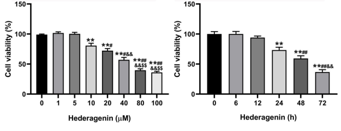
Cell Counting Kit (CCK)-8 method was performed to evaluate cell viability treated with Hederagenin (1, 5, 10, 20, 40, 80, 100 μM, 6, 12, 24, 48, 72 h).
Hederagenin purchased from MedChemExpress. Usage Cited in: Bone Joint Res. 2025 Jun 13;14(6):516-526. [Abstract]
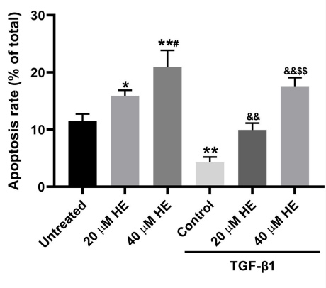
Apoptosis rate of fibroblasts was measured using flow cytometry treated with Hederageni (HE) (20, 40 μM).
Hederagenin purchased from MedChemExpress. Usage Cited in: Bone Joint Res. 2025 Jun 13;14(6):516-526. [Abstract]
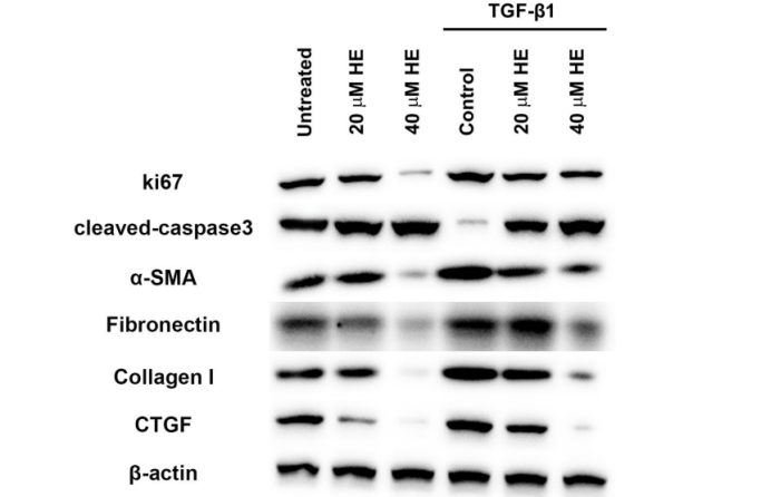
The expressions of target proteins were evaluated by western blotting treated withHederagenin (HE) (20, 40 μM).
Hederagenin purchased from MedChemExpress. Usage Cited in: Bone Joint Res. 2025 Jun 13;14(6):516-526. [Abstract]
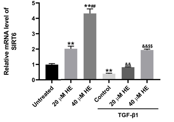
Hederagenin (HE) (20, 40 μM) treatment increased SIRT6 mRNA expression in a dose-dependent manner in fibroblasts with or without TGF-β1 stimulation by RT-qPCR.
-
J Proteome Res
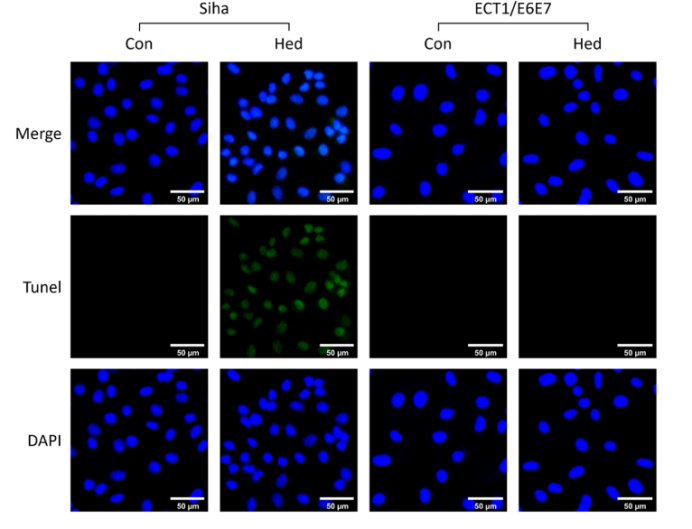
Elucidating the Molecular Mechanisms of Hederagenin-Regulated Mitophagy in Cervical Cancer SiHa Cells through an Integrative Approach Combining Proteomics and Advanced Network Association Algorithm. [Abstract]2025 Mar 26. PMID: 40135937
Hederagenin purchased from MedChemExpress. Usage Cited in: J Proteome Res. 2025 Mar 26. [Abstract]
SiHa and ECT1/E6E7 for TUNEL/DAPI staining treated with Hederagenin (Hed).
-
Biochem Biophys Res Commun
Hederagenin promotes lung cancer cell death by activating CHAC1-dependent ferroptosis pathway. [Abstract]2024 Jul 23:718:150085. PMID: 38735142 -
Biol Pharm Bull
He-Wei-Decoction Ameliorates Chronic Atrophic Gastritis via Modulation of the TLR4/NF-κB Signaling Pathway. [Abstract]2025;48(10):1514-1525. PMID: 41083380 -
Ann Med Surg (Lond)
Network pharmacology prediction and experiment validation of anti-liver cancer activity of Curcumae Rhizoma and Hedyotis diffusa Willd. [Abstract]2024 Apr 23;86(6):3337-3348. PMID: 38846818 -
Bioengineered
The protective effect of hederagenin on renal fibrosis by targeting muscarinic acetylcholine receptor. [Abstract]2022 Apr;13(4):8689-8698. PMID: 35322725


Solvent & Solubility
In Vitro:
DMSO : 50 mg/mL (105.78 mM; Need ultrasonic; Hygroscopic DMSO has a significant impact on the solubility of product, please use newly opened DMSO)
Please refer to the solubility information to select the appropriate solvent. Once prepared, please aliquot and store the solution to prevent product inactivation from repeated freeze-thaw cycles.
Storage method and period of stock solution: -80°C, 2 years; -20°C, 1 year. When stored at -80°C, please use it within 2 years. When stored at -20°C, please use it within 1 year.
Please refer to the solubility information to select the appropriate solvent. Once prepared, please aliquot and store the solution to prevent product inactivation from repeated freeze-thaw cycles.
Storage method and period of stock solution: -80°C, 2 years; -20°C, 1 year. When stored at -80°C, please use it within 2 years. When stored at -20°C, please use it within 1 year.
Concentration (start) × Volume (start) = Concentration (final) × Volume (final)
In Vivo:
Select the appropriate dissolution method based on your experimental animal and administration route.
- For the following dissolution methods, please ensure to first prepare a clear stock solution using an In Vitro approach and then sequentially add co-solvents:
- To ensure reliable experimental results, the clarified stock solution can be appropriately stored based on storage conditions. As for the working solution for In Vivo experiments, it is recommended to prepare freshly and use it on the same day.
- The percentages shown for the solvents indicate their volumetric ratio in the final prepared solution. If precipitation or phase separation occurs during preparation, heat and/or sonication can be used to aid dissolution.
Add each solvent one by one: 10% DMSO 90% (20% SBE-β-CD in Saline)
Solubility: ≥ 2.5 mg/mL (5.29 mM); Clear solution
This protocol yields a clear solution of ≥ 2.5 mg/mL (saturation unknown).
Taking 1 mL working solution as an example, add 100 μL DMSO stock solution (25.0 mg/mL) to 900 μL 20% SBE-β-CD in Saline, and mix evenly.
Preparation of 20% SBE-β-CD in Saline (4°C, storage for one week): 2 g SBE-β-CD powder is dissolved in 10 mL Saline, completely dissolve until clear.
In Vivo Dissolution Calculator
Please enter the basic information of animal experiments:
-
-
-
-
Recommended: Prepare an additional quantity of animals to account for potential losses during experiments.
Please enter your animal formula composition:
-
%DMSO +
Recommended: Keep the proportion of DMSO in working solution below 2% if your animal is weak.
-
%+
-
+%Tween-80 + +
-
%Saline +
The co-solvents required include: DMSO, . All of co-solvents are available by MedChemExpress (MCE). , Tween 80. All of co-solvents are available by MedChemExpress (MCE).
Working solution concentration: 0.22 mg/mL
Method for preparing stock solution: mg drug dissolved in μL DMSO. Stock solution concentration: mg/mL.
1. Take μL DMSO stock solution;
2. Add μL .
μL , mix evenly;
3. Then add μL Tween 80, mix evenly;
4. Then add μL
Please ensure that the stock solution in the first step is dissolved to a clear state, and add co-solvents in sequence. You can use ultrasonic heating (ultrasonic cleaner, recommended frequency 20-40 kHz), vortexing, etc. to assist dissolution.

Protocols
-
RNA extraction experimental
By lysing cells, releasing RNA, and removing impurities such as proteins and DNA, high-purity RNA products are finally obtained. The commonly used traditional method is the guanidine isothiocyanate/phenol/chloroform method (Trizol), which is suitable for a variety of animal materials including animal tissues, microorganisms, cultured cells, etc., and most plant materials.
-
Mitochondrial membrane-potential fluorescent assay
Mitochondrial membrane potential fluorescent assays estimate ΔΨm in living cells using lipophilic cationic dyes such as TMRM, TMRE, rhodamine 123, and JC-1, which accumulate in mitochondria according to membrane polarization; loss of signal after FCCP or CCCP treatment is interpreted as mitochondrial depolarization. TMRM/TMRE and rhodamine 123 are commonly used for semi-quantitative live-cell microscopy or flow cytometry, while JC-1 can report a shift from red aggregate fluorescence to green monomer fluorescence during depolarization; interpretation requires controls because dye concentration, quenching mode, cell type, dye efflux, and mitochondrial mass can affect fluorescence independently of ΔΨm.
-
Apoptosis
Apoptosis, also called programmed cell death, is generally characterized by distinct morphological characteristics.
-
TUNEL staining for apoptotic DNA fragmentation
TUNEL staining detects DNA strand breaks by using terminal deoxynucleotidyl transferase to add labeled nucleotides to exposed 3′-OH DNA termini, generating either microscopic staining in fixed cells or tissue sections, or fluorescence/cytometric signal in cell suspensions. TUNEL positivity reflects DNA fragmentation but should not be interpreted alone as definitive apoptosis, because TUNEL can also label necrotic, autolytic, mechanically damaged, or DNA-repair-associated DNA breaks.
-
Annexin V plus membrane-impermeant dye apoptosis staining
Annexin V-based apoptosis assays rely on the detection of phosphatidylserine (PS) externalization from the inner leaflet of the plasma membrane to the outer leaflet, an early biochemical hallmark of apoptosis. Fluorescently labeled Annexin V binds PS in a calcium-dependent manner, enabling identification of early apoptotic cells by flow cytometry or fluorescence microscopy. When combined with a membrane-impermeant DNA-binding dye (e. g. , propidium iodide), this approach allows discrimination between viable (Annexin V−/dye−), early apoptotic (Annexin V+/dye−), and late apoptotic or necrotic (Annexin V+/dye+) cell populations by assessing membrane integrity and PS exposure.
-
ROS/oxidative-stress fluorescent staining
ROS/oxidative-stress fluorescent staining uses cell-permeant fluorogenic probes that become fluorescent after oxidation inside cells or tissues; commonly used examples include DCFH-DA/DCFDA for broad cellular oxidant detection, DHE for superoxide-related signal detection, MitoSOX for mitochondrial superoxide-related signal detection, and CellROX probes for oxidative-stress-associated fluorescence readouts. The assay detects probe oxidation rather than a single ROS species unless the probe and analysis method have been chemically validated for that species. DCFH-DA enters cells, is deacetylated by intracellular esterases to DCFH, and produces fluorescent DCF after oxidation, so the readout is used as an operational measure of total cellular oxidative stress rather than a species-specific ROS measurement. DHE and MitoSOX can report superoxide-related oxidation, but red fluorescence alone can include non-specific ethidium-like oxidation products; HPLC or optimized spectral approaches are
-
Fluorescent plasma-membrane potential dye assay
Fluorescent plasma-membrane potential dye assays measure changes in cell membrane potential using voltage-sensitive dyes whose fluorescence changes when cells depolarize or hyperpolarize. Anionic bis-oxonol dyes such as DiBAC4(3) enter depolarized cells more readily and show increased fluorescence after intracellular binding, while hyperpolarization reduces dye accumulation and fluorescence. FMP/FLIPR membrane-potential dyes are used for faster, homogeneous microplate assays of ion-channel or receptor-mediated membrane-potential changes.
-
Mitochondrial membrane-potential and mitochondrial mass staining
Mitochondrial membrane potential staining measures the electrochemical polarization across the mitochondrial inner membrane in live cells using lipophilic cationic fluorescent probes; early rhodamine-based work showed that selective mitochondrial dye accumulation is lost when the mitochondrial transmembrane potential is dissipated. JC-1 reports mitochondrial polarization by shifting from green monomer fluorescence to red J-aggregate fluorescence as dye concentration increases within energized mitochondria; therefore, the red/green fluorescence ratio is used as a relative readout of mitochondrial membrane potential. TMRE or TMRM staining provides a single-channel relative readout because these cationic rhodamine esters accumulate in polarized mitochondria, and lower fluorescence indicates reduced mitochondrial polarization when acquisition and dye-loading conditions are controlled. Mitochondrial mass staining is commonly performed with MitoTracker Green FM or related MitoTracker dyes as
-
Apoptosis Solutions
Apoptosis is a regulated, generally non-lytic cell-death pathway that removes unwanted, damaged, infected, or abnormal cells through coordinated morphological changes, caspase activation, DNA fragmentation, and membrane remodeling. The intrinsic apoptosis pathway is controlled mainly by mitochondrial outer membrane permeabilization, BCL-2 family proteins, cytochrome c release, apoptosome formation, caspase-9 activation, and downstream executioner caspase-3/7 activation. The extrinsic apoptosis pathway is initiated by death receptors such as Fas, TNFR, and TRAIL receptors, which recruit adaptor proteins and activate caspase-8 before engaging executioner caspases or mitochondrial amplification through BID cleavage. Apoptosis is linked to many phenotypes, including cancer cell killing, tissue homeostasis, immune regulation, neurodegeneration, infection response, and treatment-induced cytotoxicity; unresolved questions include how apoptosis interacts with necroptosis, pyroptosis, ferroptos
-
Research Protocol for Inflammation-related Diseases
The NLRP3 inflammasome is a cytosolic innate immune signaling platform that integrates priming signals and danger-signal activation to promote caspase-1 activation, maturation of IL-1β and IL-18, and gasdermin D-mediated pyroptotic cell death. The core experimental logic is to determine whether inflammatory disease phenotypes are driven by increased NLRP3 expression, ASC-containing inflammasome assembly, caspase-1 cleavage, GSDMD cleavage, and extracellular release of IL-1β/IL-18 rather than by nonspecific cell injury alone. The pathway is strongly linked to inflammation-related disease phenotypes because monosodium urate crystals activate NALP3/NLRP3 inflammasome signaling in gout-like crystal inflammation, cholesterol crystals activate NLRP3 inflammasomes in atherogenesis models, and DSS-induced intestinal inflammation has been reported to involve NLRP3 inflammasome activity. However, experimental colitis studies also show context-dependent protective effects of NLRP3 inflammasome co
-
Subcutaneous Cell-Line-Derived Xenograft
Subcutaneous cell-line-derived xenograft (CDX) models are established by implanting cultured human cancer cell lines into immunodeficient mice, where the injected cells form localized tumors that can be monitored in vivo as a measure of tumorigenic potential, growth kinetics, and treatment response. These models are widely used in oncology research because they allow reproducible tumor formation and enable comparative assessment of tumor growth between different cell lines or genetic manipulations in a controlled in vivo microenvironment. Subcutaneous implantation of cancer cells in immunodeficient mice is a standard approach for evaluating tumor growth behavior and therapeutic response across multiple cancer types, including prostate, esophageal, pancreatic, and colon cancer models.


Purity & Documentation
-
Data Sheet (279 KB)
-
SDS (393 KB)
- English - EN (393 KB)
- Français - FR (393 KB)
- Deutsch - DE (393 KB)
- Norwegian - NO (393 KB)
- Español - ES (393 KB)
- Swedish - SV (393 KB)
- Italian - IT (393 KB)
- Korean - KR (393 KB)
- Portuguese - PT (393 KB)
-
Handling Instructions (2659 KB)
References
[1]. Su-Hong Lu et al. Experimental Study of Antiatherosclerosis Effects with Hederagenin in Rats. Evid Based Complement Alternat Med, 2015, Oct 19 [Content Brief]
[2]. Diego Rodríguez-Hernández et al. Hederagenin as a triterpene template for the development of new antitumor compounds. Eur J Med Chem, 2015 Nov 13, 105:57-62 [Content Brief]
[3]. Chul Won Lee et al. Hederagenin, a major component of Clematis mandshurica Ruprecht root, attenuates inflammatory responses in RAW 264.7 cells and in mice. Int Immunopharmacol, 2015 Dec, 29(2):528-37. [Content Brief]
[4]. Liu BX, et al. Hederagenin from the leaves of ivy (Hedera helix L.) induces apoptosis in human LoVo colon cells through the mitochondrial pathway. BMC Complement Altern Med. 2014 Oct 24;14:412. [Content Brief]
[5]. Kim GJ, et al. Hederagenin Supplementation Alleviates the Pro-Inflammatory and Apoptotic Response to Alcohol in Rats. Nutrients. 2017 Jan 6;9(1):41. [Content Brief]
[6]. Wang K, et al. Hederagenin potentiated cisplatin- and paclitaxel-mediated cytotoxicity by impairing autophagy in lung cancer cells. Cell Death Dis. 2020 Aug 13;11(8):611. [Content Brief]
[7]. Chen L, et al. Natural Product-Inspired PROTACs for Leukemia Therapy: Hederagenin-Driven Targeted Degradation of SKP2. J Med Chem. 2026;69(4):4579-4601. [Content Brief]


Complete Stock Solution Preparation Table
Please refer to the solubility information to select the appropriate solvent. Once prepared, please aliquot and store the solution to prevent product inactivation from repeated freeze-thaw cycles.
Storage method and period of stock solution: -80°C, 2 years; -20°C, 1 year. When stored at -80°C, please use it within 2 years. When stored at -20°C, please use it within 1 year.
| Optional Solvent | Concentration Solvent Mass | 1 mg | 5 mg | 10 mg | 25 mg |
|---|---|---|---|---|---|
| DMSO | 1 mM | 2.1155 mL | 10.5775 mL | 21.1551 mL | 52.8877 mL |
| 5 mM | 0.4231 mL | 2.1155 mL | 4.2310 mL | 10.5775 mL | |
| 10 mM | 0.2116 mL | 1.0578 mL | 2.1155 mL | 5.2888 mL | |
| 15 mM | 0.1410 mL | 0.7052 mL | 1.4103 mL | 3.5258 mL | |
| 20 mM | 0.1058 mL | 0.5289 mL | 1.0578 mL | 2.6444 mL | |
| 25 mM | 0.0846 mL | 0.4231 mL | 0.8462 mL | 2.1155 mL | |
| 30 mM | 0.0705 mL | 0.3526 mL | 0.7052 mL | 1.7629 mL | |
| 40 mM | 0.0529 mL | 0.2644 mL | 0.5289 mL | 1.3222 mL | |
| 50 mM | 0.0423 mL | 0.2116 mL | 0.4231 mL | 1.0578 mL | |
| 60 mM | 0.0353 mL | 0.1763 mL | 0.3526 mL | 0.8815 mL | |
| 80 mM | 0.0264 mL | 0.1322 mL | 0.2644 mL | 0.6611 mL | |
| 100 mM | 0.0212 mL | 0.1058 mL | 0.2116 mL | 0.5289 mL |


Hederagenin Related Classifications
HY-N0256 Related Classifications
 Powered by Bioz