



I-CBP112
Based on 5 publication(s) in Google Scholar
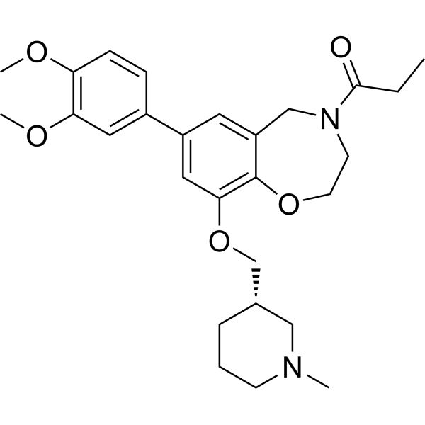
I-CBP112, a chemical probe, is a specific and potent acetyl-lysine competitive protein-protein interaction inhibitor, that inhibits the CBP/p300 bromodomains, enhances acetylation by p300.
For research use only. We do not sell to patients.
- Purity : 98.43%
- CAS No.: 1640282-31-0
- Formula: C27H36N2O5
- Molecular Weight:468.59
-
Storage:Powder -20°C, 3 years , 4°C, 2 years ; In solvent -80°C, 2 years , -20°C, 1 year

To place orders, for customer services and technical support, please contact: MedChemExpress USA
Tel: 609-228-6898 E-mail: [email protected] [email protected]


-
 Biological Activity
-
Chemical Information
-
Solvent & Solubility
- Protocols
- Purity & Documentation
- References
-
 Help & FAQs


-
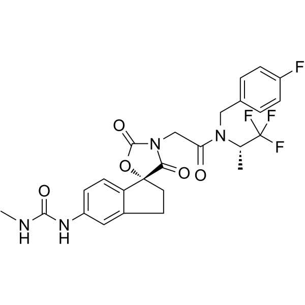
HY-107455
A-485p300/CBP Inhibitor -
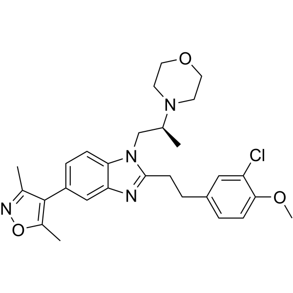
HY-15826
SGC-CBP30CBP/p300 Inhibitor -
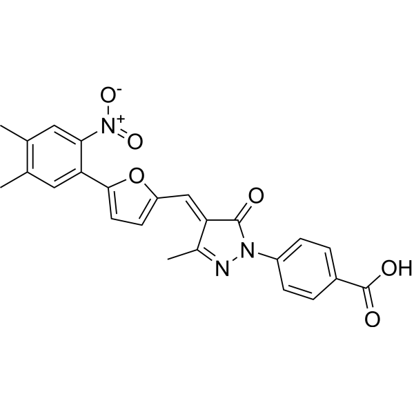
HY-13823
C646p300/CBP HAT Inhibitor -
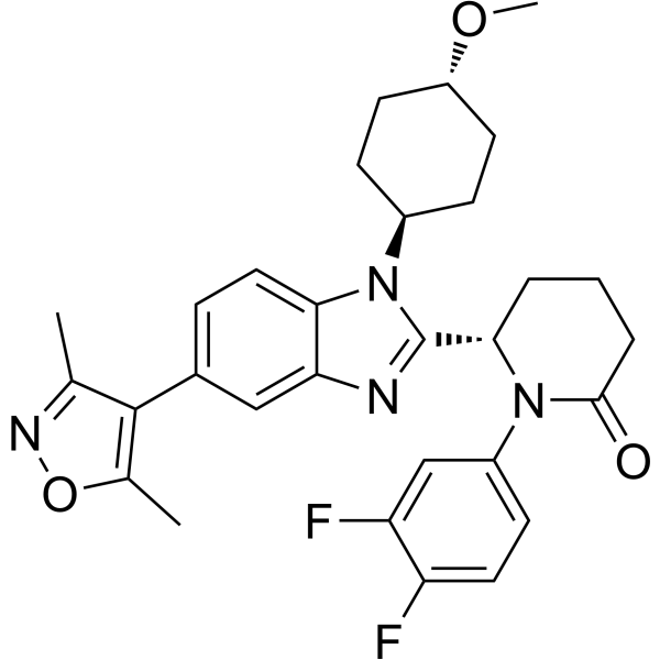
HY-111784
Inobrodibp300/CBP Inhibitor -
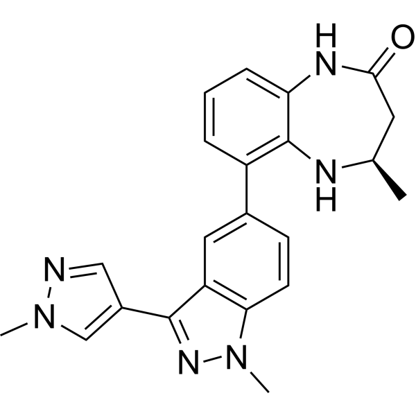
HY-100482
CPI-637CBP/EP300 Inhibitor -
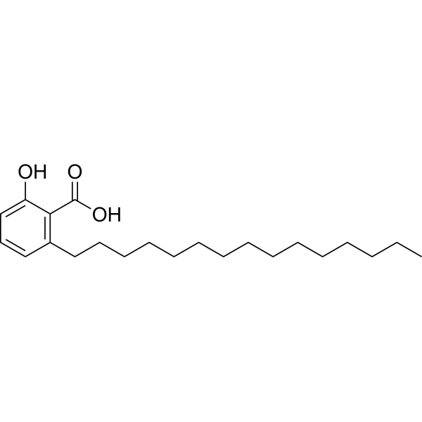
HY-N2020
Anacardic AcidHistone Acetyltransferase Inhibitor -
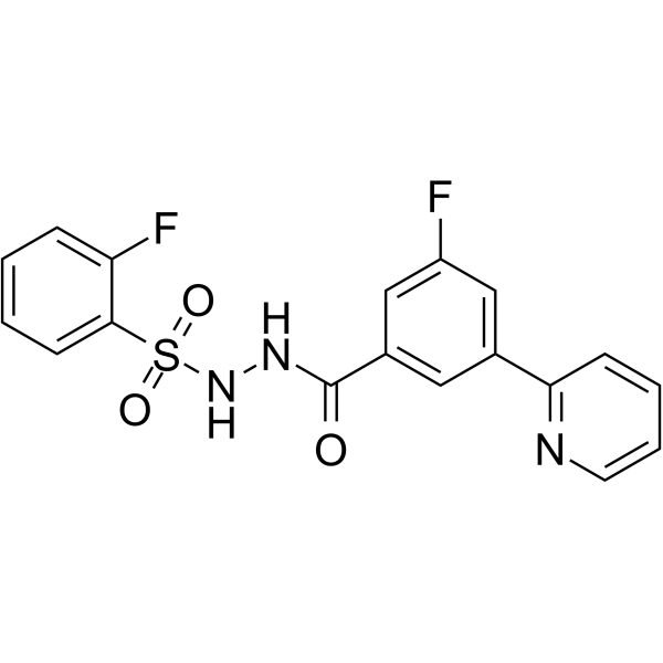
HY-102058
WM-1119KAT6A Inhibitor -

HY-107455R
A-485 (Standard)Reference Standard -
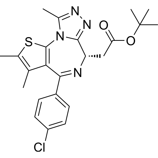
HY-13030
(+)-JQ-1BET Inhibitor -
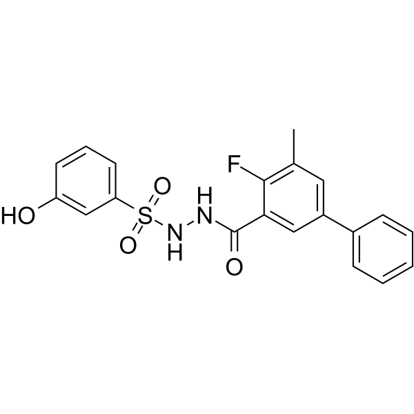
HY-134901
WM-3835HBO1 Inhibitor -
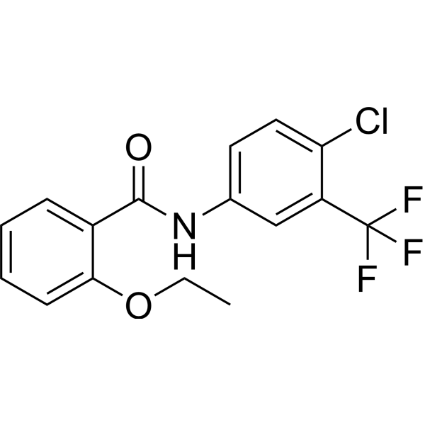
HY-134964
CTBp300 Activator -
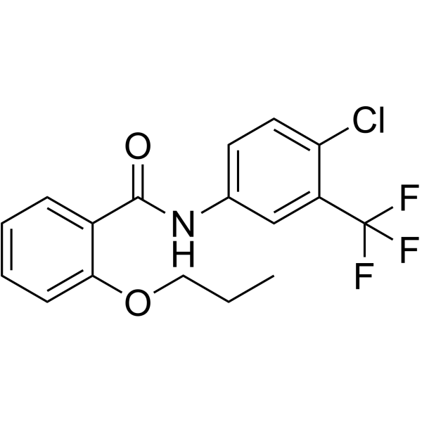
HY-116673
TTK21CBP/p300 Activator
Publications Citing Use of MedChemExpress (MCE) I-CBP112
More

Biological Activity
Description
IC50 & Target
|
CBP/p300 |
Cellular Effect
|
Cell Line

|
Type | Value | Description | References |
|---|---|---|---|---|
| HEK293 | IC50 |
>10 μM
Compound: 4
|
Inhibition of Halo-tagged histone H3.3 binding to NanoLuc luciferase conjugated CBP (unknown origin) expressed in HEK293 cells after overnight incubation by BRET assay
Inhibition of Halo-tagged histone H3.3 binding to NanoLuc luciferase conjugated CBP (unknown origin) expressed in HEK293 cells after overnight incubation by BRET assay
|
[PMID: 27682507] |
| HL-60 | GI50 |
>50 μM
Compound: 20; I-CBP 112
|
Growth inhibition of human HL60 cells after 72 hrs by CellTiter 96S AQeous non-radioactive cell proliferation assay
Growth inhibition of human HL60 cells after 72 hrs by CellTiter 96S AQeous non-radioactive cell proliferation assay
|
[PMID: 33275431] |
| MCF7 | GI50 |
>50 μM
Compound: 20; I-CBP 112
|
Growth inhibition of human MCF7 cells after 72 hrs by CellTiter 96S AQeous non-radioactive cell proliferation assay
Growth inhibition of human MCF7 cells after 72 hrs by CellTiter 96S AQeous non-radioactive cell proliferation assay
|
[PMID: 33275431] |
| SK-MEL-5 | GI50 |
>50 μM
Compound: 20; I-CBP 112
|
Growth inhibition of human SK-MEL-5 cells after 72 hrs by CellTiter 96S AQeous non-radioactive cell proliferation assay
Growth inhibition of human SK-MEL-5 cells after 72 hrs by CellTiter 96S AQeous non-radioactive cell proliferation assay
|
[PMID: 33275431] |
In Vitro
I-CBP112 significantly enhances acetylation by p300 at the histone H3K18 and H3K23 sites. I-CBP112 stimulated H3K18ac by ~3-fold, I-CBP112 induced enhances acetylation of these same sites by CBP as well as at H4K5. The EC50’s of activation of I-CBP112 on p300- and CBP-mediated H3K18 acetylation are ~2 μM[1]. Exposure of human and mouse leukemic cell lines to I-CBP112 results in substantially impaired colony formation and induces cellular differentiation without significant cytotoxicity. Exposure of the BioMAP primary cell panel to I-CBP112 results in a unique response on cytokine and marker protein expression[2].
MedChemExpress (MCE) has not independently confirmed the accuracy of these methods. They are for reference only. Further protocols information, click here.
In Vivo
MedChemExpress (MCE) has not independently confirmed the accuracy of these methods. They are for reference only.


Chemical Information
-
CAS No. 1640282-31-0
-
Appearance Solid
-
Molecular Weight 468.59
-
Formula C27H36N2O5
-
Color White to off-white
-
SMILES
COC1=C(OC)C=C(C2=CC(OC[C@H]3CCCN(C)C3)=C(OCCN(C(CC)=O)C4)C4=C2)C=C1
-
Shipping
Room temperature in continental US; may vary elsewhere.
-
Storage
Powder -20°C 3 years 4°C 2 years In solvent -80°C 2 years -20°C 1 year


Publications (5)
-
Journal Impact Factor
-
Most Recent
-
Cell
Fibroblasts of disparate developmental origins harbor anatomically variant scarring potential. [Abstract]2026 Feb 5;189(3):783-799.e20. PMID: 41576949 -
Cell Res
2021 Mar;31(3):291-311. PMID: 33299139 -
Nat Commun
Human iPSC-based Modeling of Pulmonary Fibrosis Reveals p300/CBP Inhibition Suppresses Alveolar Transitional Cell State. [Abstract]2026 Feb 12;17(1):1214. PMID: 41680175 -
iScience
Transcriptional synergy in human aortic endothelial cells is vulnerable to combination p300/CBP and BET bromodomain inhibition. [Abstract]2024 May 16;27(6):110011. PMID: 38868181 -


Solvent & Solubility
In Vitro:
1M HCl : 100 mg/mL (213.41 mM; ultrasonic and adjust pH to 1 with HCl)
DMSO : ≥ 32 mg/mL (68.29 mM; Hygroscopic DMSO has a significant impact on the solubility of product, please use newly opened DMSO)
* "≥" means soluble, but saturation unknown.
Please refer to the solubility information to select the appropriate solvent. Once prepared, please aliquot and store the solution to prevent product inactivation from repeated freeze-thaw cycles.
Storage method and period of stock solution: -80°C, 2 years; -20°C, 1 year. When stored at -80°C, please use it within 2 years. When stored at -20°C, please use it within 1 year.
Please refer to the solubility information to select the appropriate solvent. Once prepared, please aliquot and store the solution to prevent product inactivation from repeated freeze-thaw cycles.
Storage method and period of stock solution: -80°C, 2 years; -20°C, 1 year. When stored at -80°C, please use it within 2 years. When stored at -20°C, please use it within 1 year.
Concentration (start) × Volume (start) = Concentration (final) × Volume (final)
In Vivo:
Select the appropriate dissolution method based on your experimental animal and administration route.
- For the following dissolution methods, please ensure to first prepare a clear stock solution using an In Vitro approach and then sequentially add co-solvents:
- To ensure reliable experimental results, the clarified stock solution can be appropriately stored based on storage conditions. As for the working solution for In Vivo experiments, it is recommended to prepare freshly and use it on the same day.
- The percentages shown for the solvents indicate their volumetric ratio in the final prepared solution. If precipitation or phase separation occurs during preparation, heat and/or sonication can be used to aid dissolution.
Add each solvent one by one: 10% DMSO 40% PEG300 5% Tween-80 45% Saline
Solubility: ≥ 2.5 mg/mL (5.34 mM); Clear solution
This protocol yields a clear solution of ≥ 2.5 mg/mL (saturation unknown).
Taking 1 mL working solution as an example, add 100 μL DMSO stock solution (25.0 mg/mL) to 400 μL PEG300, and mix evenly; then add 50 μL Tween-80 and mix evenly; then add 450 μL Saline to adjust the volume to 1 mL.
Preparation of Saline: Dissolve 0.9 g sodium chloride in ddH₂O and dilute to 100 mL to obtain a clear Saline solution.
Add each solvent one by one: 10% DMSO 90% (20% SBE-β-CD in Saline)
Solubility: ≥ 2.5 mg/mL (5.34 mM); Clear solution
This protocol yields a clear solution of ≥ 2.5 mg/mL (saturation unknown).
Taking 1 mL working solution as an example, add 100 μL DMSO stock solution (25.0 mg/mL) to 900 μL 20% SBE-β-CD in Saline, and mix evenly.
Preparation of 20% SBE-β-CD in Saline (4°C, storage for one week): 2 g SBE-β-CD powder is dissolved in 10 mL Saline, completely dissolve until clear.
In Vivo Dissolution Calculator
Please enter the basic information of animal experiments:
-
-
-
-
Recommended: Prepare an additional quantity of animals to account for potential losses during experiments.
Please enter your animal formula composition:
-
%DMSO +
Recommended: Keep the proportion of DMSO in working solution below 2% if your animal is weak.
-
%+
-
+%Tween-80 + +
-
%Saline +
The co-solvents required include: DMSO, . All of co-solvents are available by MedChemExpress (MCE). , Tween 80. All of co-solvents are available by MedChemExpress (MCE).
Working solution concentration: 0.22 mg/mL
Method for preparing stock solution: mg drug dissolved in μL DMSO. Stock solution concentration: mg/mL.
1. Take μL DMSO stock solution;
2. Add μL .
μL , mix evenly;
3. Then add μL Tween 80, mix evenly;
4. Then add μL
Please ensure that the stock solution in the first step is dissolved to a clear state, and add co-solvents in sequence. You can use ultrasonic heating (ultrasonic cleaner, recommended frequency 20-40 kHz), vortexing, etc. to assist dissolution.

Protocols
-
Pull-down
The pull-down assay is an in vitro technique used to detect physical interactions between two or more proteins and an invaluable tool for confirming a predicted protein-protein interaction or identifying novel interacting partners. This method typically involves the use of affinity purification with various wash and elution steps.
-
Immunoprecipitation
Immunoprecipitation (IP) is an experimental method that uses the principle of antibody specific binding to purify and enrich target proteins.
-
Protocol for Bimolecular Fluorescence Complementation (BiFC) Assay
Bimolecular fluorescence complementation detects protein-protein proximity in living or fixed cells by fusing two candidate interaction partners to nonfluorescent N- and C-terminal fragments of a fluorescent protein; when the partners interact or remain close enough, the fluorescent fragments complement, mature, and generate a fluorescent signal at the site of the protein complex. The BiFC readout is fluorescence intensity and subcellular localization of the reconstituted fluorophore, which reflects formation or stabilization of a protein complex rather than direct biochemical binding kinetics; BiFC is therefore useful for mapping where interactions occur in cancer cells, neurons, macrophages, organoid-derived cells, or drug-screening systems, but results should be validated by independent assays such as co-IP or Western blot. BiFC signal formation is delayed by fluorophore maturation and can stabilize otherwise transient complexes, so it is not a real-time reversible interaction assay
-
Co-Immunoprecipitation
Co-immunoprecipitation technology can verify protein interaction based on the specific immune reaction between antibodies and antigens.
-
Protocol for Yeast Two-Hybrid (Y2H) Assay
The yeast two-hybrid assay detects binary protein-protein interactions by separating a transcription factor into a DNA-binding domain fused to a "bait" protein and a transcriptional activation domain fused to a "prey" protein; if bait and prey interact in yeast, the transcription factor is reconstituted and activates reporter genes such as HIS3, ADE2, lacZ, MEL1, or other selectable/readable reporters. The readout is yeast growth on selective medium and/or reporter activity, which reflects proximity-dependent transcriptional activation in the yeast nucleus rather than direct biochemical binding in the original mammalian, tumor, neuronal, macrophage, or organoid context. Because yeast two-hybrid can generate false positives and false negatives, interaction claims should be validated using independent assays such as co-immunoprecipitation, Western blot, immunofluorescence colocalization, BiFC, pull-down, or mammalian two-hybrid assays.


Purity & Documentation
-
Data Sheet (281 KB)
-
SDS (596 KB)
- English - EN (596 KB)
- Français - FR (596 KB)
- Deutsch - DE (596 KB)
- Norwegian - NO (596 KB)
- Español - ES (596 KB)
- Swedish - SV (596 KB)
- Italian - IT (596 KB)
- Korean - KR (596 KB)
- Portuguese - PT (596 KB)
-
Handling Instructions (2659 KB)
References
[1]. Zucconi BE, et al. Modulation of p300/CBP Acetylation of Nucleosomes by Bromodomain LigandI-CBP112. Biochemistry. 2016 Jul 12;55(27):3727-34. [Content Brief]
[2]. Picaud S, et al. Generation of a Selective Small Molecule Inhibitor of the CBP/p300 Bromodomain for Leukemia Therapy. Cancer Res. 2015 Dec 1;75(23):5106-19. [Content Brief]


Complete Stock Solution Preparation Table
Please refer to the solubility information to select the appropriate solvent. Once prepared, please aliquot and store the solution to prevent product inactivation from repeated freeze-thaw cycles.
Storage method and period of stock solution: -80°C, 2 years; -20°C, 1 year. When stored at -80°C, please use it within 2 years. When stored at -20°C, please use it within 1 year.
| Optional Solvent | Concentration Solvent Mass | 1 mg | 5 mg | 10 mg | 25 mg |
|---|---|---|---|---|---|
| DMSO / 1M HCl | 1 mM | 2.1341 mL | 10.6703 mL | 21.3406 mL | 53.3515 mL |
| 5 mM | 0.4268 mL | 2.1341 mL | 4.2681 mL | 10.6703 mL | |
| 10 mM | 0.2134 mL | 1.0670 mL | 2.1341 mL | 5.3352 mL | |
| 15 mM | 0.1423 mL | 0.7114 mL | 1.4227 mL | 3.5568 mL | |
| 20 mM | 0.1067 mL | 0.5335 mL | 1.0670 mL | 2.6676 mL | |
| 25 mM | 0.0854 mL | 0.4268 mL | 0.8536 mL | 2.1341 mL | |
| 30 mM | 0.0711 mL | 0.3557 mL | 0.7114 mL | 1.7784 mL | |
| 40 mM | 0.0534 mL | 0.2668 mL | 0.5335 mL | 1.3338 mL | |
| 50 mM | 0.0427 mL | 0.2134 mL | 0.4268 mL | 1.0670 mL | |
| 60 mM | 0.0356 mL | 0.1778 mL | 0.3557 mL | 0.8892 mL | |
| 1M HCl | 80 mM | 0.0267 mL | 0.1334 mL | 0.2668 mL | 0.6669 mL |
| 100 mM | 0.0213 mL | 0.1067 mL | 0.2134 mL | 0.5335 mL |


I-CBP112 Related Classifications
HY-19541 Related Classifications
 Powered by Bioz