



(±)-Stylopine
Based on 2 publication(s) in Google Scholar
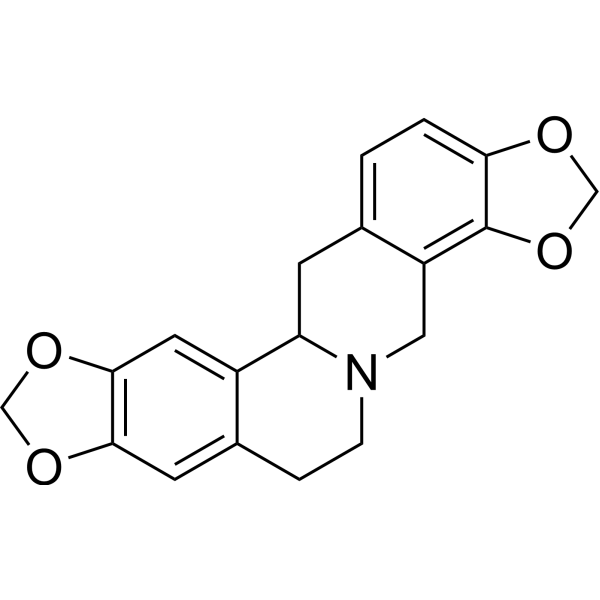
(±)-Stylopine (Tetrahydrocoptisine) is an alkaloid compound. (±)-Stylopine can be isolated from the tubers of the plant Corydalis. (±)-Stylopine inhibits TNF-α, IL-6, and NO production, and attenuates phosphorylation of p38 MAPK, ERK1/2. (±)-Stylopine inhibits NF-κB expression. (±)-Stylopine exhibits anti-inflammatory activity. (±)-Stylopine has protective effects against foot edema, gastric ulcers, anxiety, depression, and acute lung injury.
For research use only. We do not sell to patients.
- Purity : 99.86%
- CAS No.: 4312-32-7
- Formula: C19H17NO4
- Molecular Weight:323.34
-
Storage:
4°C, sealed storage, away from moisture and light
* In solvent : -80°C, 6 months; -20°C, 1 month (sealed storage, away from moisture and light)

To place orders, for customer services and technical support, please contact: MedChemExpress USA
Tel: 609-228-6898 E-mail: [email protected] [email protected]


-
 Biological Activity
-
Chemical Information
-
Solvent & Solubility
- Protocols
- Purity & Documentation
- References
-
 Help & FAQs


-
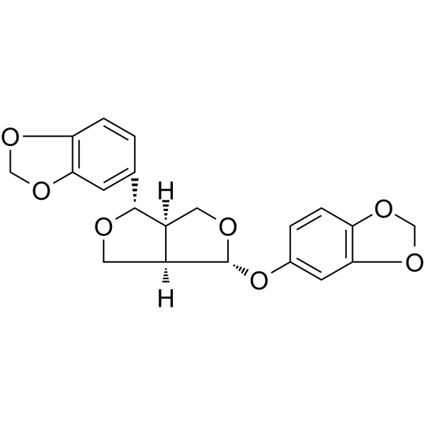
HY-N0809
SesamolinMAPK Inhibitor -
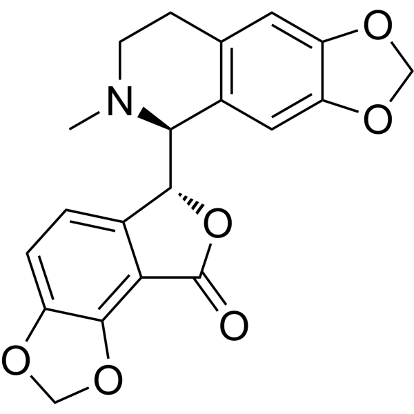
HY-N0219
BicucullineGABAA Receptor Antagonist -
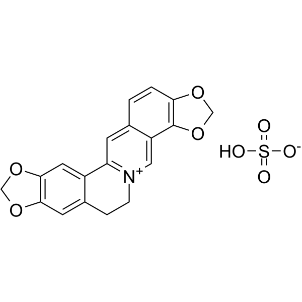
HY-N0430A
Coptisine SulfateIDO Inhibitor -
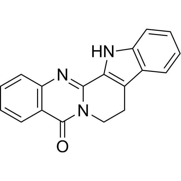
HY-N0147
RutaecarpineCOX Inhibitor -
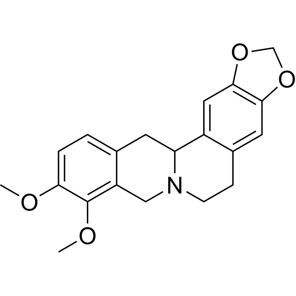
HY-N0925
TetrahydroberberineDopamine Receptor Antagonist -
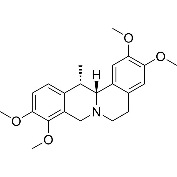
HY-N0923
CorydalineCholinesterase (ChE) Inhibitor -

HY-N0924R
(±)-Stylopine (Standard)Alkaloid -
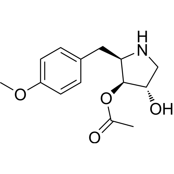
HY-18982
AnisomycinProtein Synthesis Inhibitor -
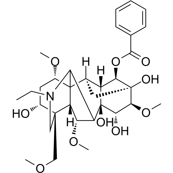
HY-N0217
BenzoylaconineAnti-inflammatory Agent -
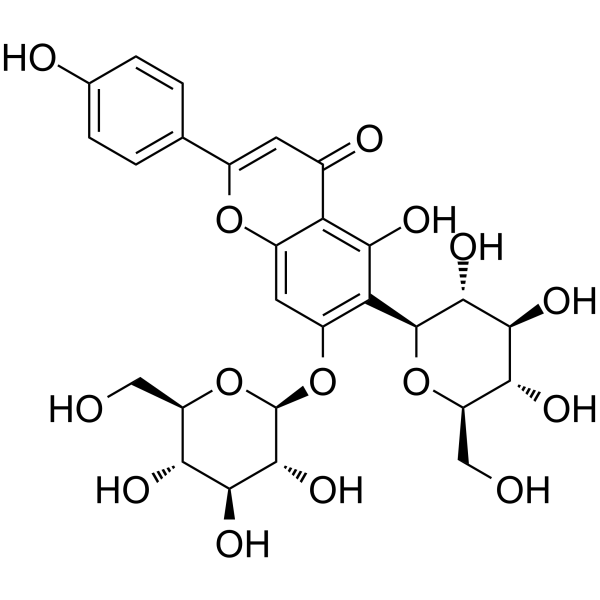
HY-N5083
SaponarinFlavonoid -
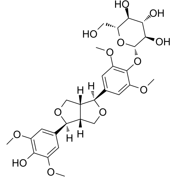
HY-N2807
Acanthoside BBioactive Lignan -
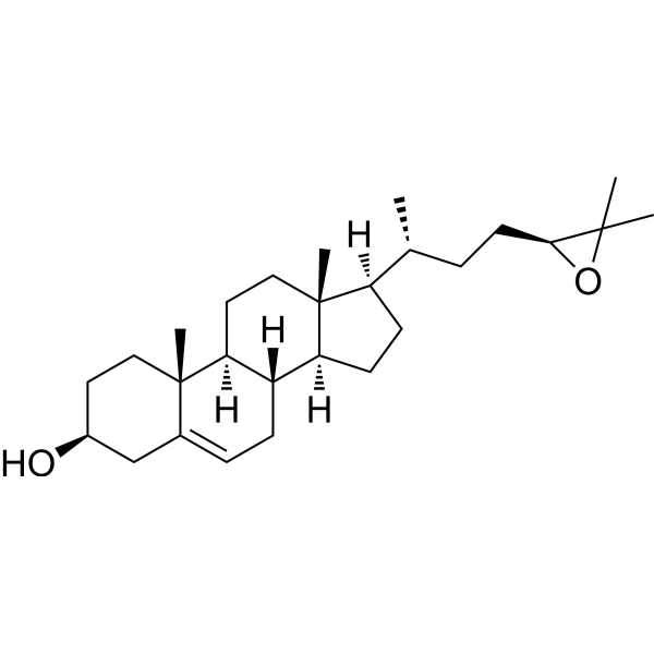
HY-W040150
(24S,25)-EpoxycholesterolLXR Agonist
Publications Citing Use of MedChemExpress (MCE) (±)-Stylopine
More

Biological Activity
Description
Cellular Effect
|
Cell Line

|
Type | Value | Description | References |
|---|---|---|---|---|
| GES1 | IC50 |
>150 μM
Compound: 1
|
Antiproliferative activity against human GES1 cells assessed as reduction in cell viability after 24 hrs by MTT assay
Antiproliferative activity against human GES1 cells assessed as reduction in cell viability after 24 hrs by MTT assay
|
[PMID: 35286954] |
| HGC-27 | IC50 |
55.06 μM
Compound: 1
|
Antiproliferative activity against human HGC-27 cells assessed as reduction in cell viability after 24 hrs by MTT assay
Antiproliferative activity against human HGC-27 cells assessed as reduction in cell viability after 24 hrs by MTT assay
|
[PMID: 35286954] |
| MGC-803 | IC50 |
107.3 μM
Compound: 1
|
Antiproliferative activity against human MGC-803 cells assessed as reduction in cell viability after 24 hrs by MTT assay
Antiproliferative activity against human MGC-803 cells assessed as reduction in cell viability after 24 hrs by MTT assay
|
[PMID: 35286954] |
In Vitro
(±)-Stylopine (10-4 -1 μg/mL; 24 h pretreatment before 12 h LPS stimulation) significantly inhibits LPS-induced TNF-α, IL-6 production[1].
MedChemExpress (MCE) has not independently confirmed the accuracy of these methods. They are for reference only. Further protocols information, click here.
-
Cell Line:Mouse peritoneal macrophages
-
Concentration:10-4 μg/mL, 10-2 μg/mL, 1 μg/mL
-
Incubation Time:24 h
-
Result:Reduced phosphorylation of p38MAPK and ERK1/2.
-
Cell Line:Mouse peritoneal macrophages
-
Concentration:0-4 μg/mL, 10-3 μg/mL, 10-2 μg/mL, 10-1 μg/mL, 1 μg/mL
-
Incubation Time:24 h
-
Result:Downregulated TNF-α and IL-6 mRNA.
In Vivo
(±)-Stylopine (10-20 mg/kg; i.p.; 3 days) attenuates ethanol-induced gastric ulcer in mice, reduces NO, TNF-α, IL-6 levels, MPO activity and NF-κB expression in gastric tissue and serum[2].
(±)-Stylopine (18.4-36.8 mg/kg; i.p.; 14 days) alleviates LPS-induced neuroinflammation in mice, improves anxiety, depression and anhedonia, reduces neurodegeneration, inhibits KMO expression, NO and lipid peroxidation[3].
(±)-Stylopine (10-20 mg/kg; i.p.; 30 min before LPS injection) reduces mortality, ameliorates lung injury, inhibits inflammatory cell infiltration, protein leakage, TNF-α, IL-6 production and NF-κB activation in LPS-induced acute lung injury in rats[4].
MedChemExpress (MCE) has not independently confirmed the accuracy of these methods. They are for reference only.
-
Animal Model:Male Sprague-Dawley rats (220-250 g) with Carrageenan-induced paw edema; Male Kunming mice (20-25 g) with Xylene-induced ear edema and LPS-induced systemic inflammation[1]
-
Dosage:7 mg/kg, 21 mg/kg (for Carrageenan); 10 mg/kg, 30 mg/kg (for Xylene); 10 mg/kg, 20 mg/kg, 30 mg/kg (for LPS)
-
Administration:i.p., 30 min before Carrageenan injection (for Carrageenan); i.p. (for Xylene); i.p., at 12 h and 1 h before LPS injection (for LPS)
-
Result:Reduced paw edema (56.25% and 75.35% inhibition at 5 h) and TNF-α, IL-6 in paw tissue.
Decreased ear edema weight.
Inhibited serum TNF-α in a dose- and time-dependent manner.


Chemical Information
-
CAS No. 4312-32-7
-
Appearance Solid
-
Molecular Weight 323.34
-
Formula C19H17NO4
-
Color White to off-white
-
SMILES
C1(C(CCN2CC3=C4OCOC4=CC=C3CC21)=C5)=CC6=C5OCO6
-
Synonyms
Tetrahydrocoptisine
-
Structure Classification
-
Initial Source
-
Shipping
Room temperature in continental US; may vary elsewhere.
-
Storage
4°C, sealed storage, away from moisture and light
* In solvent : -80°C, 6 months; -20°C, 1 month (sealed storage, away from moisture and light)


Publications (2)
-
Journal Impact Factor
-
Most Recent
-
BMC Complement Med Ther
Hepatotoxicity prediction for traditional Chinese medicine: a two-step in silico framework integrating network and machine learning approaches. [Abstract]2026 Apr 2;26(1):177. PMID: 41923057 -
Planta Med
Novel Approaches for the Analysis and Isolation of Benzylisoquinoline Alkaloids in Chelidonium majus. [Abstract]2024 Jun;90(7-08):523-533. PMID: 38843792


Solvent & Solubility
In Vitro:
DMF : 4 mg/mL (12.37 mM; ultrasonic and warming and heat to 60°C)
DMSO : 2.5 mg/mL (7.73 mM; ultrasonic and warming and heat to 60°C; Hygroscopic DMSO has a significant impact on the solubility of product, please use newly opened DMSO)
Acetone : 1 mg/mL (3.09 mM; Need ultrasonic)
Please refer to the solubility information to select the appropriate solvent. Once prepared, please aliquot and store the solution to prevent product inactivation from repeated freeze-thaw cycles.
Storage method and period of stock solution: -80°C, 6 months; -20°C, 1 month (sealed storage, away from moisture and light). When stored at -80°C, please use it within 6 months. When stored at -20°C, please use it within 1 month.
Please refer to the solubility information to select the appropriate solvent. Once prepared, please aliquot and store the solution to prevent product inactivation from repeated freeze-thaw cycles.
Storage method and period of stock solution: -80°C, 6 months; -20°C, 1 month (sealed storage, away from moisture and light). When stored at -80°C, please use it within 6 months. When stored at -20°C, please use it within 1 month.
Concentration (start) × Volume (start) = Concentration (final) × Volume (final)
In Vivo:
For the following dissolution methods, please prepare the working solution directly:
It is recommended to prepare fresh solutions and use them promptly within a short period of time.
The percentages shown for the solvents indicate their volumetric ratio in the final prepared solution. If precipitation or phase separation occurs during preparation, heat and/or sonication can be used to aid dissolution.
Add each solvent one by one: 20% HP-β-CD in Saline
Solubility: 12.5 mg/mL (38.66 mM); Suspended solution; Need ultrasonic

Protocols
-
Kinase activity and phosphorylation assays
Kinase activity assays measure the ability of kinases to transfer phosphate groups from ATP to specific substrates, while phosphorylation assays detect the presence and levels of phosphorylated proteins. Common methods include radiolabeled ATP incorporation (e. g. ,), ADP release detection via bioluminescence (e. g. ,[3]), enzyme-linked immunosorbent assays (ELISA) for phospho-specific epitopes (e. g. ,[6]), and microtiter-based formats for high-throughput screening (e. g. ,[8]). The ADP-Glo assay quantifies kinase activity by measuring ADP produced during phosphorylation using a luciferase-based system. Radiometric assays involve autoradiography or scintillation counting after incorporation of 32P-labeled ATP into substrate proteins. ELISA-based approaches rely on phospho-specific antibodies to detect activated kinases in cell lysates or purified samples.
-
Western Blot
Western blotting (WB) is a commonly used experimental method in molecular biology, biochemistry, and immunogenetics for identifying and quantifying target proteins. It combines gel electrophoresis with immunoassay, enabling researchers to analyze protein expression, post-translational modifications, and molecular weight.
-
RNA extraction experimental
By lysing cells, releasing RNA, and removing impurities such as proteins and DNA, high-purity RNA products are finally obtained. The commonly used traditional method is the guanidine isothiocyanate/phenol/chloroform method (Trizol), which is suitable for a variety of animal materials including animal tissues, microorganisms, cultured cells, etc., and most plant materials.
-
Cotton Pellet Granuloma
Cotton pellet granuloma is a classical in vivo chronic inflammation model used to evaluate the anti-inflammatory potential of test substances by measuring their ability to inhibit granuloma tissue formation around an implanted foreign body (cotton pellet) in rodents. The method is based on the biological response to a sterile implanted material, which induces proliferative phase inflammation characterized by fibroblast proliferation and collagen-rich granuloma formation, and the final readout reflects the extent of chronic inflammatory tissue growth surrounding the pellet. In multiple preclinical pharmacological evaluations, inhibition of cotton pellet-induced granuloma formation has been used as an indicator of anti-inflammatory activity in both synthetic and natural product screening contexts.
-
Carrageenan-Induced Paw Edema
Carrageenan-induced paw edema is an acute inflammation model in which intraplantar injection of carrageenan induces localized inflammatory swelling characterized by vascular permeability, leukocyte infiltration, and production of inflammatory mediators such as prostaglandins and cytokines, making it widely used to evaluate anti-inflammatory agents in vivo. The resulting paw volume or thickness increase is quantified over time as a direct readout of inflammatory intensity and drug efficacy, typically reflecting cyclooxygenase-mediated prostaglandin-driven edema formation and immune cell recruitment in peripheral tissue[20].
-
Protocol for Kinase activity and phosphorylation assays
Kinase activity assays measure transfer of phosphate from ATP to a protein or peptide substrate, generating phosphorylated substrate, ADP, or incorporated radiolabeled phosphate as the readout; phosphorylation assays measure site-specific phosphorylation in cells or tissues as a proxy for kinase-pathway activation, inhibition, or substrate regulation. Phosphorylation can be detected by phospho-specific Western blot, immunoprecipitation kinase assay, phospho-immunofluorescence, phospho-flow cytometry, luminescent ADP detection, radiolabeled ATP incorporation, or reporter-based pathway assays, and these readouts can be applied to cancer cells, primary neurons, mouse tumors, organoids, inflammatory macrophages, ferroptosis studies, and mitophagy studies when the kinase target is biologically relevant.
-
Research Protocol for Inflammation-related Diseases
The NLRP3 inflammasome is a cytosolic innate immune signaling platform that integrates priming signals and danger-signal activation to promote caspase-1 activation, maturation of IL-1β and IL-18, and gasdermin D-mediated pyroptotic cell death. The core experimental logic is to determine whether inflammatory disease phenotypes are driven by increased NLRP3 expression, ASC-containing inflammasome assembly, caspase-1 cleavage, GSDMD cleavage, and extracellular release of IL-1β/IL-18 rather than by nonspecific cell injury alone. The pathway is strongly linked to inflammation-related disease phenotypes because monosodium urate crystals activate NALP3/NLRP3 inflammasome signaling in gout-like crystal inflammation, cholesterol crystals activate NLRP3 inflammasomes in atherogenesis models, and DSS-induced intestinal inflammation has been reported to involve NLRP3 inflammasome activity. However, experimental colitis studies also show context-dependent protective effects of NLRP3 inflammasome co
-
Inhalation Toxicity Study
Inhalation toxicity studies expose rodents to a controlled aerosol, vapor, gas, or smoke atmosphere and assess respiratory and systemic toxicity using exposure-atmosphere characterization, clinical observations, body and organ weights, bronchoalveolar lavage fluid, histopathology, blood chemistry, hematology, and, when included, molecular endpoints such as transcriptomics, proteomics, lipidomics, or tissue burden analysis. The primary biological readouts are airway irritation, pulmonary inflammation, cytotoxicity, altered surfactant or lipid homeostasis, impaired particle clearance, and tissue remodeling, reflected by BALF cell differentials, BALF protein, LDH, phosphatase activities, cytokines, lung weight, microscopic respiratory-tract lesions, and retained lung burden.


Purity & Documentation
-
Data Sheet (282 KB)
-
SDS (393 KB)
- English - EN (393 KB)
- Français - FR (393 KB)
- Deutsch - DE (393 KB)
- Norwegian - NO (393 KB)
- Español - ES (393 KB)
- Swedish - SV (393 KB)
- Italian - IT (393 KB)
- Korean - KR (393 KB)
- Portuguese - PT (393 KB)
-
Handling Instructions (2659 KB)
References
[1]. Li W, et al. Anti-inflammatory effect of tetrahydrocoptisine from Corydalis impatiens is a function of possible inhibition of TNF-α, IL-6 and NO production in lipopolysaccharide-stimulated peritoneal macrophages through inhibiting NF-κB activation and MAPK pathway. Eur J Pharmacol. 2013 Sep 5;715(1-3):62-71. [Content Brief]
[2]. Li W, et al. Protective effect of tetrahydrocoptisine against ethanol-induced gastric ulcer in mice. Toxicol Appl Pharmacol. 2013 Oct 1;272(1):21-9. [Content Brief]
[3]. Khatun A, et al. In-Silico and In-Vivo Characterization of Anti-Neuro inflammatory potential of Tetrahydrocoptisine by using LPS-Induced model in mice. Neuroscience. 2025 Jan 26;565:232-246. [Content Brief]
[4]. Li W, et al. Tetrahydrocoptisine protects rats from LPS-induced acute lung injury. Inflammation. 2014 Dec;37(6):2106-15. [Content Brief]


Complete Stock Solution Preparation Table
Please refer to the solubility information to select the appropriate solvent. Once prepared, please aliquot and store the solution to prevent product inactivation from repeated freeze-thaw cycles.
Storage method and period of stock solution: -80°C, 6 months; -20°C, 1 month (sealed storage, away from moisture and light). When stored at -80°C, please use it within 6 months. When stored at -20°C, please use it within 1 month.
| Optional Solvent | Concentration Solvent Mass | 1 mg | 5 mg | 10 mg | 25 mg |
|---|---|---|---|---|---|
| Acetone / DMSO / DMF | 1 mM | 3.0927 mL | 15.4636 mL | 30.9272 mL | 77.3180 mL |
| DMSO / DMF | 5 mM | 0.6185 mL | 3.0927 mL | 6.1854 mL | 15.4636 mL |
| DMF | 10 mM | 0.3093 mL | 1.5464 mL | 3.0927 mL | 7.7318 mL |


(±)-Stylopine Related Classifications
HY-N0924 Related Classifications
 Powered by Bioz