



Abietic acid
Based on 3 publication(s) in Google Scholar
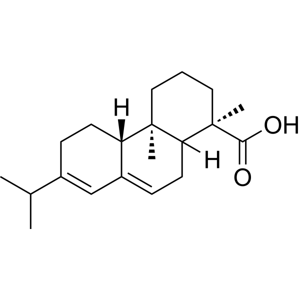
Abietic acid, an orally active diterpene isolated from Colophony, displays significant anti-proliferative, anti-inflammatory, anti-obesity effect, bacteriostatic, cell cycle arresting and pro-apoptotic activities. Abietic acid inhibits lipoxygenase activity for allergy. Abietic acid enhances cell migration and tube formation in HUVECs. Abietic acid induces significant angiogenic potential, which is associated with upregulation of extracellular signal-regulated kinase (ERK) and p38 expression. Abietic acid attenuates sepsis-induced lung injury by inhibiting nuclear factor kappa-light-chain-enhancer of activated B cells (NF-κB) pathway to inhibit M1 macrophage polarization. Abietic acid exhibits a positive effect against liver injury by attenuating inflammation and ferroptosis. Abietic acid shows accelerated wound closure in a mouse model of cutaneous wounds. Abietic acid significantly reduces the proliferation and growth of NSCLC cells by IKKβ inhibition.Additionally, Abietic acid ameliorates psoriasis-like inflammation and modulates gut microbiota in mice. Abietic acid is promising for research in non-small-cell lung cancer (NSCLC), liver injury-related deseases and psoriasis.
For research use only. We do not sell to patients.
- Purity : 92.70%
- CAS No.: 514-10-3
- Formula: C20H30O2
- Molecular Weight:302.45
-
Storage:Powder -20°C, 3 years , 4°C, 2 years ; In solvent -80°C, 6 months , -20°C, 1 month

To place orders, for customer services and technical support, please contact: MedChemExpress USA
Tel: 609-228-6898 E-mail: [email protected] [email protected]


-
 Biological Activity
-
Chemical Information
-
Solvent & Solubility
- Protocols
- Purity & Documentation
- References
-
 Help & FAQs


-
HY-N0872
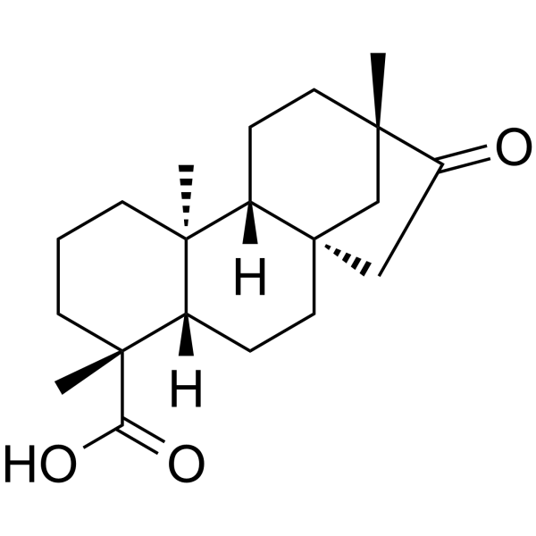
IsosteviolDNA Inhibitor -
HY-N0156
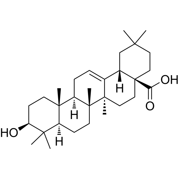
Oleanolic acidAutophagy Inducer -
HY-N0198
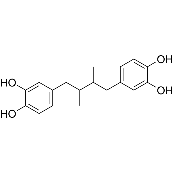
Nordihydroguaiaretic acidAutophagy Inducer -
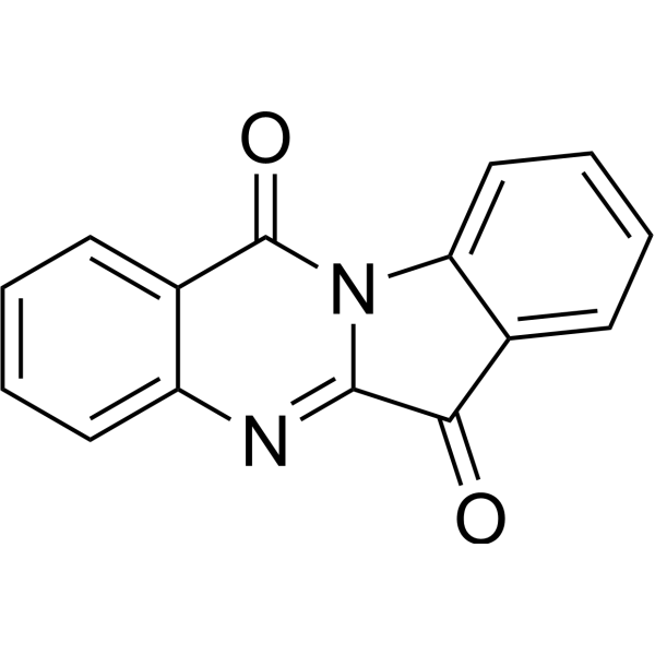
HY-N6607
TryptanthrinLT Inhibitor -
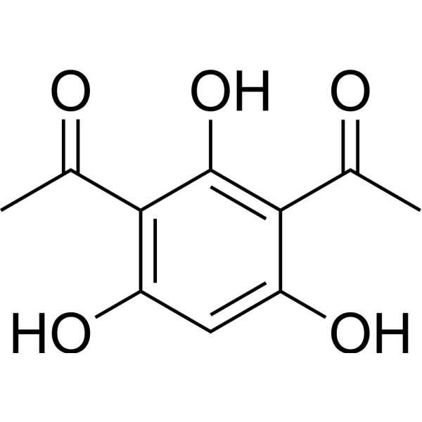
HY-118448
2,4-DiacetylphloroglucinolAntibiotic -
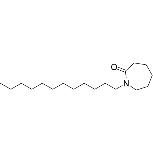
HY-W009326
LaurocapramAbsorption Enhancer -
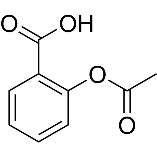
HY-14654
AspirinCOX Inhibitor -

HY-N0198R
Nordihydroguaiaretic acid (Standard)Autophagy Inducer -
HY-B0355
Ginkgolide AGABA Antagonist -

HY-N6607R
Tryptanthrin (Standard)LT Inhibitor -
HY-N0570
HydroxytyrosolPhenolic Compound -
HY-14654R
Aspirin (Standard)
Publications Citing Use of MedChemExpress (MCE) Abietic acid
More

Biological Activity
Description
Cellular Effect
|
Cell Line

|
Type | Value | Description | References |
|---|---|---|---|---|
| A549 | IC50 |
>100 μM
Compound: 1
|
Cytotoxic activity against human A549 cells assessed as reduction in cell viability after 72 hrs by SRB assay
Cytotoxic activity against human A549 cells assessed as reduction in cell viability after 72 hrs by SRB assay
|
[PMID: 28011223] |
| HeLa | CC50 |
14.9 μg/mL
Compound: 1
|
Cytotoxicity against human HeLa cells after 48 hrs by MTT assay
Cytotoxicity against human HeLa cells after 48 hrs by MTT assay
|
[PMID: 19217699] |
| PC-3 | IC50 |
>100 μM
Compound: 1
|
Cytotoxic activity against human PC3 cells assessed as reduction in cell viability after 72 hrs by SRB assay
Cytotoxic activity against human PC3 cells assessed as reduction in cell viability after 72 hrs by SRB assay
|
[PMID: 28011223] |
| SK-OV-3 | IC50 |
90.4 μM
Compound: 1
|
Cytotoxic activity against human SKOV3 cells assessed as reduction in cell viability after 72 hrs by SRB assay
Cytotoxic activity against human SKOV3 cells assessed as reduction in cell viability after 72 hrs by SRB assay
|
[PMID: 28011223] |
| Vero | CC50 |
52.5 μg/mL
Compound: 1
|
Cytotoxicity against african green monkey Vero cells after 48 hrs by MTT assay
Cytotoxicity against african green monkey Vero cells after 48 hrs by MTT assay
|
[PMID: 19217699] |
In Vitro
Abietic acid (0.8 μM, 24 h) inhibits the cell proliferation in in HUVECs, NSCLC and mouse macrophages, but increases tube formation, cell migration and the expression of p-p38 and p-ERK in HUVECs[2][3][6].
Abietic acid (0-50 μM, 24h) effectively arrested the cells in the G0/G1 phase through reducing the expression of cell cycle-related proteins[3].
Abietic acid (0-100 μM, 0.5 h or 1 h) binds directly to IKKβ and suppresses the IKKβ/NF-κB signaling pathway in NSCLC cells[3].
The structure of hepatocytes in acetaminophen (APAP) (HY-66005) (300 mg/kg, i.p., 13 h) + abietic acid (10, 20, 40mg /kg, i.p., 13 h) group is similar to that in APAP group, the area of inflammatory infiltration and tissue necrosis is significantly reduced, and the nucleolus is obvious in the liver of mice[5].
Abietic acid (20, 40, 80 μM, 13 h)significantly inhibits APAP-induced TNF-α, IL-1β expression and MDA and Fe2+ production, but significantly increases the expression of Nrf2 and HO-1 in liver cells[5].
MedChemExpress (MCE) has not independently confirmed the accuracy of these methods. They are for reference only. Further protocols information, click here.
-
Cell Line:Human umbilical vein vascular endothelial cells (HUVECs)
-
Concentration:0.4-25 μM
-
Incubation Time:24 h
-
Result:Inhibited the proliferation of HUVECs in a dose-dependent manner.
-
Cell Line:HUVECs
-
Concentration:0.4 and 0.8 µM
-
Incubation Time:24 h
-
Result:Resulted in HUVECs migration increases of 9.23 and 25.17% at 0.4 and 0.8 µM.
-
Cell Line:NSCLC cells
-
Concentration:0-50 μM
-
Incubation Time:24 h
-
Result:Effectively arrested PC-9 and H1975 cells at the G0/G1 phase and down-regulated the expression of cyclin D1 and cdk4 in NSCLC cells.
-
Cell Line:Mouse macrophages
-
Concentration:low concentrations (20, 40 and 80 µmol/L) and high concentrations (160 and 320 µmol/L)
-
Incubation Time:26 h
-
Result:Decreased the viability of mouse macrophages at high concentrations and the concentration of IL-1β, TNF-α, IL-6 and MIP-2 which were induced by LPS in the cell culture medium of mouse macrophages.
In Vivo
Abietic acid (40 mg/kg, i.p., 6 or 24 h) improved survival and attenuates sepsis induced lung injury in mice[6].
Abietic acid (1.0 mg/kg, p.o., daily for 7 days) ameliorates the symptom of imiquimod (IMQ) (HY-B0180)-induced psoriasis-like inflammation and reconstructs the microbiota community composition in mice[7].
MedChemExpress (MCE) has not independently confirmed the accuracy of these methods. They are for reference only.
-
Animal Model:Male ICR mouse model of cutaneous wounds (4-5 weeks of age)[2]
-
Dosage:0.8 µM
-
Administration:treat wound daily, captured images on day 0, 2, 4, 6, 8, and 10
-
Result:Had an effect on wound closure in male ICR mouse model of cutaneous wounds.
-
Animal Model:Male C57BL/6 mice (24-30 g, 8-10 weeks old)[6]
-
Dosage:40 mg/kg
-
Administration:i.p., 6 or 24 h
-
Result:Improved survival and attenuated sepsis induced lung injury in mice.
-
Animal Model:IMQ-induced psoriasis-like inflammation mice (7 weeks old, 16-18 g)
-
Dosage:1.0 mg/kg
-
Administration:p.o., daily for 7 days
-
Result:Attenuated the imiquimod-induced psoriasis-like lesions and decreased PASI score of skin lesions in mice.


Chemical Information
-
CAS No. 514-10-3
-
Appearance Solid
-
Molecular Weight 302.45
-
Formula C20H30O2
-
Color White to off-white
-
SMILES
C[C@@]1(C2[C@@](CCC1)(C)[C@@]3([H])CCC(C(C)C)=CC3=CC2)C(O)=O
-
Structure Classification
-
Initial Source
-
Shipping
Room temperature in continental US; may vary elsewhere.
-
Storage
Powder -20°C 3 years 4°C 2 years In solvent -80°C 6 months -20°C 1 month


Publications (3)
-
Journal Impact Factor
-
Most Recent
-
Cell Rep
2025 Apr 16;44(5):115572. PMID: 40249703 -
-
Immunol Invest
NLRP12 Mediates Macrophage Polarization and Drives Host Defense Against Severe Klebsiella Pneumoniae Pneumonia. [Abstract]2025 Oct 23:1-17. PMID: 41128475


Solvent & Solubility
In Vitro:
DMSO : 100 mg/mL (330.63 mM; Need ultrasonic; Hygroscopic DMSO has a significant impact on the solubility of product, please use newly opened DMSO)
Please refer to the solubility information to select the appropriate solvent. Once prepared, please aliquot and store the solution to prevent product inactivation from repeated freeze-thaw cycles.
Storage method and period of stock solution: -80°C, 6 months; -20°C, 1 month. When stored at -80°C, please use it within 6 months. When stored at -20°C, please use it within 1 month.
Please refer to the solubility information to select the appropriate solvent. Once prepared, please aliquot and store the solution to prevent product inactivation from repeated freeze-thaw cycles.
Storage method and period of stock solution: -80°C, 6 months; -20°C, 1 month. When stored at -80°C, please use it within 6 months. When stored at -20°C, please use it within 1 month.
Concentration (start) × Volume (start) = Concentration (final) × Volume (final)
In Vivo:
Select the appropriate dissolution method based on your experimental animal and administration route.
- For the following dissolution methods, please ensure to first prepare a clear stock solution using an In Vitro approach and then sequentially add co-solvents:
- To ensure reliable experimental results, the clarified stock solution can be appropriately stored based on storage conditions. As for the working solution for In Vivo experiments, it is recommended to prepare freshly and use it on the same day.
- The percentages shown for the solvents indicate their volumetric ratio in the final prepared solution. If precipitation or phase separation occurs during preparation, heat and/or sonication can be used to aid dissolution.
Add each solvent one by one: 10% DMSO 40% PEG300 5% Tween-80 45% Saline
Solubility: ≥ 2.5 mg/mL (8.27 mM); Clear solution
This protocol yields a clear solution of ≥ 2.5 mg/mL (saturation unknown).
Taking 1 mL working solution as an example, add 100 μL DMSO stock solution (25.0 mg/mL) to 400 μL PEG300, and mix evenly; then add 50 μL Tween-80 and mix evenly; then add 450 μL Saline to adjust the volume to 1 mL.
Preparation of Saline: Dissolve 0.9 g sodium chloride in ddH₂O and dilute to 100 mL to obtain a clear Saline solution.
Add each solvent one by one: 10% DMSO 90% (20% SBE-β-CD in Saline)
Solubility: ≥ 2.5 mg/mL (8.27 mM); Clear solution
This protocol yields a clear solution of ≥ 2.5 mg/mL (saturation unknown).
Taking 1 mL working solution as an example, add 100 μL DMSO stock solution (25.0 mg/mL) to 900 μL 20% SBE-β-CD in Saline, and mix evenly.
Preparation of 20% SBE-β-CD in Saline (4°C, storage for one week): 2 g SBE-β-CD powder is dissolved in 10 mL Saline, completely dissolve until clear.
In Vivo Dissolution Calculator
Please enter the basic information of animal experiments:
-
-
-
-
Recommended: Prepare an additional quantity of animals to account for potential losses during experiments.
Please enter your animal formula composition:
-
%DMSO +
Recommended: Keep the proportion of DMSO in working solution below 2% if your animal is weak.
-
%+
-
+%Tween-80 + +
-
%Saline +
The co-solvents required include: DMSO, . All of co-solvents are available by MedChemExpress (MCE). , Tween 80. All of co-solvents are available by MedChemExpress (MCE).
Working solution concentration: 0.22 mg/mL
Method for preparing stock solution: mg drug dissolved in μL DMSO. Stock solution concentration: mg/mL.
1. Take μL DMSO stock solution;
2. Add μL .
μL , mix evenly;
3. Then add μL Tween 80, mix evenly;
4. Then add μL
Please ensure that the stock solution in the first step is dissolved to a clear state, and add co-solvents in sequence. You can use ultrasonic heating (ultrasonic cleaner, recommended frequency 20-40 kHz), vortexing, etc. to assist dissolution.

Protocols
-
RNA extraction experimental
By lysing cells, releasing RNA, and removing impurities such as proteins and DNA, high-purity RNA products are finally obtained. The commonly used traditional method is the guanidine isothiocyanate/phenol/chloroform method (Trizol), which is suitable for a variety of animal materials including animal tissues, microorganisms, cultured cells, etc., and most plant materials.
-
Cell migration
Cell migration is a method that plays an important role in wound healing, cell differentiation, embryonic development, etc.
-
Apoptosis
Apoptosis, also called programmed cell death, is generally characterized by distinct morphological characteristics.
-
Flow cytometric DNA-content cell-cycle staining
Flow cytometric DNA-content cell-cycle staining measures the fluorescence intensity of DNA-bound fluorochromes in single cells or nuclei to estimate DNA content distributions, allowing assignment of populations to G0/G1, S, and G2/M phases by DNA histogram deconvolution. Propidium iodide (PI) intercalates into DNA, and PI fluorescence is proportional to cellular DNA content when staining is performed under conditions that make DNA accessible and minimize non-DNA signal. Cells with G2/M DNA content are expected to show approximately twice the fluorescence intensity of G0/G1 cells, while S-phase cells occupy intermediate fluorescence values. PI-based DNA-content analysis can also detect cells with fractional DNA content, often reported as sub-G1, when DNA fragmentation and extraction during staining reduce retained DNA signal in apoptotic cells. DAPI is an alternative DNA fluorochrome for univariate DNA-content analysis, while bivariate approaches combining DNA content with proliferation
-
Chemotaxis Gradient Chamber Assay 1
The chemotaxis gradient chamber assay is based on the principle of observing directional cell migration in response to a stable, linear or quasi-linear concentration gradient of a chemoattractant formed between two fluid reservoirs separated by a narrow observation chamber. Cells placed within the chamber respond to the gradient by polarized movement toward higher chemoattractant concentrations, allowing quantification of chemotactic behavior in real time under microscopy. The classic Zigmond chamber design enables simultaneous visualization of gradient formation and individual cell trajectories, making it suitable for studying leukocyte chemotaxis and other motile cell types in vitro.
-
Under-Agarose Cell Migration Assay
The under-agarose cell migration assay is a classical in vitro chemotaxis method designed to evaluate directed cell movement through a semi-solid agarose matrix toward soluble chemoattractant gradients, enabling visualization and quantification of leukocyte or motile cell migration in a confined 2D-like environment. In this system, cells and chemoattractants are placed in separate wells cut into an agarose gel, allowing diffusion-driven gradient formation that guides directional migration, which is typically assessed by measuring migration distance, cell morphology changes, and accumulation toward the chemoattractant source. This assay has been widely used to study neutrophil and leukocyte chemotaxis as a simple alternative to filter-based migration systems and allows direct microscopic observation of migrating cells under near-physiological confinement conditions.
-
Transwell/Boyden Chamber Migration Assay
The Transwell/Boyden chamber migration assay measures cell movement through a porous membrane separating an upper and lower chamber, usually after a chemoattractant gradient is established by placing cells in the upper chamber and chemoattractant-containing medium in the lower chamber. The readout is generated by quantifying cells that traverse the membrane and appear on the lower membrane surface or in the lower chamber, depending on whether the cell type is adherent or non-adherent. This assay reflects chemotactic or haptotactic migration rather than matrix invasion unless an extracellular-matrix barrier is added to the membrane.
-
Apoptosis Solutions
Apoptosis is a regulated, generally non-lytic cell-death pathway that removes unwanted, damaged, infected, or abnormal cells through coordinated morphological changes, caspase activation, DNA fragmentation, and membrane remodeling. The intrinsic apoptosis pathway is controlled mainly by mitochondrial outer membrane permeabilization, BCL-2 family proteins, cytochrome c release, apoptosome formation, caspase-9 activation, and downstream executioner caspase-3/7 activation. The extrinsic apoptosis pathway is initiated by death receptors such as Fas, TNFR, and TRAIL receptors, which recruit adaptor proteins and activate caspase-8 before engaging executioner caspases or mitochondrial amplification through BID cleavage. Apoptosis is linked to many phenotypes, including cancer cell killing, tissue homeostasis, immune regulation, neurodegeneration, infection response, and treatment-induced cytotoxicity; unresolved questions include how apoptosis interacts with necroptosis, pyroptosis, ferroptos
-
3D Collagen/Hydrogel Matrix Migration Assay
The 3D collagen/hydrogel matrix migration assay is based on embedding cells within or on top of a fibrillar collagen type I-rich three-dimensional matrix to model in vivo-like extracellular matrix (ECM) architecture, enabling analysis of cell migration through a physically and biochemically relevant scaffold. In contrast to 2D migration systems, cells in 3D matrices interact with fibrillar collagen networks, requiring coordinated adhesion remodeling and proteolytic or non-proteolytic deformation mechanisms to move through confined spaces, thereby providing a more physiologically relevant readout of invasive and migratory behavior in tissue-like environments. Cell movement in 3D collagen matrices is typically quantified by tracking single-cell trajectories, invasion depth, or matrix penetration over time, reflecting combined effects of cytoskeletal dynamics, cell-ECM adhesion turnover, and ECM remodeling. These systems are widely used to study tumor cell invasion and stromal cell motili
-
Ferroptosis Solutions
Ferroptosis is an iron-dependent, non-apoptotic form of regulated cell death characterized by lethal lipid peroxidation and sensitivity to suppression by iron chelators or lipophilic radical-trapping antioxidants. The core pathway links cystine uptake through system Xc−, glutathione availability, GPX4-dependent detoxification of phospholipid hydroperoxides, iron-dependent oxidative reactions, and polyunsaturated-phospholipid metabolism into a cell-death program that is biochemically and morphologically distinct from apoptosis, necrosis, and autophagy. The ferroptosis pathway is experimentally linked to phenotype through chemical and genetic perturbation. Erastin induces ferroptosis by inhibiting cystine uptake through system Xc− and weakening antioxidant defenses, while GPX4 inhibition or depletion causes lipid peroxide accumulation and ferroptotic cancer-cell death. ACSL4 and oxidizable arachidonoyl- or adrenoyl-containing phosphatidylethanolamines shape ferroptosis sensitivity by con
-
Inhalation Toxicity Study
Inhalation toxicity studies expose rodents to a controlled aerosol, vapor, gas, or smoke atmosphere and assess respiratory and systemic toxicity using exposure-atmosphere characterization, clinical observations, body and organ weights, bronchoalveolar lavage fluid, histopathology, blood chemistry, hematology, and, when included, molecular endpoints such as transcriptomics, proteomics, lipidomics, or tissue burden analysis. The primary biological readouts are airway irritation, pulmonary inflammation, cytotoxicity, altered surfactant or lipid homeostasis, impaired particle clearance, and tissue remodeling, reflected by BALF cell differentials, BALF protein, LDH, phosphatase activities, cytokines, lung weight, microscopic respiratory-tract lesions, and retained lung burden.
-
Cell-Exclusion Zone Migration Assay
The Cell-Exclusion Zone (CEZ) migration assay is an in vitro 2D cell migration method in which a defined cell-free area is created using removable physical barriers such as silicone stoppers, allowing cells to be seeded around the barrier and subsequently migrate into the cleared zone after barrier removal. This approach enables quantification of collective cell migration by monitoring repopulation of the initially cell-free region over time using microscopy-based imaging. Compared with scratch-based wound healing assays, barrier-based exclusion methods are designed to avoid mechanical damage to the extracellular matrix and reduce injury-induced effects on boundary cells, thereby improving interpretability of migration behavior in vitro. The assay readout is typically the progressive reduction in the cell-free area or the number of cells invading the exclusion zone, reflecting coordinated cell motility relevant to physiological processes such as wound healing, epithelial repair, and ca
-
Chemotaxis Gradient Chamber Assay 2
Chemotaxis gradient chamber assays measure directional cell migration in response to a soluble chemical gradient by imaging cells as they move across a defined observation region; the readout is generated from time-lapse cell trajectories, displacement toward the gradient, forward migration index, trajectory plots, rose/polar plots, and statistical tests of non-random directionality. The Dunn chamber is a direct-viewing glass chamber in which cells migrate across a bridge between control and chemoattractant wells, allowing observation of cells in a linear concentration gradient; related direct-viewing formats include the Insall chamber, which supports defined unidirectional gradients and high numerical-aperture microscopy, and the μ-Slide Chemotaxis chamber, which supports long-term live-cell imaging and gradient characterization with fluorescent dye.
-
Scratch/Wound-Healing Migration Assay
The scratch/wound-healing migration assay measures collective migration of adherent cells into an experimentally created cell-free gap in a confluent monolayer. The readout is generated by imaging the gap immediately after scratching and at later time points, then quantifying reduction in wound area, wound width, or percentage closure as cells move into the denuded region. Gap closure reflects cell migration but may also include cell proliferation, so interpretation should distinguish migration-focused conditions from proliferation-driven closure when possible, such as by using short assay windows, serum-controlled conditions, cell counting, or proliferation controls reported in published protocols.
-
BrdU Incorporation Assay
Bromodeoxyuridine (BrdU) incorporation assay is based on the principle that BrdU, a thymidine analog, is incorporated into newly synthesized DNA during the S phase of the cell cycle, thereby serving as a marker of DNA replication and cellular proliferation. Incorporated BrdU can be detected using anti-BrdU antibodies following DNA denaturation, enabling visualization or quantification of proliferating cells through immunochemical detection methods such as immunofluorescence or immunohistochemistry.
-
Neural Crest/Neuronal Cell Migration Explant Assay
Neural crest (NC) and neuronal cell migration explant assays are in vitro systems in which neural tube-derived tissues are cultured to allow premigratory or newly emigrated neural crest cells to undergo epithelial-to-mesenchymal transition (EMT), migrate away from the explant, and form a measurable radial outgrowth that reflects migratory capacity and environmental responsiveness. These assays typically quantify migration by measuring the expansion of cell outgrowth from neural tube or neural plate border explants over time, often comparing early and later timepoints to derive a migration index such as a radius ratio, which reflects net cell dispersal from the explant core. Neural tube explant cultures preserve key aspects of neural crest behavior, including EMT, migration, and early differentiation, making them suitable for assessing intrinsic migratory ability and extrinsic cue dependence. However, studies emphasize that migratory outgrowth from neural tube explants may include non-n
-
Research Protocol for Microbiome Analysis
Microbiome analysis characterizes microbial communities in biological or environmental samples by measuring community composition, diversity, taxonomic structure, functional potential, and associations with host or environmental phenotypes. 16S rRNA gene amplicon sequencing is commonly used for bacterial and archaeal taxonomic profiling, while shotgun metagenomics provides higher taxonomic resolution and direct functional information, including microbial genes, pathways, viruses, fungi, and antimicrobial-resistance genes when sequencing depth and host-DNA contamination are adequately controlled. Microbiome results are strongly affected by sample collection, storage, DNA extraction, contamination, sequencing method, reference database, and bioinformatic pipeline; therefore, standardized protocols, negative controls, mock communities, and transparent analysis workflows are required. Unresolved issues include low-biomass contamination, compositional-data bias, inconsistent species-level c
-
Protocol for Cell Cycle
Cell-cycle analysis by flow cytometry measures DNA content in single cells to estimate the fraction of cells in G0/G1, S, and G2/M phases. Propidium iodide intercalates into DNA, and after RNA removal with RNase, fluorescence intensity reflects cellular DNA content: 2N cells are assigned to G0/G1, cells between 2N and 4N to S phase, and 4N cells to G2/M. DNA-content analysis alone cannot reliably separate G0 from G1 or G2 from M. Ki-67 can distinguish quiescent G0 cells from cycling cells, EdU or BrdU incorporation marks active DNA synthesis in S phase, and phospho-histone H3 staining identifies mitotic cells within the 4N population.
-
Research Protocol for Inflammation-related Diseases
The NLRP3 inflammasome is a cytosolic innate immune signaling platform that integrates priming signals and danger-signal activation to promote caspase-1 activation, maturation of IL-1β and IL-18, and gasdermin D-mediated pyroptotic cell death. The core experimental logic is to determine whether inflammatory disease phenotypes are driven by increased NLRP3 expression, ASC-containing inflammasome assembly, caspase-1 cleavage, GSDMD cleavage, and extracellular release of IL-1β/IL-18 rather than by nonspecific cell injury alone. The pathway is strongly linked to inflammation-related disease phenotypes because monosodium urate crystals activate NALP3/NLRP3 inflammasome signaling in gout-like crystal inflammation, cholesterol crystals activate NLRP3 inflammasomes in atherogenesis models, and DSS-induced intestinal inflammation has been reported to involve NLRP3 inflammasome activity. However, experimental colitis studies also show context-dependent protective effects of NLRP3 inflammasome co
-
Research Protocol for Metabolic Diseases
AMP-activated protein kinase, AMPK, is a conserved cellular energy sensor that responds to reduced cellular energy status and coordinates metabolism by increasing ATP-generating catabolic pathways while suppressing ATP-consuming anabolic processes. In metabolic disease research, the AMPK pathway is experimentally relevant because it regulates hepatic lipid synthesis, fatty acid oxidation, glucose production, skeletal-muscle glucose disposal, mTORC1-linked biosynthesis, autophagy, mitochondrial homeostasis, and whole-body energy balance. The central pathway logic is that energy stress, metformin, exercise-like stimulation, or direct AMPK activators increase AMPKα Thr172 phosphorylation and downstream substrate phosphorylation, including ACC and RAPTOR. Phosphorylation of ACC suppresses lipogenesis and supports fatty acid oxidation, whereas phosphorylation of RAPTOR suppresses mTORC1 signaling and links cellular energy status to growth and protein synthesis control. The pathway is linked
-
Cell Viability Determination by MTT Colorimetric Assay
The following protocol uses the MTT colorimetric assay as a classic literature-established method for assessing cell viability/metabolic activity in cultured mammalian cells. MTT[3-(4,5-dimethylthiazol-2-yl)-2,5-diphenyltetrazolium bromide] is reduced by metabolically active cells to a colored formazan product; the amount of formazan is quantified spectrophotometrically and provides an indirect measure of metabolically active viable cells. Importantly, MTT reduction reflects cellular oxidoreductase/metabolic activity rather than an absolute direct count of living cells, so changes in cellular metabolism can alter the signal independently of cell number.
-
How to Choose the Right Model Animal
Choosing the right model animal is a validity-driven decision in which the species, strain, sex, age, genetic background, disease-induction method, outcome measures, and welfare burden must match the scientific question rather than laboratory tradition or convenience. A model should be selected by judging face validity, construct validity, and predictive validity: whether it resembles the human phenotype, whether it reproduces relevant mechanisms, and whether results are likely to predict human biology or treatment response. Animal studies often fail to translate because of species differences, weak disease resemblance, poor experimental design, inadequate reporting, publication bias, and underuse of randomization, blinding, and sample-size justification. Unresolved questions include how to rank competing models objectively, how much human-disease complexity must be reproduced for a given objective, and when non-animal systems such as organoids, ex vivo tissue, or computational models
-
LPS-Induced Endotoxemia/Systemic Inflammation
Lipopolysaccharide (LPS)-induced endotoxemia is a widely used in vivo model of acute systemic inflammation in which LPS, a Gram-negative bacterial endotoxin, activates innate immune signaling primarily through TLR4, leading to rapid and transient induction of pro-inflammatory cytokines such as TNF-α, IL-6, and IL-1β in circulation and tissues. This cytokine surge is commonly used as a measurable readout of systemic inflammatory activation and immune dysregulation, and is typically assessed within hours after intraperitoneal LPS administration in mouse models of endotoxemia. The model captures key features of systemic inflammatory response syndrome, including cytokine release, immune cell activation, and downstream tissue responses, and has been used to evaluate anti-inflammatory interventions such as cytokine modulation, lipid mediators, and immune cell-targeting therapies.
-
Imiquimod-Induced Psoriasiform Dermatitis
Imiquimod (IMQ)-induced psoriasiform dermatitis is a widely used murine model in which topical application of IMQ, a Toll-like receptor 7 (TLR7) agonist, triggers innate immune activation in the skin and induces a psoriasis-like inflammatory cascade characterized by epidermal hyperplasia, immune cell infiltration, and cytokine production dominated by the IL-23/IL-17 axis. This inflammatory response is mediated through activation of dendritic cells and downstream induction of IL-23, IL-17A, IL-22, and related pro-inflammatory mediators, recapitulating key features of human plaque psoriasis and enabling mechanistic and therapeutic studies. The model is commonly induced using Aldara (5% IMQ cream) applied topically to murine skin, resulting in rapid onset of erythema, scaling, and thickening that can be quantified as disease severity indices and validated histologically.
-
Primary monocyte-to-macrophage differentiation
Primary human monocytes can be differentiated ex vivo into monocyte-derived macrophages by culturing purified blood monocytes for approximately 5-7 days in macrophage-supporting cytokine conditions; M-CSF commonly yields CD14^high/CD163^high macrophages, while GM-CSF yields a phenotypically distinct macrophage population, so the cytokine condition should be chosen according to the downstream model. The readout of successful differentiation is a combined change in morphology, adherence, surface phenotype, and function: differentiated macrophages become adherent, enlarge, acquire macrophage-associated markers such as CD14, CD68, CD163, CD206, or HLA-DR depending on culture condition, and show increased phagocytic capacity compared with starting monocytes.


Purity & Documentation
-
Data Sheet (284 KB)
-
SDS (537 KB)
- English - EN (537 KB)
- Français - FR (537 KB)
- Deutsch - DE (537 KB)
- Norwegian - NO (537 KB)
- Español - ES (537 KB)
- Swedish - SV (537 KB)
- Italian - IT (537 KB)
- Korean - KR (537 KB)
- Portuguese - PT (537 KB)
-
Handling Instructions (2659 KB)
References
[1]. Ulusu NN, et al. Abietic acid inhibits lipoxygenase activity. Phytother Res. 2002 Feb;16(1):88-90. [Content Brief]
[2]. Park JY, et al. Abietic acid isolated from pine resin (Resina Pini) enhances angiogenesis in HUVECs and accelerates cutaneous wound healing in mice[J]. J Ethnopharmacol. 2017 May 5;203:279-287. [Content Brief]
[3]. Liu X, et al. Abietic acid suppresses non-small-cell lung cancer cell growth via blocking IKKβ/NF-κB signaling[J]. Onco Targets Ther. 2019 Jun 20;12:4825-4837. [Content Brief]
[4]. Fernández MA, et al. Anti-inflammatory activity of abietic acid, a diterpene isolated from Pimenta racemosa var. grissea. J Pharm Pharmacol. 2001 Jun;53(6):867-72. [Content Brief]
[5]. An Y, et al. Abietic acid inhibits acetaminophen-induced liver injury by alleviating inflammation and ferroptosis through regulating Nrf2/HO-1 axis[J]. Int Immunopharmacol. 2023 May;118:110029. [Content Brief]
[6]. Fang H, et al. Abietic acid attenuates sepsis-induced lung injury by inhibiting nuclear factor kappa-light-chain-enhancer of activated B cells (NF-κB) pathway to inhibit M1 macrophage polarization[J]. Exp Anim. 2022 Nov 10;71(4):481-490. [Content Brief]
[7]. Li XQ, et al. Abietic acid ameliorates psoriasis-like inflammation and modulates gut microbiota in mice[J]. J Ethnopharmacol. 2021 May 23;272:113934. [Content Brief]


Complete Stock Solution Preparation Table
Please refer to the solubility information to select the appropriate solvent. Once prepared, please aliquot and store the solution to prevent product inactivation from repeated freeze-thaw cycles.
Storage method and period of stock solution: -80°C, 6 months; -20°C, 1 month. When stored at -80°C, please use it within 6 months. When stored at -20°C, please use it within 1 month.
| Optional Solvent | Concentration Solvent Mass | 1 mg | 5 mg | 10 mg | 25 mg |
|---|---|---|---|---|---|
| DMSO | 1 mM | 3.3063 mL | 16.5317 mL | 33.0633 mL | 82.6583 mL |
| 5 mM | 0.6613 mL | 3.3063 mL | 6.6127 mL | 16.5317 mL | |
| 10 mM | 0.3306 mL | 1.6532 mL | 3.3063 mL | 8.2658 mL | |
| 15 mM | 0.2204 mL | 1.1021 mL | 2.2042 mL | 5.5106 mL | |
| 20 mM | 0.1653 mL | 0.8266 mL | 1.6532 mL | 4.1329 mL | |
| 25 mM | 0.1323 mL | 0.6613 mL | 1.3225 mL | 3.3063 mL | |
| 30 mM | 0.1102 mL | 0.5511 mL | 1.1021 mL | 2.7553 mL | |
| 40 mM | 0.0827 mL | 0.4133 mL | 0.8266 mL | 2.0665 mL | |
| 50 mM | 0.0661 mL | 0.3306 mL | 0.6613 mL | 1.6532 mL | |
| 60 mM | 0.0551 mL | 0.2755 mL | 0.5511 mL | 1.3776 mL | |
| 80 mM | 0.0413 mL | 0.2066 mL | 0.4133 mL | 1.0332 mL | |
| 100 mM | 0.0331 mL | 0.1653 mL | 0.3306 mL | 0.8266 mL |


Abietic acid Related Classifications
HY-N6871 Related Classifications
 Powered by Bioz