



Alpelisib GMP is Alpelisib (HY-15244) produced by using GMP guidelines. GMP small molecules works appropriately as an auxiliary reagent for cell therapy manufacture. Alpelisib (BYL-719) is an orally active PI3Kα-selective inhibitor that blocks the conversion of PIP2 to PIP3, thereby inhibiting pathways including PI3K/AKT/mTOR, MAPK/ERK, Notch and JAK-STAT. Alpelisib also induces apoptosis, G0/G1 phase arrest and senescence; it significantly inhibits the proliferation, self-renewal, stemness and epithelial-mesenchymal transition (EMT) of tumor cells, reduces cancer stem cell populations and decreases the expression of stem cell markers. Alpelisib not only enhances the sensitivity to Eribulin (HY-13442) and exerts a synergistic effect with Paclitaxel (HY-B0015), but may also induce drug resistance by upregulating the SGK3/GSK3β/β-catenin signaling pathway. Alpelisib can be applied to research related to breast cancer, gastric cancer and lipomas associated with PTEN hamartoma tumor syndrome.
For research use only. We do not sell to patients.
- CAS No.: 1217486-61-7
- Formula: C19H22F3N5O2S
- Molecular Weight:441.47
-
Storage:
Please store the product under the recommended conditions in the Certificate of Analysis.


To place orders, for customer services and technical support, please contact: MedChemExpress USA
Tel: 609-228-6898 E-mail: [email protected] [email protected]


-
 Biological Activity
-
Chemical Information
- Protocols
- Purity & Documentation
- References
-
 Help & FAQs


-
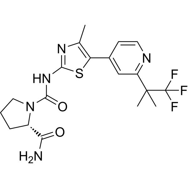
HY-15244
AlpelisibPI3Kα Inhibitor -
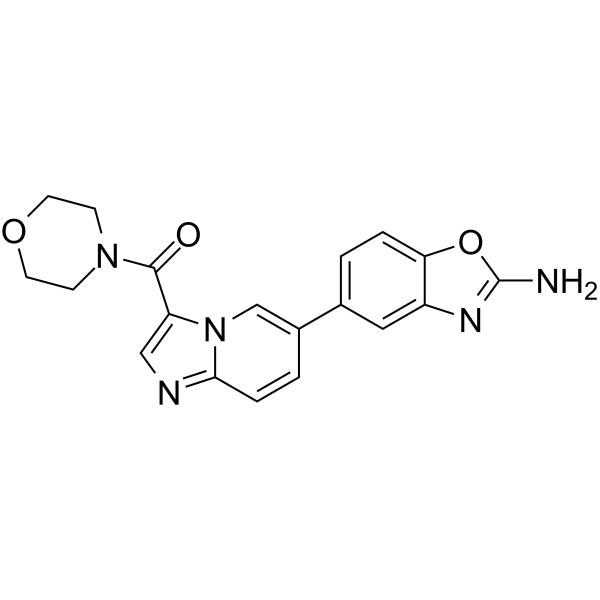
HY-12285
Serabelisibp110α Inhibitor -

HY-15244R
Alpelisib (Standard)Reference Standard -
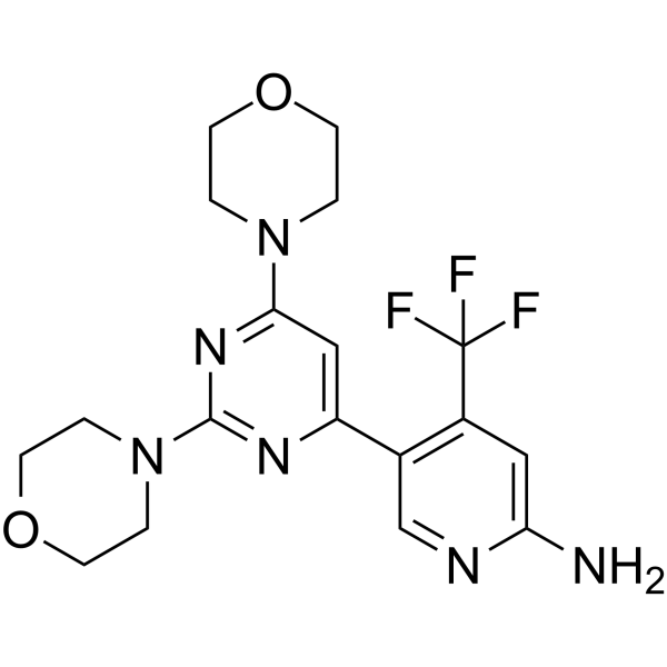
HY-70063
BuparlisibPI3K Inhibitor -
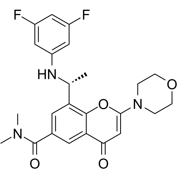
HY-12330
AZD8186PI3K Inhibitor -
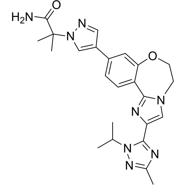
HY-13898
TaselisibPIK3CA Inhibitor -
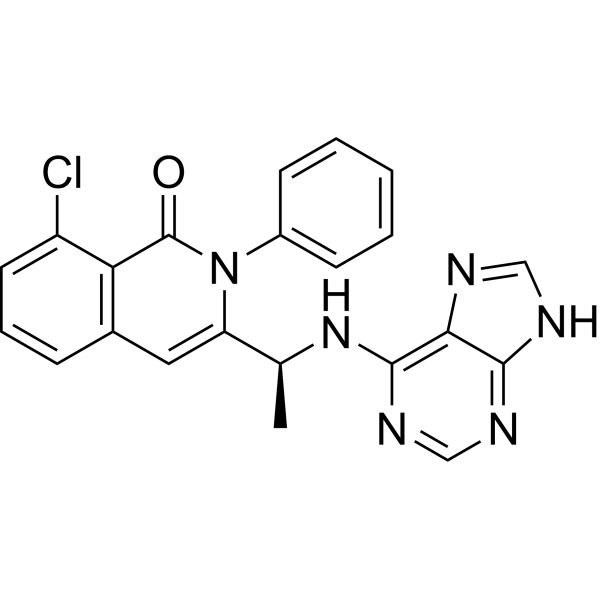
HY-17044
DuvelisibPI3Kδ Inhibitor -
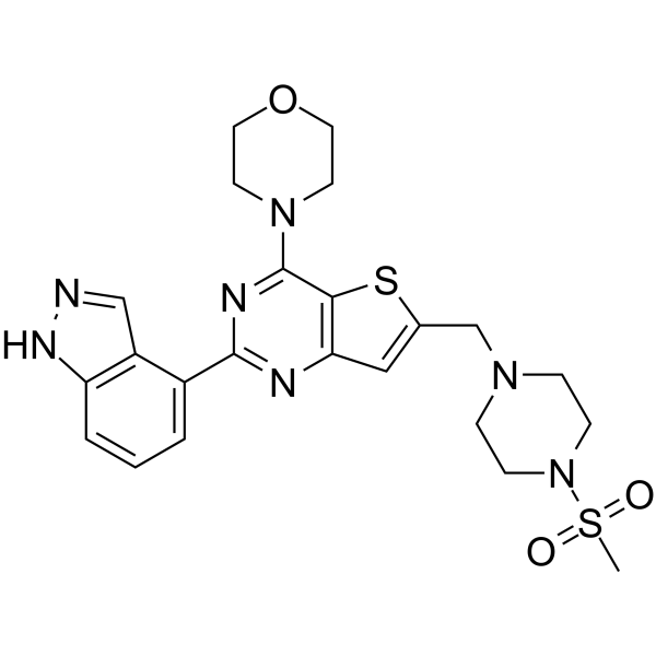
HY-50094
PictilisibPI3Kα/δ Inhibitor -
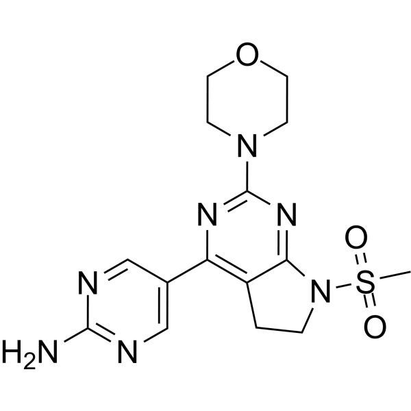
HY-15466
IzorlisibClass I PI3K Inhibitor -
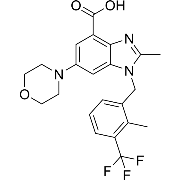
HY-15245
GSK2636771PI3Kβ Inhibitor -
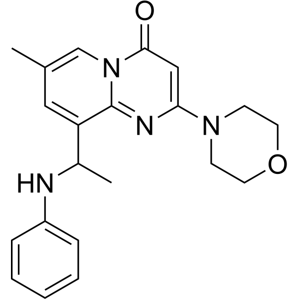
HY-10114
TGX-221Keap1-Nrf2 Interaction Inhibitor -
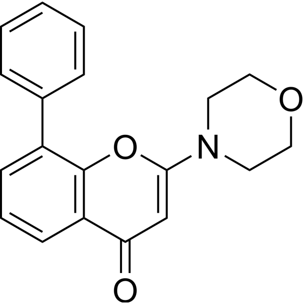
HY-10108
LY294002PI3K Inhibitor


Biological Activity
Description
IC50 & Target
IC50: 5 nM (p110α), 250 nM (p110γ), 290 nM (p110δ), 1200 nM (p110β)[1]
Cellular Effect
|
Cell Line

|
Type | Value | Description | References |
|---|---|---|---|---|
| A549 | IC50 |
4 μM
Compound: 1
|
Antiproliferative activity against human A549 cells incubated for 96 hrs by MTT assay
Antiproliferative activity against human A549 cells incubated for 96 hrs by MTT assay
|
[PMID: 40047238] |
| AGS | IC50 |
0.52 μM
Compound: Alpelisib
|
Antiproliferative activity against human AGS cells assessed as inhibition of cell viability incubated for 3 days by MTT/CTG assay
Antiproliferative activity against human AGS cells assessed as inhibition of cell viability incubated for 3 days by MTT/CTG assay
|
[PMID: 40518729] |
| DU-145 | IC50 |
4 μM
Compound: 1
|
Antiproliferative activity against human DU-145 cells incubated for 96 hrs by MTT assay
Antiproliferative activity against human DU-145 cells incubated for 96 hrs by MTT assay
|
[PMID: 40047238] |
| HCT-116 | IC50 |
3.73 μM
Compound: 1
|
Antiproliferative activity against human HCT-116 cells incubated for 96 hrs by MTT assay
Antiproliferative activity against human HCT-116 cells incubated for 96 hrs by MTT assay
|
[PMID: 40047238] |
| HGC-27 | IC50 |
1.92 μM
Compound: Alpelisib
|
Antiproliferative activity against human HGC-27 cells assessed as inhibition of cell viability incubated for 3 days by MTT/CTG assay
Antiproliferative activity against human HGC-27 cells assessed as inhibition of cell viability incubated for 3 days by MTT/CTG assay
|
[PMID: 40518729] |
| HT-29 | IC50 |
1.94 μM
Compound: 1
|
Antiproliferative activity against human HT-29 cells incubated for 96 hrs by MTT assay
Antiproliferative activity against human HT-29 cells incubated for 96 hrs by MTT assay
|
[PMID: 40047238] |
| Kasumi 1 | IC50 |
0.44 μM
Compound: Alpelisib
|
Antiproliferative activity against human Kasumi 1 cells assessed as cell growth inhibition incubated for 72 hrs by MTT assay
Antiproliferative activity against human Kasumi 1 cells assessed as cell growth inhibition incubated for 72 hrs by MTT assay
|
[PMID: 37126967] |
| L-363 | IC50 |
0.26 μM
Compound: 1; BYL-719
|
Antiproliferative activity against human L-363 cells
Antiproliferative activity against human L-363 cells
|
[PMID: 37652098] |
| L-363 | IC50 |
0.17 μM
Compound: Alpelisib
|
Antiproliferative activity against human L-363 cells assessed as reduction in cell growth incubated for 72 hrs by MTT assay
Antiproliferative activity against human L-363 cells assessed as reduction in cell growth incubated for 72 hrs by MTT assay
|
[PMID: 39605166] |
| LNCaP | IC50 |
15.8 μM
Compound: Alpelisib
|
Antiproliferative activity against human LNCaP cells assessed as inhibition of cell viability incubated for 3 days by MTT/CTG assay
Antiproliferative activity against human LNCaP cells assessed as inhibition of cell viability incubated for 3 days by MTT/CTG assay
|
[PMID: 40518729] |
| MCF7 | IC50 |
530 nM
Compound: 2; BYL719
|
Antiproliferation activity against human MCF7 cells assessed as reduction in cell viability incubated for 7 days by Cell-titer Glo reagent based assay
Antiproliferation activity against human MCF7 cells assessed as reduction in cell viability incubated for 7 days by Cell-titer Glo reagent based assay
|
[PMID: 33356246] |
| MCF7 | IC50 |
0.43 μM
Compound: Alpelisib
|
Antiproliferative activity against human MCF7 cells assessed as inhibition of cell growth incubated for 72 hrs by CCK8 assay
Antiproliferative activity against human MCF7 cells assessed as inhibition of cell growth incubated for 72 hrs by CCK8 assay
|
[PMID: 35834807] |
| MCF7 | IC50 |
0.25 μM
Compound: 1; BYL-719
|
Antiproliferative activity against human MCF7 cells
Antiproliferative activity against human MCF7 cells
|
[PMID: 37652098] |
| MCF7 | IC50 |
0.6 μM
Compound: Alpelisib
|
Antiproliferative activity against human MCF7 cells assessed as inhibition of cell growth incubated for 7 days by CCK8 assay
Antiproliferative activity against human MCF7 cells assessed as inhibition of cell growth incubated for 7 days by CCK8 assay
|
[PMID: 39159497] |
| MCF7 | IC50 |
1.2 μM
Compound: Alpelisib
|
Antiproliferative activity against human MCF7 cells assessed as reduction in cell viability incubated for 48 hrs by MTT assay
Antiproliferative activity against human MCF7 cells assessed as reduction in cell viability incubated for 48 hrs by MTT assay
|
[PMID: 39605166] |
| MCF7 | IC50 |
3.34 μM
Compound: 1
|
Antiproliferative activity against human MCF7 cells incubated for 96 hrs by MTT assay
Antiproliferative activity against human MCF7 cells incubated for 96 hrs by MTT assay
|
[PMID: 40047238] |
| MDA-MB-231 | IC50 |
62.9 μM
Compound: Alpesilib
|
Antiproliferative activity against human MDA-MB-231 cells assessed as cell viability after 24 hrs
Antiproliferative activity against human MDA-MB-231 cells assessed as cell viability after 24 hrs
|
[PMID: 33139111] |
| MDA-MB-231 | IC50 |
3.12 μM
Compound: 1
|
Antiproliferative activity against human MDA-MB-231 cells incubated for 96 hrs by MTT assay
Antiproliferative activity against human MDA-MB-231 cells incubated for 96 hrs by MTT assay
|
[PMID: 40047238] |
| MDA-MB-453 | IC50 |
0.77 μM
Compound: Alpelisib
|
Antiproliferative activity against human MDA-MB-453 cells assessed as inhibition of cell viability incubated for 3 days by MTT/CTG assay
Antiproliferative activity against human MDA-MB-453 cells assessed as inhibition of cell viability incubated for 3 days by MTT/CTG assay
|
[PMID: 40518729] |
| MGC-803 | IC50 |
0.43 μM
Compound: Alpelisib
|
Antiproliferative activity against human MGC-803 cells assessed as inhibition of cell growth incubated for 72 hrs by CCK8 assay
Antiproliferative activity against human MGC-803 cells assessed as inhibition of cell growth incubated for 72 hrs by CCK8 assay
|
[PMID: 35834807] |
| MV4-11 | IC50 |
3.5 μM
Compound: Alpelisib
|
Antiproliferative activity against human MV4-11 cells assessed as reduction in cell viability incubated for 72 hrs by CCK-8 assay
Antiproliferative activity against human MV4-11 cells assessed as reduction in cell viability incubated for 72 hrs by CCK-8 assay
|
[PMID: 38048697] |
| MV4-11 | IC50 |
0.19 μM
Compound: Alpelisib
|
Antiproliferative activity against human MV4-11 cells assessed as reduction in cell viability incubated for 72 hrs in presence of chidamide by CCK-8 assay
Antiproliferative activity against human MV4-11 cells assessed as reduction in cell viability incubated for 72 hrs in presence of chidamide by CCK-8 assay
|
[PMID: 38048697] |
| NCI-H446 | IC50 |
0.68 μM
Compound: 1
|
Antiproliferative activity against human NCI-H446 cells incubated for 96 hrs by MTT assay
Antiproliferative activity against human NCI-H446 cells incubated for 96 hrs by MTT assay
|
[PMID: 40047238] |
| PC-3 | IC50 |
3.53 μM
Compound: 1
|
Antiproliferative activity against human PC-3 cells incubated for 96 hrs by MTT assay
Antiproliferative activity against human PC-3 cells incubated for 96 hrs by MTT assay
|
[PMID: 40047238] |
| PC-3 | IC50 |
13.19 μM
Compound: Alpelisib
|
Antiproliferative activity against human PC-3 cells assessed as inhibition of cell viability incubated for 3 days by MTT/CTG assay
Antiproliferative activity against human PC-3 cells assessed as inhibition of cell viability incubated for 3 days by MTT/CTG assay
|
[PMID: 40518729] |
| Pfeiffer | IC50 |
50 μM
Compound: Alpelisib
|
Antiproliferative activity against human Pfeiffer cells assessed as inhibition of cell viability incubated for 3 days by MTT/CTG assay
Antiproliferative activity against human Pfeiffer cells assessed as inhibition of cell viability incubated for 3 days by MTT/CTG assay
|
[PMID: 40518729] |
| Rat1 | IC50 |
1.2 μM
Compound: 8, NVP-BYL719
|
Inhibition of N-terminal myristoylated P110delta (unknown origin)-mediated AKT phosphorylation at Ser473 expressed in rat Rat1 cells by ELISA
Inhibition of N-terminal myristoylated P110delta (unknown origin)-mediated AKT phosphorylation at Ser473 expressed in rat Rat1 cells by ELISA
|
[PMID: 23726034] |
| Rat1 | IC50 |
2.2 μM
Compound: 8, NVP-BYL719
|
Inhibition of N-terminal myristoylated P110beta (unknown origin)-mediated AKT phosphorylation at Ser473 expressed in rat Rat1 cells by ELISA
Inhibition of N-terminal myristoylated P110beta (unknown origin)-mediated AKT phosphorylation at Ser473 expressed in rat Rat1 cells by ELISA
|
[PMID: 23726034] |
| Rat1 | IC50 |
0.074 μM
Compound: 8, NVP-BYL719
|
Inhibition of N-terminal myristoylated P110alpha (unknown origin)-mediated AKT phosphorylation at Ser473 expressed in rat Rat1 cells by ELISA
Inhibition of N-terminal myristoylated P110alpha (unknown origin)-mediated AKT phosphorylation at Ser473 expressed in rat Rat1 cells by ELISA
|
[PMID: 23726034] |
| Rat1 | IC50 |
0.074 μM
Compound: 1, BYL719
|
Inhibition of myristoylated human P110alpha expressed in Rat1 cells assessed as inhibition of Akt phosphorylation at Serine 473 by Western blot analysis
Inhibition of myristoylated human P110alpha expressed in Rat1 cells assessed as inhibition of Akt phosphorylation at Serine 473 by Western blot analysis
|
[PMID: 26164189] |
| Rat1 | IC50 |
2.2 μM
Compound: 1, BYL719
|
Inhibition of myristoylated human P110beta expressed in Rat1 cells assessed as inhibition of Akt phosphorylation at Serine 473 by Western blot analysis
Inhibition of myristoylated human P110beta expressed in Rat1 cells assessed as inhibition of Akt phosphorylation at Serine 473 by Western blot analysis
|
[PMID: 26164189] |
| Rat1 | IC50 |
1.2 μM
Compound: 1, BYL719
|
Inhibition of myristoylated human P110delta expressed in Rat1 cells assessed as inhibition of Akt phosphorylation at Serine 473 by Western blot analysis
Inhibition of myristoylated human P110delta expressed in Rat1 cells assessed as inhibition of Akt phosphorylation at Serine 473 by Western blot analysis
|
[PMID: 26164189] |
| Rat1 | IC50 |
0.074 μM
Compound: Alpelisib
|
Inhibition of PI3Kalpha in rat Rat1 cells assessed as reduction of Akt phosphorylation at Ser473 in presence of 0.5% fetal calf serum
Inhibition of PI3Kalpha in rat Rat1 cells assessed as reduction of Akt phosphorylation at Ser473 in presence of 0.5% fetal calf serum
|
[PMID: 26206504] |
| Rat1 | IC50 |
2.2 μM
Compound: Alpelisib
|
Inhibition of PI3Kbeta in rat Rat1 cells assessed as reduction of Akt phosphorylation at Ser473 in presence of 0.5% fetal calf serum
Inhibition of PI3Kbeta in rat Rat1 cells assessed as reduction of Akt phosphorylation at Ser473 in presence of 0.5% fetal calf serum
|
[PMID: 26206504] |
| Rat1 | IC50 |
1.2 μM
Compound: Alpelisib
|
Inhibition of PI3Kgamma in rat Rat1 cells assessed as reduction of Akt phosphorylation at Ser473 in presence of 0.5% fetal calf serum
Inhibition of PI3Kgamma in rat Rat1 cells assessed as reduction of Akt phosphorylation at Ser473 in presence of 0.5% fetal calf serum
|
[PMID: 26206504] |
| SJRH30 | IC50 |
7.6 μM
Compound: BYL719
|
Antiproliferative activity against human Rh30 cells assessed as reduction in cell viability after 72 hrs by sulforhodamine B assay
Antiproliferative activity against human Rh30 cells assessed as reduction in cell viability after 72 hrs by sulforhodamine B assay
|
[PMID: 33109399] |
| T47D | IC50 |
420 nM
Compound: 2; BYL719
|
Antiproliferation activity against human T47D cells assessed as reduction in cell viability incubated for 7 days by Cell-titer Glo reagent based assay
Antiproliferation activity against human T47D cells assessed as reduction in cell viability incubated for 7 days by Cell-titer Glo reagent based assay
|
[PMID: 33356246] |
| T47D | IC50 |
0.1 μM
Compound: Alpelisib
|
Antiproliferative activity against human T47D cells expressing PIK3CA mutant assessed as cell growth inhibition incubated for 72 hrs by MTT assay
Antiproliferative activity against human T47D cells expressing PIK3CA mutant assessed as cell growth inhibition incubated for 72 hrs by MTT assay
|
[PMID: 37126967] |
| T47D | IC50 |
2.3 μM
Compound: 1; BYL-719
|
Antiproliferative activity against human T47D cells
Antiproliferative activity against human T47D cells
|
[PMID: 37652098] |
| T47D | IC50 |
0.4 μM
Compound: Alpelisib
|
Antiproliferative activity against human T47D cells assessed as inhibition of cell growth incubated for 7 days by CCK8 assay
Antiproliferative activity against human T47D cells assessed as inhibition of cell growth incubated for 7 days by CCK8 assay
|
[PMID: 39159497] |
| T47D | IC50 |
0.36 μM
Compound: Alpelisib
|
Antiproliferative activity against human T47D cells assessed as inhibition of cell viability incubated for 3 days by MTT/CTG assay
Antiproliferative activity against human T47D cells assessed as inhibition of cell viability incubated for 3 days by MTT/CTG assay
|
[PMID: 40518729] |
In Vitro
Alpelisib GMP (0-5 μM; 14 d) significantly inhibits the clonal growth of MCF-7 and T47D breast cancer cells in 2D colony formation assays[1].
Alpelisib GMP (5 μM; 5 d) reduces the mammosphere formation efficiency of MCF-7 and T47D breast cancer stem cell-like (BCSC-like) cells in a dose-dependent manner[1].
Alpelisib GMP (0-10 μM; 10 d) significantly reduces the spheroid diameter of MCF-7 and T47D breast cancer stem cell-like (BCSC-like) cells in 3D culture systems, and inhibits their stem cell properties and drug resistance[1].
Alpelisib GMP (1 μM; 24 h) significantly reduces the protein levels of the stem cell markers Nanog, Sox2, and OCT3/4 in MCF-7 and T47D breast cancer stem-like cells (BCSC-like cells)[1].
Alpelisib (10 μM; 10 d) inhibits adipogenesis, thereby attenuating adipocyte differentiation of primary LipPD1 lipoma cells in 2D culture systems and reducing the volume of 3D LipPD1 lipospheres[2].
Combination treatment with Alpelisib GMP (10 μM; 4 d) and a 1 μM SGK3 inhibitor (VPS34-IN1 (HY-12795) or SGK3-IN) produces enhanced antiproliferative activity in Alpelisib GMP-resistant MCF7R and T47DR breast cancer cells, with a synergistic effect observed for the Alpelisib+SGK3-IN combination[3].
MedChemExpress (MCE) has not independently confirmed the accuracy of these methods. They are for reference only. Further protocols information, click here.
-
Cell Line:human MCF-7 breast cancer monolayer cells, MCF-7 breast cancer stem cell (BCSC)-enriched mammosphere cells, human T47D breast cancer monolayer cells, T47D BCSC-enriched mammosphere cells
-
Concentration:Serial dilutions
-
Incubation Time:96 h
-
Result:Inhibited cell viability in a dose-dependent manner.
Achieved an IC50 of 0.225 μM in MCF-7 monolayer cells.
Achieved an IC50 of 0.453 μM in MCF-7 BCSC-enriched mammosphere cells.
Achieved an IC50 of 3.055 μM in T47D monolayer cells.
Achieved an IC50 of 5.105 μM in T47D BCSC-enriched mammosphere cells.
-
Cell Line:LipPD1, LipPD2, LipPD3 primary lipoma cells
-
Concentration:1-100 µM
-
Incubation Time:24-144 h
-
Result:Attenuated growth of all three lipoma cell cultures alone and in combination with rapamycin.
Maintained stable cell count for 6 days in LipPD1 cells treated with 100 µM.
Exhibited IC50 values for 72 h Hoechst assays of 15.91 µM (LipPD1), 10.06 µM (LipPD2), and 15.79 µM (LipPD3).
Reduced the fraction of Ki-67 positive LipPD1 cells in a concentration-dependent manner: to 0.75 fold (1 µM, p=0.074), 0.55 fold (10 µM, p=0.018), and 0.22 fold (100 µM, p=0.017).
-
Cell Line:LipPD1, LipPD2, LipPD3, Lip3, Lip4 primary lipoma cells
-
Concentration:50 µM
-
Incubation Time:24, 72 h
-
Result:Observed no cell death after 72 h 50 µM treatment in LipPD1 and Lip3 cells.
Detected a slight reduction in viable cells in Lip4 cells.
Detected no additional LDH release in LipPD1, LipPD2, or LipPD3 cells after 24 h or 72 h 50 µM treatment, indicating no induction of cell death.
-
Cell Line:LipPD1 primary lipoma cells
-
Concentration:10-100 µM
-
Incubation Time:24, 48 h
-
Result:Reduced AKT activation (phospho-Thr 308) in 50 µM treated cells.
Reduced mTOR activation (phospho-Ser 2448) in 10 µM and 50 µM treated cells.
Significantly reduced phosphorylation of S6 (phospho-Ser 235/236) for all tested concentrations.
Reduced the fraction of pS6 positive LipPD1 cells to 0.57 fold (10 µM) and 0.01 fold (100 µM).
-
Cell Line:LipPD1 primary lipoma cells
-
Concentration:10 µM
-
Incubation Time:10 days
-
Result:Reduced lipid accumulation in 2D culture, with the fraction of adipocytes decreasing from 54.8% to 29.9%.
Downregulated mRNA expression of differentiation markers: PPARγ to 0.55 fold, adiponectin to 0.54 fold, aP2 to 0.54 fold, and FASN to 0.58 fold.
Reduced 3D spheroid size to 0.78 fold after 4 days and 0.79 fold after 10 days, with significant differences from day 4 onward, while control spheroid size increased to 1.25 fold after 10 days.
In Vivo
Alpelisib GMP (50 mg/kg; i.p.; daily) exerts partial antitumor activity against alpelisib-resistant T47DR breast cancer xenografts, with maximum efficacy achieved when combined with SGK3 knockdown[3].
Alpelisib GMP (25 mg/kg; p.o.; daily; 4 weeks) significantly inhibits tumor growth, reduces tumor cell proliferation, and increases tumor cell apoptosis in a PIK3CA-mutant gastric cancer xenograft model with 100% survival of treated mice over 4 weeks[4].
MedChemExpress (MCE) has not independently confirmed the accuracy of these methods. They are for reference only.
-
Animal Model:BALB/c nude (6-week-old female)[3]
-
Dosage:50 mg/kg
-
Administration:i.p.; daily
-
Result:Exerted partial inhibition of MCF7R xenograft tumor growth.
Achieved maximum tumor growth inhibition when combined with SGK3 knockdown, with significantly reduced final tumor volumes and weights compared to vehicle control or SGK3 knockdown alone.\nExerted partial inhibition of T47DR xenograft tumor growth.
Achieved maximum tumor growth inhibition when combined with SGK3 knockdown, with significantly reduced final tumor volumes and weights compared to vehicle control or SGK3 knockdown alone.
-
Animal Model:Balb/c athymic nude mice (female, 5 weeks old, 26-28 g, subcutaneous xenograft of luciferase-expressing PIK3CA-mutant MKN1 human gastric cancer cells)[4]
-
Dosage:25 mg/kg
-
Administration:p.o.; daily; 4 weeks
-
Result:Significantly retarded tumor growth compared to the control group.
Decreased Ki-67 expression (proliferation marker).
Increased TUNEL expression (apoptosis marker).
Maintained stable body weight over the 4-week period.
Achieved 100% survival of treated mice over the 4-week period.


Chemical Information
-
CAS No. 1217486-61-7
-
Molecular Weight 441.47
-
Formula C19H22F3N5O2S
-
SMILES
CC(N=C(S1)NC(N2CCC[C@H]2C(N)=O)=O)=C1C3=CC(C(C)(C(F)(F)F)C)=NC=C3
-
Shipping
Room temperature in continental US; may vary elsewhere.
-
Storage
Please store the product under the recommended conditions in the Certificate of Analysis.

Protocols
-
RNA extraction experimental
By lysing cells, releasing RNA, and removing impurities such as proteins and DNA, high-purity RNA products are finally obtained. The commonly used traditional method is the guanidine isothiocyanate/phenol/chloroform method (Trizol), which is suitable for a variety of animal materials including animal tissues, microorganisms, cultured cells, etc., and most plant materials.
-
Apoptosis
Apoptosis, also called programmed cell death, is generally characterized by distinct morphological characteristics.
-
TUNEL staining for apoptotic DNA fragmentation
TUNEL staining detects DNA strand breaks by using terminal deoxynucleotidyl transferase to add labeled nucleotides to exposed 3′-OH DNA termini, generating either microscopic staining in fixed cells or tissue sections, or fluorescence/cytometric signal in cell suspensions. TUNEL positivity reflects DNA fragmentation but should not be interpreted alone as definitive apoptosis, because TUNEL can also label necrotic, autolytic, mechanically damaged, or DNA-repair-associated DNA breaks.
-
Flow cytometric DNA-content cell-cycle staining
Flow cytometric DNA-content cell-cycle staining measures the fluorescence intensity of DNA-bound fluorochromes in single cells or nuclei to estimate DNA content distributions, allowing assignment of populations to G0/G1, S, and G2/M phases by DNA histogram deconvolution. Propidium iodide (PI) intercalates into DNA, and PI fluorescence is proportional to cellular DNA content when staining is performed under conditions that make DNA accessible and minimize non-DNA signal. Cells with G2/M DNA content are expected to show approximately twice the fluorescence intensity of G0/G1 cells, while S-phase cells occupy intermediate fluorescence values. PI-based DNA-content analysis can also detect cells with fractional DNA content, often reported as sub-G1, when DNA fragmentation and extraction during staining reduce retained DNA signal in apoptotic cells. DAPI is an alternative DNA fluorochrome for univariate DNA-content analysis, while bivariate approaches combining DNA content with proliferation
-
Annexin V plus membrane-impermeant dye apoptosis staining
Annexin V-based apoptosis assays rely on the detection of phosphatidylserine (PS) externalization from the inner leaflet of the plasma membrane to the outer leaflet, an early biochemical hallmark of apoptosis. Fluorescently labeled Annexin V binds PS in a calcium-dependent manner, enabling identification of early apoptotic cells by flow cytometry or fluorescence microscopy. When combined with a membrane-impermeant DNA-binding dye (e. g. , propidium iodide), this approach allows discrimination between viable (Annexin V−/dye−), early apoptotic (Annexin V+/dye−), and late apoptotic or necrotic (Annexin V+/dye+) cell populations by assessing membrane integrity and PS exposure.
-
Apoptosis Solutions
Apoptosis is a regulated, generally non-lytic cell-death pathway that removes unwanted, damaged, infected, or abnormal cells through coordinated morphological changes, caspase activation, DNA fragmentation, and membrane remodeling. The intrinsic apoptosis pathway is controlled mainly by mitochondrial outer membrane permeabilization, BCL-2 family proteins, cytochrome c release, apoptosome formation, caspase-9 activation, and downstream executioner caspase-3/7 activation. The extrinsic apoptosis pathway is initiated by death receptors such as Fas, TNFR, and TRAIL receptors, which recruit adaptor proteins and activate caspase-8 before engaging executioner caspases or mitochondrial amplification through BID cleavage. Apoptosis is linked to many phenotypes, including cancer cell killing, tissue homeostasis, immune regulation, neurodegeneration, infection response, and treatment-induced cytotoxicity; unresolved questions include how apoptosis interacts with necroptosis, pyroptosis, ferroptos
-
Notch Pathway Solutions
The Notch pathway is a contact-dependent signaling pathway that controls cell-fate decisions, differentiation, proliferation, and tissue patterning through interactions between membrane-bound Notch receptors and membrane-bound ligands on neighboring cells. Canonical Notch signaling is activated when ligand engagement triggers proteolytic release of the Notch intracellular domain, which enters the nucleus and regulates transcription together with DNA-binding transcriptional complexes. In the canonical mechanism, ligand-dependent Notch activation leads to release of the intracellular Notch domain, and presenilin-dependent γ-secretase activity is required for production of the active intracellular signaling fragment. The released intracellular domain functions as a nuclear signal that converts Notch receptor activation at the membrane into transcriptional regulation of target programs such as HES/HEY-family genes and other context-dependent downstream targets. The literature links Notch p
-
Protocol for Cell Cycle
Cell-cycle analysis by flow cytometry measures DNA content in single cells to estimate the fraction of cells in G0/G1, S, and G2/M phases. Propidium iodide intercalates into DNA, and after RNA removal with RNase, fluorescence intensity reflects cellular DNA content: 2N cells are assigned to G0/G1, cells between 2N and 4N to S phase, and 4N cells to G2/M. DNA-content analysis alone cannot reliably separate G0 from G1 or G2 from M. Ki-67 can distinguish quiescent G0 cells from cycling cells, EdU or BrdU incorporation marks active DNA synthesis in S phase, and phospho-histone H3 staining identifies mitotic cells within the 4N population.
-
hPSC maintenance and expansion
This protocol maintains and expands human pluripotent stem cells under feeder-free, chemically defined conditions using E8 medium and vitronectin-coated culture surfaces; the readout is sustained adherent colony growth with undifferentiated morphology and retained pluripotency-marker expression during serial passaging. E8-based hPSC culture relies on defined soluble factors and matrix-dependent adhesion rather than feeder cells; vitronectin supports hPSC attachment through integrin-mediated interactions, and EDTA passaging dissociates colonies as small aggregates without enzymatic digestion, centrifugation, or routine ROCK-inhibitor treatment.
-
Breast Cancer Modeling
Breast cancer is a heterogeneous cancer, and it has been distinguished into four subtypes: luminal A, luminal B, HER2-positive and basal-like. Molecular mutations, epigenetic alterations, hormone exposure and immune microenvironment are related to the progression of breast cancer.
-
Senescence-associated β-galactosidase staining
Senescence-associated β-galactosidase staining detects β-galactosidase activity that is histochemically visible at pH 6. 0 in senescent cells, where X-gal cleavage produces an insoluble blue precipitate observable by bright-field microscopy. This activity reflects increased lysosomal β-galactosidase/lysosomal mass rather than a senescence-essential enzyme, because GLB1 depletion or genetic lysosomal β-galactosidase deficiency can abolish SA-β-gal staining while cells still undergo senescence. SA-β-gal was originally reported in senescent but not presenescent fibroblasts and keratinocytes, absent from quiescent fibroblasts and terminally differentiated keratinocytes, and increased with donor age in human skin samples. Because SA-β-gal can also appear in some non-senescent or tissue-specific contexts, interpretation should be paired with experimental controls and, when possible, independent senescence markers.
-
Cell Viability Determination by MTT Colorimetric Assay
The following protocol uses the MTT colorimetric assay as a classic literature-established method for assessing cell viability/metabolic activity in cultured mammalian cells. MTT[3-(4,5-dimethylthiazol-2-yl)-2,5-diphenyltetrazolium bromide] is reduced by metabolically active cells to a colored formazan product; the amount of formazan is quantified spectrophotometrically and provides an indirect measure of metabolically active viable cells. Importantly, MTT reduction reflects cellular oxidoreductase/metabolic activity rather than an absolute direct count of living cells, so changes in cellular metabolism can alter the signal independently of cell number.


Purity & Documentation
References
[1]. Yu L, et al. Effects of BYL-719 (alpelisib) on human breast cancer stem cells to overcome drug resistance in human breast cancer. Front Pharmacol. 2024;15:1443422. Published 2024 Oct 14. [Content Brief]
[2]. Kirstein AS, et al. The Novel Phosphatidylinositol-3-Kinase (PI3K) Inhibitor Alpelisib Effectively Inhibits Growth of PTEN-Haploinsufficient Lipoma Cells. Cancers (Basel). 2019;11(10):1586. Published 2019 Oct 17. [Content Brief]
[3]. Kang T, et al. The SGK3/GSK3β/β-catenin signaling promotes breast cancer stemness and confers resistance to alpelisib therapy. Int J Biol Sci. 2025;21(6):2462-2475. Published 2025 Mar 19. [Content Brief]
[4]. Kim KJ, et al. PI3K-targeting strategy using alpelisib to enhance the antitumor effect of paclitaxel in human gastric cancer. Sci Rep. 2020;10(1):12308. Published 2020 Jul 23. [Content Brief]

Calculators
Concentration (start) × Volume (start) = Concentration (final) × Volume (final)


Alpelisib (GMP) Related Classifications
HY-15244G Related Classifications
 Powered by Bioz