



Urolithin A
Based on 50 publication(s) in Google Scholar
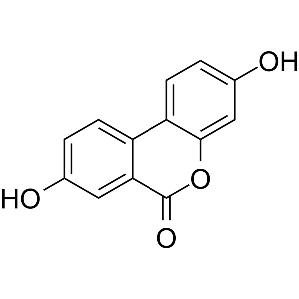
Urolithin A, a gut-microbial metabolite of ellagic acid, exerts anti-inflammatory, antiproliferative, and antioxidant properties. Urolithin A induces autophagy and apoptosis, suppresses cell cycle progression, and inhibits DNA synthesis.
Nur für Forschungszwecke. Wir verkaufen nicht an Patienten.
- Reinheit : 99.71%
- CAS. Nr.: 1143-70-0
- Formel: C13H8O4
- Molecular Weight:228.20
-
Speicherung:Powder -20°C, 3 years , 4°C, 2 years ; In solvent -80°C, 1 year , -20°C, 6 months

To place orders, for customer services and technical support, please contact: MedChemExpress USA
Tel: 609-228-6898 E-mail: [email protected] [email protected]


-
 Biologische Aktivität
-
Chemical Information
-
Lösungsmittel & Löslichkeit
- Protokoll
- Reinheit & Dokumentation
- Verweise
-
 Help & FAQs


-
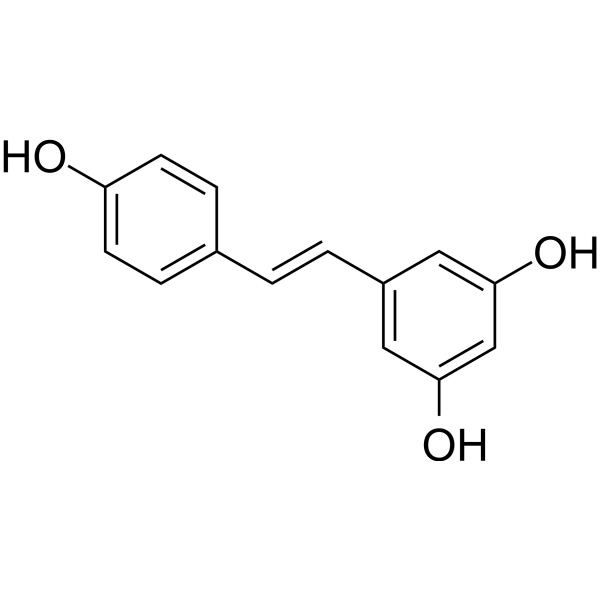
HY-16561
ResveratrolPolyphenolic Phytoalexin -
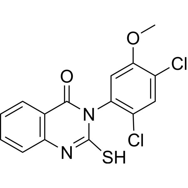
HY-15886
Mdivi-1Drp1 Inhibitor -
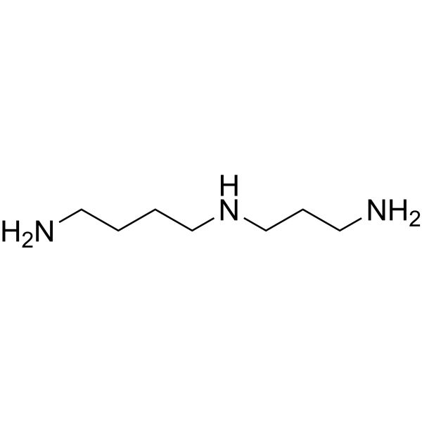
HY-B1776
SpermidinePolyamine Compound -

HY-10219
RapamycinmTOR Inhibitor -
HY-18085
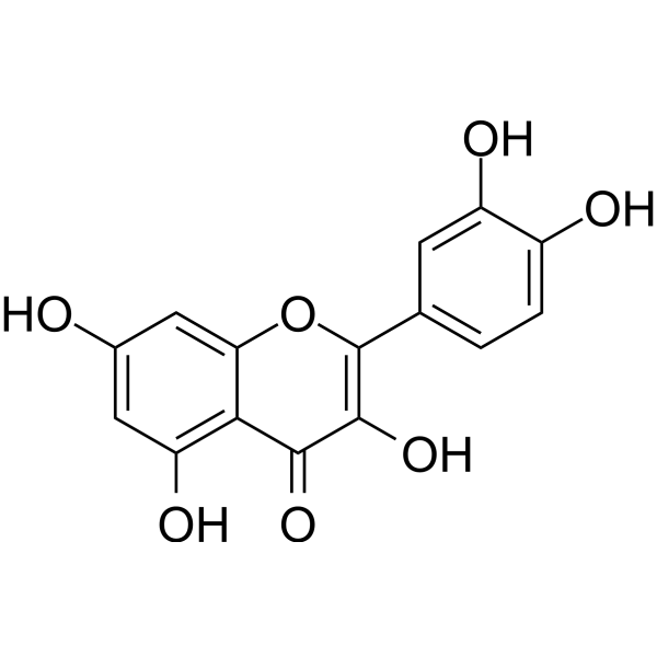
QuercetinFlavonoid Antioxidant -
HY-F0004
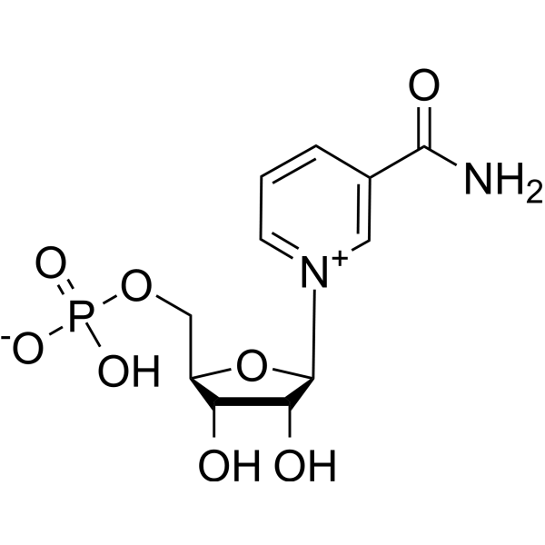
FosribnicotinamideNAD+ Intermediate -
HY-126307
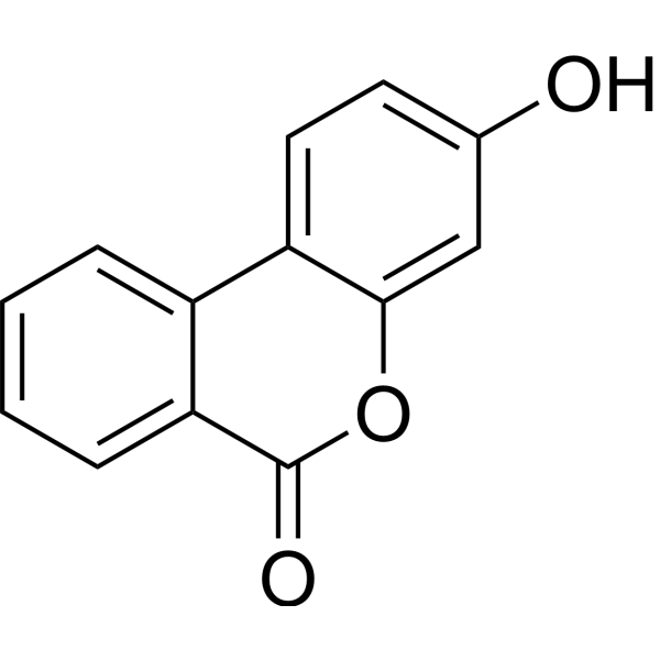
Urolithin BNF-κB Inhibitor -
HY-B0627
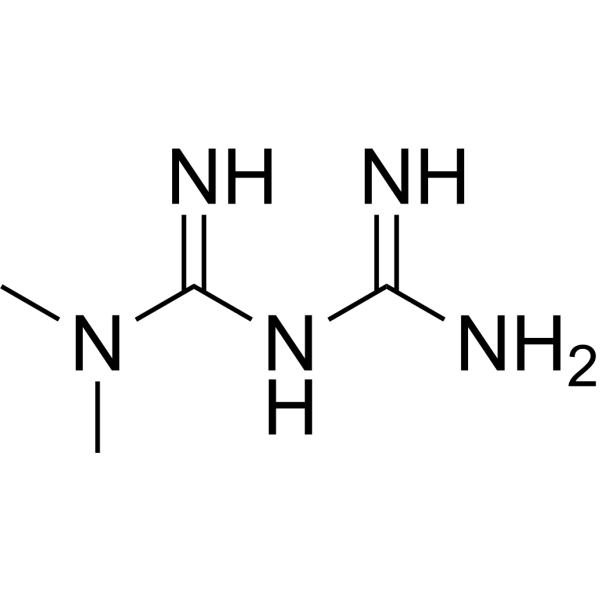
MetforminAntihyperglycemic Agent -
HY-100941
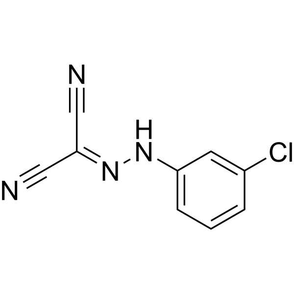
CCCPOXPHOS Uncoupler -
HY-B0183
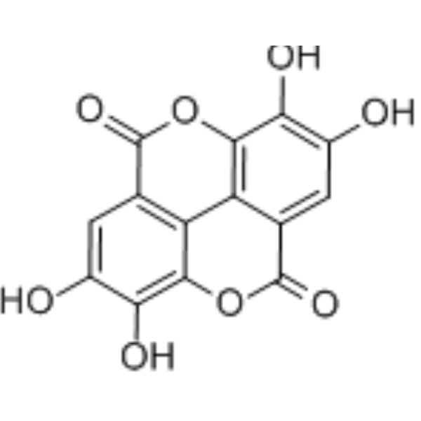
Ellagic acidSHP2 Inhibitor -

HY-100579
Ferrostatin-1Ferroptosis Inhibitor -
HY-19312
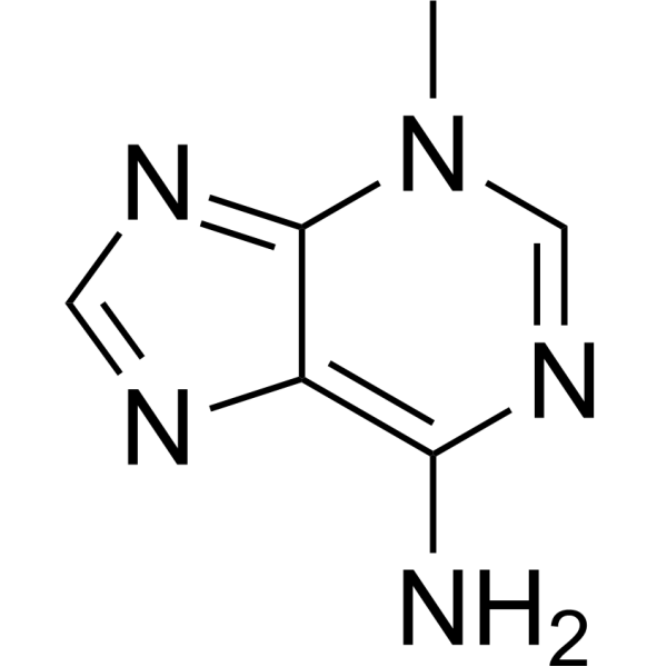
3-MethyladenineAutophagy/PI3K Inhibitor
Publications Citing Use of MedChemExpress (MCE) Urolithin A
More- Mater Today. 2026 Jan 7;92:453-472.
- Autophagy. 2026 Jul 31:1-19.
- Autophagy. 2026 Mar 15:1-22. [Abstract]
- Nat Commun. 2025 May 22;16(1):4746. [Abstract]
- Mol Cell. 2026 Feb 24:S1097-2765(26)00071-7. [Abstract]
- J Nanobiotechnology. 2024 Dec 18;22(1):758. [Abstract]
- J Nanobiotechnology. 2022 Mar 19;20(1):149. [Abstract]
- Cell Death Dis. 2023 May 24;14(5):339. [Abstract]
- J Neuroinflammation. 2025 Feb 28;22(1):55. [Abstract]
- Phytomedicine. 2026 Apr:153:157985. [Abstract]
- J Hazard Mater. 2024 Dec 5:480:135938. [Abstract]
- Environ Int. 2026 May:211:110262. [Abstract]
- J Headache Pain. 2023 Sep 5;24(1):122. [Abstract]
- Free Radic Biol Med. 2025 Aug 14:240:296-313. [Abstract]
- Chin Med. 2026 Aug 4;21(1):207.
- Redox Rep. 2026 Dec;31(1):2622255. [Abstract]
- Food Biosci. 2024 Feb, 57, 103549.
- Food Biosci. 2023 Jul 31, 102983.
- ACS Biomater Sci Eng. 2025 Jul 14;11(7):4254-4265. [Abstract]
- Radiother Oncol. 2024 Jan:190:110028. [Abstract]
- Commun Biol. 2022 Jun 22;5(1):616. [Abstract]
- Int Immunopharmacol. 2025 May 16:155:114604. [Abstract]
- Int Immunopharmacol. 2025 May 16:155:114572. [Abstract]
- Int J Mol Sci. 2025 Mar 26;26(7):3037. [Abstract]
- Int Immunopharmacol. 2025 Feb 20:148:114057. [Abstract]
- Int Immunopharmacol. 2024 Sep 19;142(Pt B):113151. [Abstract]
- Int J Mol Sci. 2024 Apr 29;25(9):4853. [Abstract]
- Sci Rep. 2026 Apr 1;16(1):15438. [Abstract]
- Toxics. 2025 Apr 23;13(5):332. [Abstract]
- World J Stem Cells. 2021 Dec 26;13(12):1928-1946. [Abstract]
- J Inflamm Res. 2025 Oct 8:18:13965-13984. [Abstract]
- Biology (Basel). 2025 Jul 8;14(7):829. [Abstract]
- J Biol Chem. 2023 Aug;299(8):105015. [Abstract]
- Cell Stress Chaperones. 2021 May;26(3):473-493. [Abstract]
- mSphere. 2025 May 27;10(5):e0006125. [Abstract]
- Naunyn Schmiedebergs Arch Pharmacol. 2024 Sep;397(9):6633-6645. [Abstract]
- Cancer Res Commun. 2023 Jul 12;3(7):1224-1236. [Abstract]
- Mol Immunol. 2025 Oct:186:48-62. [Abstract]
- Cell Biochem Funct. 2024 Apr;42(3):e4019. [Abstract]
- Arch Biochem Biophys. 2023 Jul 15:743:109644. [Abstract]
- J Neurosci Res. 2025 Jun;103(6):e70054. [Abstract]
- Sports Med Health Sci. 2024 Apr 4;7(1):16-27. [Abstract]
- Vet Parasitol. 2025 Jan:333:110360. [Abstract]
- Connect Tissue Res. 2026 May;67(3):243-257. [Abstract]
- Neurochirurgie. 2023 Aug 18;69(5):101480. [Abstract]
- Science Essence Journal. 2025.
- SSRN. 2025 Aug 7.
- bioRxiv. 2025 May 19:2025.05.16.654477. [Abstract]
- Phytomed Plus. 2023 Nov 4, 100495.
- Research Square Preprint. 2021 Oct.
 Customer Validation & Images

-
IF
-
In Vivo Efficacy Study
-
IF
-
WB
-
In Vivo Imaging
Alle DNA/RNA Synthesis Isoform-spezifische Produkte anzeigen
MoreAlle Endogenous Metabolite Isoform-spezifische Produkte anzeigen
More

Biologische Aktivität
Beschreibung
IC50 & Target
|
Microbial Metabolite |
In Vitro
Micromolar urolithin A concentrations induces both autophagy and apoptosis. Urolithin A suppresses cell cycle progression and inhibited DNA synthesis in human sw620 colorectal cancer cells[2].
Urolithin A shows antiproliferative effects and inhibits T24 and Caco-2 cell growth with IC50s of 43.9 and 49 μM, respectively[3].
Urolithin A exerts a dose- and time-dependent significant arrest at G2/M and S phases after treatments with 50 and 100 μM at 24 and 48 h compared to control cells. It induces cell apoptosis with 50 and 100 μM [4].
Urolithin A shows potent antiproliferative activity on HepG2 cells. When cell death is induced by Urolithin A, the expression of β-catenin, c-Myc and Cyclin D1 are decreased and TCF/LEF transcriptional activation is notably down-regulated. Urolithin A also increases protein expression of p53, p38-MAPK and caspase-3, but suppresses expression of NF-κB p65 and other inflammatory mediators[5].
MedChemExpress (MCE) has not independently confirmed the accuracy of these methods. They are for reference only.
In Vivo
MedChemExpress (MCE) has not independently confirmed the accuracy of these methods. They are for reference only.
- Annexin V plus membrane-impermeant dye apoptosis staining
- BrdU Incorporation Assay
- Protocol for Cell Cycle
- Research Protocol for Inflammation-related Diseases
- Cell Viability Determination by MTT Colorimetric Assay
- EdU Incorporation Assay (Click Chemistry-Based DNA Synthesis Measurement)
- Detection of Apoptosis
- TUNEL staining for apoptotic DNA fragmentation
- Flow cytometric DNA-content cell-cycle staining
- Autophagy
- Apoptosis Solutions
- Lysosome and acidic-vesicle live-cell staining
- Macroautophagy Solutions
Clinical Trial
| NCT Number | Sponsor | Condition | Start Date |
Phase

|
|---|---|---|---|---|
| NCT01329991 | Plexxikon| | 2011-05 | PHASE1 |


Chemical Information
-
CAS. Nr. 1143-70-0
-
Appearance Solid
-
Molecular Weight 228.20
-
Formel C13H8O4
-
SMILES
O=C1C2=CC(O)=CC=C2C3=CC=C(O)C=C3O1
-
Structure Classification
-
Initial Source
-
Versand
Room temperature in continental US; may vary elsewhere.
-
Speicherung
Powder -20°C 3 years 4°C 2 years In solvent -80°C 1 year -20°C 6 months


Publications (50)
-
Journal Impact Factor
-
Most Recent
-
-
-
Autophagy
Impaired mitophagy contributes to osteogenesis and mineralization disorders in fibrous dysplasia. [Abstract]2026 Mar 15:1-22. PMID: 41803635 -
Nat Commun
Osteocyte-derived extracellular vesicles mediate the bone-to-cartilage crosstalk and promote osteoarthritis progression. [Abstract]2025 May 22;16(1):4746. PMID: 40399261 -
Mol Cell
The human antibacterial factor APOL3 couples lysosomal damage to mitochondrial DNA efflux and type I IFN induction. [Abstract]2026 Feb 24:S1097-2765(26)00071-7. PMID: 41742416 -
J Nanobiotechnology
Osteoblastic ferroptosis inhibition by small-molecule promoting GPX4 activation for peri-prosthetic osteolysis therapy. [Abstract]2024 Dec 18;22(1):758. PMID: 39696565 -
J Nanobiotechnology
PINK1/TAX1BP1-directed mitophagy attenuates vascular endothelial injury induced by copper oxide nanoparticles. [Abstract]2022 Mar 19;20(1):149. PMID: 35305662
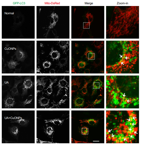
Urolithin A purchased from MedChemExpress. Usage Cited in: J Nanobiotechnology. 2022 Mar 19;20(1):149. [Abstract]
Urolithin A (100 μM; 12 h). Representative fluorescence images of EA.hy926 cell line stable expressing GFP-LC3 and Mito-DsRed.
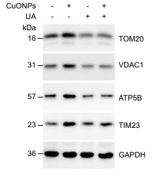
Urolithin A purchased from MedChemExpress. Usage Cited in: J Nanobiotechnology. 2022 Mar 19;20(1):149. [Abstract]
UA (100 μM; 24 h). The protein levels of TOM20, VDAC1, ATP5B and TIM23 in CuONPs-treated EA.hy926 cells were analyzed and quantified by Western blotting.
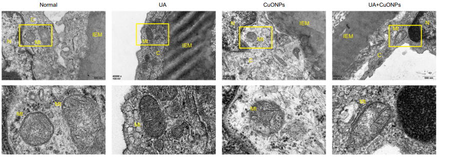
Urolithin A purchased from MedChemExpress. Usage Cited in: J Nanobiotechnology. 2022 Mar 19;20(1):149. [Abstract]
Representative TEM images of mice abdominal aorta dissected from CuONPs-treated mice. Mice were treated with UA (30 mg/kg; 10 days) by intragastric gavage and then intratracheally instilled with CuONPs (5 mg/kg) at day 7.
-
Cell Death Dis
Activation of HIF-1α C-terminal transactivation domain protects against hypoxia-induced kidney injury through hexokinase 2-mediated mitophagy. [Abstract]2023 May 24;14(5):339. PMID: 37225700 -
J Neuroinflammation
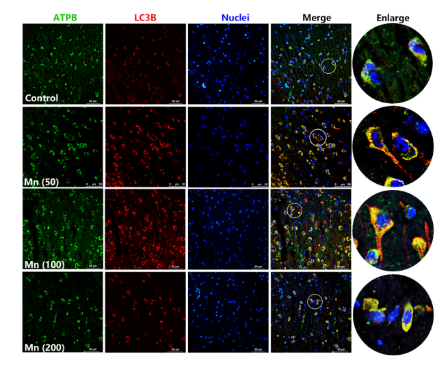
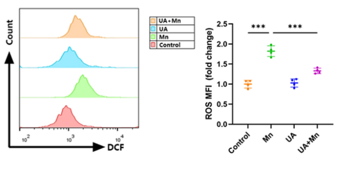
Protective role of mitophagy on microglia-mediated neuroinflammatory injury through mtDNA-STING signaling in manganese-induced parkinsonism. [Abstract]2025 Feb 28;22(1):55. PMID: 40022162
Urolithin A purchased from MedChemExpress. Usage Cited in: J Neuroinflammation. 2025 Feb 28;22(1):55. [Abstract]
UA (10 μM; 2 h) pretreatment ameliorated Mn-induced microglial mitophagy suppression.
Urolithin A purchased from MedChemExpress. Usage Cited in: J Neuroinflammation. 2025 Feb 28;22(1):55. [Abstract]
Flow cytometry shows reduced ROS levels in the striatum after UA (2.3 mg/kg; ip; 3 days) pretreatment compared with Mn-treated.
-
Phytomedicine
Kakkalide promotes spinal cord injury repair by regulating microglial M2 polarization via mitophagy. [Abstract]2026 Apr:153:157985. PMID: 41720005 -
J Hazard Mater
Environmentally relevant concentrations of perfluorobutane sulfonate impair locomotion behaviors and healthspan by downregulating mitophagy in C. elegans. [Abstract]2024 Dec 5:480:135938. PMID: 39326150 -
Environ Int
Trimethyltin chloride triggers ferroptosis in myocardial injury: mitochondria-dependent protection by luteolin. [Abstract]2026 May:211:110262. PMID: 42013553 -
J Headache Pain
Proteomics profiling reveals mitochondrial damage in the thalamus in a mouse model of chronic migraine. [Abstract]2023 Sep 5;24(1):122. PMID: 37667199 -
Free Radic Biol Med
Urolithin A protects against domoic acid-induced cognitive deficits via promoting estrogen receptor-α-mediated mitochondrial biogenesis signaling in mice. [Abstract]2025 Aug 14:240:296-313. PMID: 40818743 -
-
Redox Rep
Urolithin A alleviates vascular remodeling through mitochondrial SIRT3-mediated SOD2 deacetylation and antioxidation in hypertensive rats. [Abstract]2026 Dec;31(1):2622255. PMID: 41645805 -
-
-
ACS Biomater Sci Eng
Microbiome Metabolite-Incorporated Lipid Nanoparticles Augment CD8+ T Cell Memory Potential and Immunity for mRNA Cancer Vaccines. [Abstract]2025 Jul 14;11(7):4254-4265. PMID: 40490465 -
Radiother Oncol
Mitophagy induction improves salivary gland stem/progenitor cell function by reducing senescence after irradiation. [Abstract]2024 Jan:190:110028. PMID: 38007043 -
Commun Biol
Activating Parkin-dependent mitophagy alleviates oxidative stress, apoptosis, and promotes random-pattern skin flaps survival. [Abstract]2022 Jun 22;5(1):616. PMID: 35732814 -
Int Immunopharmacol
Urolithin a attenuates rheumatoid arthritis by inhibiting inflammation and pyroptosis in fibroblasts via the AMPK/ NF-κB signaling pathway. [Abstract]2025 May 16:155:114604. PMID: 40215775 -
Int Immunopharmacol
Urolithin A enhances diabetic wound healing: Insights from parkin-mediated mitophagy in endothelial progenitor cells. [Abstract]2025 May 16:155:114572. PMID: 40203794 -
Int J Mol Sci
Urolithin A Protects Porcine Oocytes from Artificially Induced Oxidative Stress Damage to Enhance Oocyte Maturation and Subsequent Embryo Development. [Abstract]2025 Mar 26;26(7):3037. PMID: 40243704 -
Int Immunopharmacol
Urolithin A alleviates NLRP3 inflammasome activation and pyroptosis by promoting microglial mitophagy following spinal cord injury. [Abstract]2025 Feb 20:148:114057. PMID: 39827665 -
Int Immunopharmacol
The ellagitannin metabolite urolithin C attenuated cognitive impairment by inhibiting neuroinflammation via downregulation of MAPK/NF-kB signaling pathways in aging mice. [Abstract]2024 Sep 19;142(Pt B):113151. PMID: 39303538 -
Int J Mol Sci
Mitophagy Regulates the Circadian Rhythms by Degrading NR1D1 in Simulated Microgravity and Isolation Environments. [Abstract]2024 Apr 29;25(9):4853. PMID: 38732079 -
Sci Rep
Urolithin A blocks colorectal cancer progression by AKT1 inhibition-driven immune activation. [Abstract]2026 Apr 1;16(1):15438. PMID: 41922440 -
Toxics
2025 Apr 23;13(5):332. PMID: 40423411 -
World J Stem Cells
Urolithin a alleviates oxidative stress-induced senescence in nucleus pulposus-derived mesenchymal stem cells through SIRT1/PGC-1α pathway. [Abstract]2021 Dec 26;13(12):1928-1946. PMID: 35069991 -
J Inflamm Res
Apigenin Suppresses NLRP3 Inflammasome Activation and Pyroptosis and Promotes Functional Recovery by Promoting Mitophagy in Experimental Spinal Cord Injured Rats. [Abstract]2025 Oct 8:18:13965-13984. PMID: 41084615 -
Biology (Basel)
Urolithin A Protects Ovarian Reserve Via Inhibiting PI3K/Akt Signaling and Preventing Chemotherapy-Induced Follicle Apoptosis. [Abstract]2025 Jul 8;14(7):829. PMID: 40723388 -
J Biol Chem
The FSH-mTOR-CNP signaling axis initiates follicular antrum formation by regulating tight junction, ion pumps, and aquaporins. [Abstract]2023 Aug;299(8):105015. PMID: 37414146 -
Cell Stress Chaperones
Urolithin A induces cell cycle arrest and apoptosis by inhibiting Bcl-2, increasing p53-p21 proteins and reactive oxygen species production in colorectal cancer cells. [Abstract]2021 May;26(3):473-493. PMID: 33666815 -
mSphere
Effect of urolithin A on intracellular survival of Mycobacterium tuberculosis by regulating AKT-FOXO1-mediated autophagy. [Abstract]2025 May 27;10(5):e0006125. PMID: 40207917 -
Naunyn Schmiedebergs Arch Pharmacol
Urolithin A exerts anti-tumor effects on gastric cancer via activating autophagy-Hippo axis and modulating the gut microbiota. [Abstract]2024 Sep;397(9):6633-6645. PMID: 38489081 -
Cancer Res Commun
Remodeling of Stromal Immune Microenvironment by Urolithin A Improves Survival with Immune Checkpoint Blockade in Pancreatic Cancer. [Abstract]2023 Jul 12;3(7):1224-1236. PMID: 37448553 -
Mol Immunol
Gelatin enhances bacteria-phagocytosis via a ROS-mitochondria-STING axis in differentiated human macrophage-like U937 cells. [Abstract]2025 Oct:186:48-62. PMID: 40815890 -
Cell Biochem Funct
Urolithin A affects cellular migration and modulates matrix metalloproteinase expression in colorectal cancer cells. [Abstract]2024 Apr;42(3):e4019. PMID: 38622949 -
Arch Biochem Biophys
Silibinin alleviates ferroptosis of rat islet β cell INS-1 induced by the treatment with palmitic acid and high glucose through enhancing PINK1/parkin-mediated mitophagy. [Abstract]2023 Jul 15:743:109644. PMID: 37245586 -
J Neurosci Res
Astrocytic HSP60 Deletion Induced Astrocyte Senescence and Inhibited Neuroregeneration via Modulating the S1P/Truncated-BDNF Pathway. [Abstract]2025 Jun;103(6):e70054. PMID: 40448367 -
Sports Med Health Sci
Sulforaphane, Urolithin A, and ZLN005 induce time-dependent alterations in antioxidant capacity, mitophagy, and mitochondrial biogenesis in muscle cells. [Abstract]2024 Apr 4;7(1):16-27. PMID: 39649792 -
Vet Parasitol
2025 Jan:333:110360. PMID: 39616807 -
Connect Tissue Res
Collagen I alleviates UVB-induced oxidative DNA damage and inhibits YAP-p73 apoptotic pathway in human keratinocytes HaCaT. [Abstract]2026 May;67(3):243-257. PMID: 41700367 -
Neurochirurgie
Urolithin A alleviates early brain injury after subarachnoid hemorrhage by regulating the AMPK/mTOR pathway-mediated autophagy. [Abstract]2023 Aug 18;69(5):101480. PMID: 37598622 -
-
-
bioRxiv
The antibacterial factor APOL3 couples lysosomal damage to mitochondrial DNA efflux and type I IFN induction. [Abstract]2025 May 19:2025.05.16.654477. PMID: 40475483 -
-


Lösungsmittel & Löslichkeit
In Vitro:
DMSO : 50 mg/mL (219.11 mM; Need ultrasonic; Hygroscopic DMSO has a significant impact on the solubility of product, please use newly opened DMSO)
Please refer to the solubility information to select the appropriate solvent. Once prepared, please aliquot and store the solution to prevent product inactivation from repeated freeze-thaw cycles.
Storage method and period of stock solution: -80°C, 1 year; -20°C, 6 months. When stored at -80°C, please use it within 1 year. When stored at -20°C, please use it within 6 months.
Please refer to the solubility information to select the appropriate solvent. Once prepared, please aliquot and store the solution to prevent product inactivation from repeated freeze-thaw cycles.
Storage method and period of stock solution: -80°C, 1 year; -20°C, 6 months. When stored at -80°C, please use it within 1 year. When stored at -20°C, please use it within 6 months.
Konzentration (Stammlösung) × Volumen (Stammlösung) = Konzentration (Ziellösung) × Volumen (Ziellösung)
In Vivo:
Select the appropriate dissolution method based on your experimental animal and administration route.
- For the following dissolution methods, please ensure to first prepare a clear stock solution using an In Vitro approach and then sequentially add co-solvents:
- To ensure reliable experimental results, the clarified stock solution can be appropriately stored based on storage conditions. As for the working solution for In Vivo experiments, it is recommended to prepare freshly and use it on the same day.
- The percentages shown for the solvents indicate their volumetric ratio in the final prepared solution. If precipitation or phase separation occurs during preparation, heat and/or sonication can be used to aid dissolution.
Add each solvent one by one: 10% DMSO 40% PEG300 5% Tween-80 45% Saline
Solubility: ≥ 2.5 mg/mL (10.96 mM); Clear solution
This protocol yields a clear solution of ≥ 2.5 mg/mL (saturation unknown).
Taking 1 mL working solution as an example, add 100 μL DMSO stock solution (25.0 mg/mL) to 400 μL PEG300, and mix evenly; then add 50 μL Tween-80 and mix evenly; then add 450 μL Saline to adjust the volume to 1 mL.
Preparation of Saline: Dissolve 0.9 g sodium chloride in ddH₂O and dilute to 100 mL to obtain a clear Saline solution.
Add each solvent one by one: 10% DMSO 90% (20% SBE-β-CD in Saline)
Solubility: 2.5 mg/mL (10.96 mM); Suspended solution; Need ultrasonic
This protocol yields a suspended solution of 2.5 mg/mL. Suspended solution can be used for oral and intraperitoneal injection.
Taking 1 mL working solution as an example, add 100 μL DMSO stock solution (25.0 mg/mL) to 900 μL 20% SBE-β-CD in Saline, and mix evenly.
Preparation of 20% SBE-β-CD in Saline (4°C, storage for one week): 2 g SBE-β-CD powder is dissolved in 10 mL Saline, completely dissolve until clear.
For the following dissolution methods, please prepare the working solution directly:
It is recommended to prepare fresh solutions and use them promptly within a short period of time.
The percentages shown for the solvents indicate their volumetric ratio in the final prepared solution. If precipitation or phase separation occurs during preparation, heat and/or sonication can be used to aid dissolution.
Add each solvent one by one: 50% PEG300 50% Saline
Solubility: 5 mg/mL (21.91 mM); Suspended solution; Need ultrasonic
Add each solvent one by one: 15% Cremophor EL 85% Saline
Solubility: 7.35 mg/mL (32.21 mM); Suspended solution; Need ultrasonic
Add each solvent one by one: 0.5% CMC/saline water
Solubility: 5 mg/mL (21.91 mM); Suspended solution; Need ultrasonic
In Vivo Dissolution Calculator
Please enter the basic information of animal experiments:
-
-
-
-
Recommended: Prepare an additional quantity of animals to account for potential losses during experiments.
Please enter your animal formula composition:
-
%DMSO +
Recommended: Keep the proportion of DMSO in working solution below 2% if your animal is weak.
-
%+
-
+%Tween-80 + +
-
%Saline +
The co-solvents required include: DMSO, . All of co-solvents are available by MedChemExpress (MCE). , Tween 80. All of co-solvents are available by MedChemExpress (MCE).
Working solution concentration: 0.22 mg/mL
Method for preparing stock solution: mg drug dissolved in μL DMSO. Stock solution concentration: mg/mL.
1. Take μL DMSO stock solution;
2. Add μL .
μL , mix evenly;
3. Then add μL Tween 80, mix evenly;
4. Then add μL
Please ensure that the stock solution in the first step is dissolved to a clear state, and add co-solvents in sequence. You can use ultrasonic heating (ultrasonic cleaner, recommended frequency 20-40 kHz), vortexing, etc. to assist dissolution.


Reinheit & Dokumentation
-
Data Sheet (280 KB)
-
SDS (393 KB)
- English - EN (393 KB)
- Français - FR (393 KB)
- Deutsch - DE (393 KB)
- Norwegian - NO (393 KB)
- Español - ES (393 KB)
- Swedish - SV (393 KB)
- Italian - IT (393 KB)
- Korean - KR (393 KB)
- Portuguese - PT (393 KB)
-
Handling Instructions (2659 KB)
Verweise
[1]. Gong Z, et al. Urolithin A attenuates memory impairment and neuroinflammation in APP/PS1 mice. [Content Brief]
[2]. Wang Y, et al. In vitro antiproliferative and antioxidant effects of urolithin A, the colonic metabolite of ellagic acid, on hepatocellular carcinomas HepG2 cells. Toxicol In Vitro. 2015 Aug;29(5):1107-15. [Content Brief]
[3]. Zhao W, et al. Metabolite of ellagitannins, urolithin A induces autophagy and inhibits metastasis in human sw620colorectal cancer cells. Mol Carcinog. 2018 Feb;57(2):193-200. [Content Brief]
[4]. Ishimoto H, et al. In vivo anti-inflammatory and antioxidant properties of ellagitannin metabolite urolithin A. Bioorg Med Chem Lett. 2011 Oct 1;21(19):5901-4. [Content Brief]
[5]. Qiu Z, et al. In vitro antioxidant and antiproliferative effects of ellagic acid and its colonic metabolite, urolithins, on human bladder cancer T24 cells. Food Chem Toxicol. 2013 Sep;59:428-37. [Content Brief]
[6]. González-Sarrías A, et al. Antiproliferative activity of the ellagic acid-derived gut microbiota isourolithin A and comparison with its urolithin A isomer: the role of cell metabolism.Eur J Nutr. 2017 Mar;56(2):831-841. [Content Brief]


Complete Stock Solution Preparation Table
Please refer to the solubility information to select the appropriate solvent. Once prepared, please aliquot and store the solution to prevent product inactivation from repeated freeze-thaw cycles.
Storage method and period of stock solution: -80°C, 1 year; -20°C, 6 months. When stored at -80°C, please use it within 1 year. When stored at -20°C, please use it within 6 months.
| Optional Solvent | Concentration Solvent Mass | 1 mg | 5 mg | 10 mg | 25 mg |
|---|---|---|---|---|---|
| DMSO | 1 mM | 4.3821 mL | 21.9106 mL | 43.8212 mL | 109.5530 mL |
| 5 mM | 0.8764 mL | 4.3821 mL | 8.7642 mL | 21.9106 mL | |
| 10 mM | 0.4382 mL | 2.1911 mL | 4.3821 mL | 10.9553 mL | |
| 15 mM | 0.2921 mL | 1.4607 mL | 2.9214 mL | 7.3035 mL | |
| 20 mM | 0.2191 mL | 1.0955 mL | 2.1911 mL | 5.4777 mL | |
| 25 mM | 0.1753 mL | 0.8764 mL | 1.7528 mL | 4.3821 mL | |
| 30 mM | 0.1461 mL | 0.7304 mL | 1.4607 mL | 3.6518 mL | |
| 40 mM | 0.1096 mL | 0.5478 mL | 1.0955 mL | 2.7388 mL | |
| 50 mM | 0.0876 mL | 0.4382 mL | 0.8764 mL | 2.1911 mL | |
| 60 mM | 0.0730 mL | 0.3652 mL | 0.7304 mL | 1.8259 mL | |
| 80 mM | 0.0548 mL | 0.2739 mL | 0.5478 mL | 1.3694 mL | |
| 100 mM | 0.0438 mL | 0.2191 mL | 0.4382 mL | 1.0955 mL |


Urolithin A Related Classifications
HY-100599 Related Classifications
 Powered by Bioz