



OSU-T315
Based on 12 publication(s) in Google Scholar
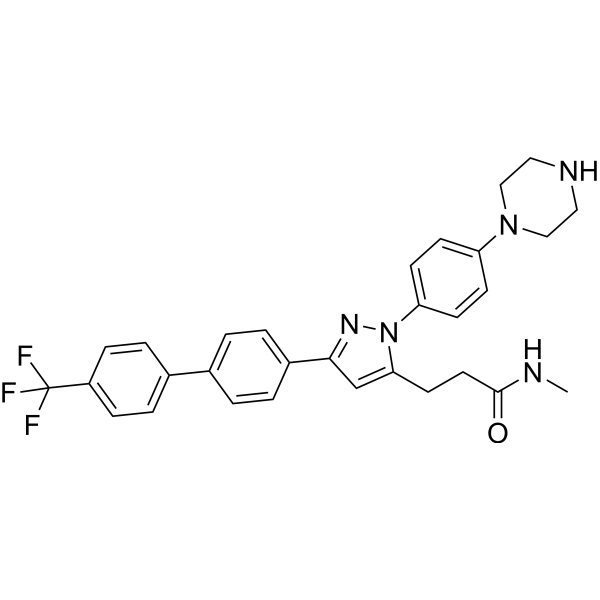
OSU-T315 (ILK-IN-1) is a small Integrin-linked kinase (ILK) inhibitor with an IC50 of 0.6 μM, inhibiting PI3K/AKT signaling by dephosphorylation of AKT-Ser473 and other ILK targets (GSK-3β and myosin light chain). OSU-T315 abrogates AKT activation by impeding AKT localization in lipid rafts and triggers caspase-dependent apoptosis in an ILK-independent manner. OSU-T315 causes cell death through apoptosis and autophagy.
Nos produits utilisent uniquement pour la recherche. Nous ne vendons pas aux patients.
- Pureté : 99.71%
- CAS No.: 2070015-22-2
- Formule: C30H30F3N5O
- Masse moléculaire:533.59
-
Stockage:Powder -20°C, 3 years , 4°C, 2 years ; In solvent -80°C, 2 years , -20°C, 1 year

To place orders, for customer services and technical support, please contact: MedChemExpress USA
Tel: 609-228-6898 E-mail: [email protected] [email protected]


-
 Activité biologique
-
Chemical Information
-
Solvant et solubilité
- Protocole
- Pureté et documentation
- Références
-
 Help & FAQs


-
HY-16141
CilengitideIntegrin Inhibitor -
HY-115677
ILK-IN-3ILK Inhibitor -
HY-19792
MertansineMicrotubule-Targeted Cytotoxin -
HY-15392
Chroman 1ROCK2 Inhibitor -
HY-12289
DefactinibFAK Inhibitor -
HY-15102
MK-0429αvβ3 Integrin Inhibitor -
HY-18676B
ILK-IN-2ILK Inhibitor -
HY-17369
Tirofiban hydrochloride monohydrateGp IIb/IIIa Receptor Antagonist -
HY-15701
Leukadherin-1CD11b/CD18 Agonist -
HY-N0416
Cucurbitacin BBioactive Tetracyclic Triterpenoid -

HY-16141R
Cilengitide (Standard)Reference Standard -

HY-N0416R
Cucurbitacin B (Standard)Bioactive Tetracyclic Triterpenoid
Publications Citing Use of MedChemExpress (MCE) OSU-T315
More- Theranostics. 2022 Jan 1;12(3):1173-1186. [Abstract]
- Br J Cancer. 2020 Aug;123(4):542-555. [Abstract]
- Colloids Surf B Biointerfaces. 2022 Feb:210:112229. [Abstract]
- Int Immunopharmacol. 2024 Nov 7;143(Pt 3):113530. [Abstract]
- Biochim Biophys Acta Mol Basis Dis. 2020 Mar 1;1866(3):165625. [Abstract]
- PLoS Pathog. 2023 Mar 17;19(3):e1011241. [Abstract]
- Cancer Sci. 2019 May;110(5):1804-1813. [Abstract]
- FASEB J. 2026 Jan 31;40(2):e71390. [Abstract]
- Mol Biol Rep. 2024 Mar 14;51(1):421. [Abstract]
- J Cardiovasc Transl Res. 2023 Apr;16(2):414-426. [Abstract]
- Genet Mol Biol. 2021 Sep 29;44(3):e20200461. [Abstract]
- Research Square Preprint. 2024 Jan 31.
 Customer Validation & Images

-
Cell Proliferation/Viability Assay
-
WB


Activité biologique
Description
IC50 & Target
IC50: 0.6μM; Integrin-linked kinase (ILK) inhibitor[1]
In Vitro
OSU-T315 (Compound 22; 0-5 μM; 24 hours) exhibits high in vitro potency against a panel of prostate and breast cancer cell lines with a IC50 range of 1-2.5 μM[1].
OSU-T315 (0-2.5 μM; 24 hours) can reduce YB-1, HER2, and EGFR expression; shows a modest suppressive effect on phosphorylated S6 levels, exhibits dose-dependent suppressive effects on the levels of phospho-ERK1/2 and phospho-p38, while that of phospho-JNK remains unaltered in PC-3 cell[1].
OSU-T315 (0-4 μM; 24 hours) causes autophagy through ILK inhibition[1].
MedChemExpress (MCE) has not independently confirmed the accuracy of these methods. They are for reference only.
-
Cell Line:PC-3 cells; MDA-MB-231 cells
-
Concentration:1 μM, 2 μM, 3 μM, 4 μM; 0.5 μM, 1 μM, 1.5 μM, 2 μM, 2.5 μM
-
Incubation Time:24 hours
-
Result:Exhibited a dose-dependent decreasing effect on the phosphorylation of pS6, ERKs, and p38 in PC-3 cells and MDA-MB-231 cells.
-
Cell Line:Prostate cancer cells: LNCaP, PC-3; breast cancer cells: MDA-MB-231, MDA-MB-468, SKBR3, MCF-7; PrEC and MEC cells
-
Concentration:0-5 μM
-
Incubation Time:24 hours
-
Result:Suppressed cancer cells viability in breast and prostate cancer cells (IC (50), 1-2.5μM).
-
Cell Line:PC-3 cells
-
Concentration:1 μM, 2 μM, 3 μM, 4 μM
-
Incubation Time:24 hours
-
Result:Induced accumulation of LC3-II and PARP cleavage.
In Vivo
No other obvious toxicity is observed in mice[1].
MedChemExpress (MCE) has not independently confirmed the accuracy of these methods. They are for reference only.
-
Animal Model:Male NCr athymic nude mice with PC-3 tumor xenografts
-
Dosage:25 mg/kg; 50 mg/kg
-
Administration:Oral gavage; single daily; 35 days
-
Result:Resulted in suppression of tumor growth relative to the vehicle control after 35 days of treatment (48% and 62% suppression for 25 and 50 mg/kg, respectively).
- Detection of Apoptosis
- TUNEL staining for apoptotic DNA fragmentation
- Cell Cytotoxicity Assay
- Mammalian live/dead viability and cytotoxicity staining
- Annexin V plus membrane-impermeant dye apoptosis staining
- Autophagy
- Apoptosis Solutions
- Lysosome and acidic-vesicle live-cell staining
- Macroautophagy Solutions


Chemical Information
-
CAS No. 2070015-22-2
-
Appearance Solid
-
Masse moléculaire 533.59
-
Formule C30H30F3N5O
-
Color White to off-white
-
SMILES
O=C(NC)CCC1=CC(C2=CC=C(C3=CC=C(C(F)(F)F)C=C3)C=C2)=NN1C4=CC=C(N5CCNCC5)C=C4
-
Livraison
Room temperature in continental US; may vary elsewhere.
-
Stockage
Powder -20°C 3 years 4°C 2 years In solvent -80°C 2 years -20°C 1 year


Publications (12)
-
Journal Impact Factor
-
Most Recent
-
Theranostics
α-Catulin promotes cancer stemness by antagonizing WWP1-mediated KLF5 degradation in lung cancer. [Abstract]2022 Jan 1;12(3):1173-1186. PMID: 35154481 -
Br J Cancer
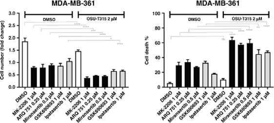
A systematic molecular and pharmacologic evaluation of AKT inhibitors reveals new insight into their biological activity. [Abstract]2020 Aug;123(4):542-555. PMID: 32439931
OSU-T315 purchased from MedChemExpress. Usage Cited in: Br J Cancer. 2020 Aug;123(4):542-555. [Abstract]
The combination of the ILK inhibitor OSU-T315 with AKT inhibitors of either class potentiated cell death in MDA-MB-361 cells
-
Colloids Surf B Biointerfaces
The osteogenic effects of porous Tantalum and Titanium alloy scaffolds with different unit cell structure. [Abstract]2022 Feb:210:112229. PMID: 34875470 -
Int Immunopharmacol
OSU-T315 overcomes immunosuppression in triple-negative breast cancer by targeting the ILK/NF-κB signaling pathway to enhance immunotherapeutic efficacy. [Abstract]2024 Nov 7;143(Pt 3):113530. PMID: 39515039 -
Biochim Biophys Acta Mol Basis Dis
Matrix stiffness modulates ILK-mediated YAP activation to control the drug resistance of breast cancer cells. [Abstract]2020 Mar 1;1866(3):165625. PMID: 31785406 -
PLoS Pathog
Negative regulation of type I interferon signaling by integrin-linked kinase permits dengue virus replication. [Abstract]2023 Mar 17;19(3):e1011241. PMID: 36930690 -
Cancer Sci
Serum deprivation-response protein regulates aldehyde dehydrogenase 1 through integrin-linked kinase signaling in endometrioid carcinoma cells. [Abstract]2019 May;110(5):1804-1813. PMID: 30907484
OSU-T315 purchased from MedChemExpress. Usage Cited in: Cancer Sci. 2019 May;110(5):1804-1813. [Abstract]
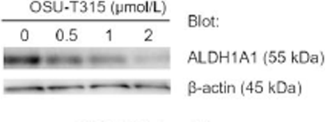
Effect of OSU-T315 on the proportion of ALDH-hi cells (left and middle) and the ALDH1A1 and SDPR protein level in EV HEC-108 cells (right). Equal protein loading is confirmed by quantifying β-actin (input control).
-
FASEB J
Unlocking the Therapeutic Potential of Integrin-Linked Kinase Inhibitors in Bioengineered 3D Breast Tumor Stroma Models. [Abstract]2026 Jan 31;40(2):e71390. PMID: 41537745 -
Mol Biol Rep
ILK inhibition reduces osteophyte formation through suppression of osteogenesis in BMSCs via Akt/GSK-3β/β-catenin pathway. [Abstract]2024 Mar 14;51(1):421. PMID: 38483756 -
J Cardiovasc Transl Res
Roles of TRPV4 in Regulating Circulating Angiogenic Cells to Promote Coronary Microvascular Regeneration. [Abstract]2023 Apr;16(2):414-426. PMID: 36103035 -
Genet Mol Biol
Periostin regulates osteogenesis of mesenchymal stem cells from ovariectomized rats through actions on the ILK/Akt/GSK-3β Axis. [Abstract]2021 Sep 29;44(3):e20200461. PMID: 34591063 -


Solvant et solubilité
In Vitro:
DMSO : ≥ 260 mg/mL (487.27 mM; Hygroscopic DMSO has a significant impact on the solubility of product, please use newly opened DMSO)
* "≥" means soluble, but saturation unknown.
Please refer to the solubility information to select the appropriate solvent. Once prepared, please aliquot and store the solution to prevent product inactivation from repeated freeze-thaw cycles.
Storage method and period of stock solution: -80°C, 2 years; -20°C, 1 year. When stored at -80°C, please use it within 2 years. When stored at -20°C, please use it within 1 year.
Please refer to the solubility information to select the appropriate solvent. Once prepared, please aliquot and store the solution to prevent product inactivation from repeated freeze-thaw cycles.
Storage method and period of stock solution: -80°C, 2 years; -20°C, 1 year. When stored at -80°C, please use it within 2 years. When stored at -20°C, please use it within 1 year.
Concentration (start) × Volume (start) = Concentration (final) × Volume (final)
In Vivo:
Select the appropriate dissolution method based on your experimental animal and administration route.
- For the following dissolution methods, please ensure to first prepare a clear stock solution using an In Vitro approach and then sequentially add co-solvents:
- To ensure reliable experimental results, the clarified stock solution can be appropriately stored based on storage conditions. As for the working solution for In Vivo experiments, it is recommended to prepare freshly and use it on the same day.
- The percentages shown for the solvents indicate their volumetric ratio in the final prepared solution. If precipitation or phase separation occurs during preparation, heat and/or sonication can be used to aid dissolution.
Add each solvent one by one: 10% DMSO 40% PEG300 5% Tween-80 45% Saline
Solubility: ≥ 2.17 mg/mL (4.07 mM); Clear solution
This protocol yields a clear solution of ≥ 2.17 mg/mL (saturation unknown).
Taking 1 mL working solution as an example, add 100 μL DMSO stock solution (21.7 mg/mL) to 400 μL PEG300, and mix evenly; then add 50 μL Tween-80 and mix evenly; then add 450 μL Saline to adjust the volume to 1 mL.
Preparation of Saline: Dissolve 0.9 g sodium chloride in ddH₂O and dilute to 100 mL to obtain a clear Saline solution.
In Vivo Dissolution Calculator
Please enter the basic information of animal experiments:
-
-
-
-
Recommended: Prepare an additional quantity of animals to account for potential losses during experiments.
Please enter your animal formula composition:
-
%DMSO +
Recommended: Keep the proportion of DMSO in working solution below 2% if your animal is weak.
-
%+
-
+%Tween-80 + +
-
%Saline +
The co-solvents required include: DMSO, . All of co-solvents are available by MedChemExpress (MCE). , Tween 80. All of co-solvents are available by MedChemExpress (MCE).
Working solution concentration: 0.22 mg/mL
Method for preparing stock solution: mg drug dissolved in μL DMSO. Stock solution concentration: mg/mL.
1. Take μL DMSO stock solution;
2. Add μL .
μL , mix evenly;
3. Then add μL Tween 80, mix evenly;
4. Then add μL
Please ensure that the stock solution in the first step is dissolved to a clear state, and add co-solvents in sequence. You can use ultrasonic heating (ultrasonic cleaner, recommended frequency 20-40 kHz), vortexing, etc. to assist dissolution.


Pureté et documentation
-
Fiche technique (279 KB)
-
SDS (393 KB)
- English - EN (393 KB)
- Français - FR (393 KB)
- Deutsch - DE (393 KB)
- Norwegian - NO (393 KB)
- Español - ES (393 KB)
- Swedish - SV (393 KB)
- Italian - IT (393 KB)
- Korean - KR (393 KB)
- Portuguese - PT (393 KB)
-
Instruction de manipulation (2659 KB)
Références
[1]. Su-Lin Lee,et al Identification and Characterization of a Novel Integrin-Linked Kinase Inhibitor.J Med Chem. 2011 Sep 22; 54(18): 6364–6374 [Content Brief]
[2]. Liu TM, et al. OSU-T315: a novel targeted therapeutic that antagonizes AKT membrane localization and activation of chronic lymphocytic leukemia cells. Blood. 2015 Jan 8;125(2):284-95. [Content Brief]


Complete Stock Solution Preparation Table
Please refer to the solubility information to select the appropriate solvent. Once prepared, please aliquot and store the solution to prevent product inactivation from repeated freeze-thaw cycles.
Storage method and period of stock solution: -80°C, 2 years; -20°C, 1 year. When stored at -80°C, please use it within 2 years. When stored at -20°C, please use it within 1 year.
| Optional Solvent | Concentration Solvent Mass | 1 mg | 5 mg | 10 mg | 25 mg |
|---|---|---|---|---|---|
| DMSO | 1 mM | 1.8741 mL | 9.3705 mL | 18.7410 mL | 46.8524 mL |
| 5 mM | 0.3748 mL | 1.8741 mL | 3.7482 mL | 9.3705 mL | |
| 10 mM | 0.1874 mL | 0.9370 mL | 1.8741 mL | 4.6852 mL | |
| 15 mM | 0.1249 mL | 0.6247 mL | 1.2494 mL | 3.1235 mL | |
| 20 mM | 0.0937 mL | 0.4685 mL | 0.9370 mL | 2.3426 mL | |
| 25 mM | 0.0750 mL | 0.3748 mL | 0.7496 mL | 1.8741 mL | |
| 30 mM | 0.0625 mL | 0.3123 mL | 0.6247 mL | 1.5617 mL | |
| 40 mM | 0.0469 mL | 0.2343 mL | 0.4685 mL | 1.1713 mL | |
| 50 mM | 0.0375 mL | 0.1874 mL | 0.3748 mL | 0.9370 mL | |
| 60 mM | 0.0312 mL | 0.1562 mL | 0.3123 mL | 0.7809 mL | |
| 80 mM | 0.0234 mL | 0.1171 mL | 0.2343 mL | 0.5857 mL | |
| 100 mM | 0.0187 mL | 0.0937 mL | 0.1874 mL | 0.4685 mL |


OSU-T315 Related Classifications
HY-18676 Related Classifications
 Powered by Bioz