



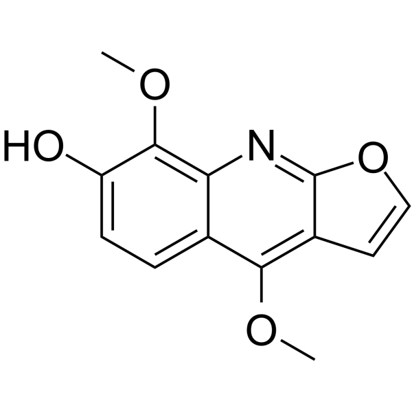
Haplopine

Based on 1 Customer Validation
Haplopine is a substance with anti-inflammatory, antioxidant and photoactivated antibacterial activities. It also acts as an inhibitor of UGT1A7 and a photoactivated restriction endonuclease inhibitor. Haplopine inhibits the mRNA/protein expression of IL-6, TSLP, GM-CSF, G-CSF, IL-4, IL-13 and COX-2, while upregulating the mRNA/protein expression of SOD, CAT and HO-1. Haplopine inhibits the glucuronidation reaction catalyzed by UGT1A7 through competitive hydrophobic binding. Haplopine exerts photoactivated restriction endonuclease inhibitory effects by binding to DNA. Haplopine exhibits photoactivated activity against methicillin-resistant Staphylococcus aureus. Haplopine alleviates symptoms of atopic dermatitis. Haplopine can be used in research related to atopic dermatitis and methicillin-resistant Staphylococcus aureus infections.
Nos produits utilisent uniquement pour la recherche. Nous ne vendons pas aux patients.
- Pureté : 99.49%
- CAS No.: 5876-17-5
- Formule: C13H11NO4
- Masse moléculaire:245.23
-
Stockage:
4°C, sealed storage, away from moisture and light
* In solvent : -80°C, 6 months; -20°C, 1 month (sealed storage, away from moisture and light)

To place orders, for customer services and technical support, please contact: MedChemExpress USA
Tel: 609-228-6898 E-mail: [email protected] [email protected]


-
 Activité biologique
-
Chemical Information
- Protocole
- Pureté et documentation
- Références
-
 Help & FAQs




Activité biologique
Description
Cellular Effect
|
Cell Line

|
Type | Value | Description | References |
|---|---|---|---|---|
| A549 | IC50 |
>10 μM
Compound: 9
|
Cytotoxicity against human A549 cells by SRB assay
Cytotoxicity against human A549 cells by SRB assay
|
[PMID: 18950230] |
| Neutrophil | IC50 |
57.32 μM
Compound: 9
|
Antiinflammatory activity in human neutrophils assessed as fMLP-induced superoxide release after 5 mins by spectrometry
Antiinflammatory activity in human neutrophils assessed as fMLP-induced superoxide release after 5 mins by spectrometry
|
[PMID: 17822293] |
In Vitro
Haplopine (12.5-25 μM; 0.5 h pre-incubation, 24 h stimulation) concentration-dependently inhibits the mRNA and protein expression of pro-inflammatory mediators IL-6, TSLP, GM-CSF and G-CSF in HaCaT cells stimulated with TNF-α/IFN-γ[1].
Haplopine (12.5-50 μM; 0.5 h pre-incubation, 24 h stimulation) inhibits the mRNA and protein expression of pro-inflammatory mediators IL-4, IL-13, and COX-2 in TNF-α/IFN-γ-stimulated Jurkat T cells[1].
Haplopine (12.5-50 μM; 0.5 h pre-incubation, 24 h stimulation) concentration-dependently restores the mRNA and protein expressions of antioxidant defense enzymes SOD, CAT and HO-1 in H2O2-stimulated Jurkat T cells[1].
Haplopine (0.5-100 μM; 30-120 min) competitively inhibits the 4-MU glucuronidation reaction catalyzed by recombinant human UGT1A7, with an IC50 of 13.4 μM, a Ki of 7.2 μM, and a calculated in vivo inhibition threshold of 0.72 μM[3].
Haplopine (100 μM; 30-120 min) inhibits the 4-MU glucuronidation reaction catalyzed by recombinant human UGT1A9 by 45% at a concentration of 100 μM[3].
Haplopine (100 μM; 30-120 min) inhibits the 4-MU glucuronidation reaction catalyzed by recombinant human UGT1A3 by 17% at a concentration of 100 μM[3].
Haplopine (100 μM; 30-120 min) inhibits 35% of the 4-MU glucuronidation reaction catalyzed by recombinant human UGT2B4 at a concentration of 100 μM[3].
Haplopine (12.5 nM/spot; overnight at 37°C) exhibits photoactivated antibacterial activity against methicillin-resistant Staphylococcus aureus after UVA irradiation, with a minimum inhibitory dose of 12.5 nM/spot, whereas it shows no activity in the dark[4].
Haplopine (100 nM/spot; overnight at 37°C) exhibits weak photoactivated antibacterial activity against Candida albicans, with a minimum inhibitory dose of 100 nM/spot under UVA irradiation, while it shows no activity in dark conditions[4].
MedChemExpress (MCE) has not independently confirmed the accuracy of these methods. They are for reference only. Further protocols information, click here.
-
Cell Line:HaCaT human keratinocyte cell line
-
Concentration:12.5 μM, 25 μM
-
Incubation Time:0.5 h pre-incubation; 24 h stimulation
-
Result:Inhibited TNF-α/IFN-γ-induced IL-6 mRNA expression by 42% at 25 μM.
Suppressed TNF-α/IFN-γ-induced increases in TSLP, GM-CSF, and G-CSF mRNA to 2.7, 1.9, and 1.9-fold versus vehicle controls, respectively, at 12.5 μM.
Reduced TNF-α/IFN-γ-induced increases in TSLP, GM-CSF, and G-CSF mRNA to 1.8, 1.2, and 1.2-fold versus vehicle controls, respectively, at 25 μM.
Reduced the protein levels of IL-6 and GM-CSF in a concentration-dependent manner, with 25 μM causing a greater reduction than 12.5 μM.
-
Cell Line:Jurkat T human lymphocyte cell line
-
Concentration:12.5 μM (protein analysis only), 25 μM, 50 μM
-
Incubation Time:0.5 h pre-incubation; 24 h stimulation
-
Result:Inhibited TNF-α/IFN-γ-induced IL-13 mRNA expression by 63% and COX-2 mRNA expression by 70% versus stimulated controls at 25 μM.
Inhibited TNF-α/IFN-γ-induced IL-13 mRNA expression by 89% and COX-2 mRNA expression by 100% versus stimulated controls at 50 μM.
Suppressed TNF-α/IFN-γ-induced IL-4 mRNA upregulation in a concentration-dependent manner.
Reduced COX-2 protein expression in a concentration-dependent manner.\nIncreased H2O2-reduced SOD mRNA activity to 0.86-fold at 25 μM and 0.91-fold at 50 μM versus vehicle controls.
Increased CAT mRNA activity to 0.84-fold versus vehicle controls at 50 μM.
Increased H2O2-reduced HO-1 mRNA levels to 0.79-fold at 25 μM and 0.88-fold at 50 μM versus vehicle controls.
Restored the reduced protein expressions of SOD, CAT, and HO-1 in a concentration-dependent manner.
In Vivo
MedChemExpress (MCE) has not independently confirmed the accuracy of these methods. They are for reference only.
-
Animal Model:Balb/c (6-week-old male, DNCB-induced atopic dermatitis)[1]
-
Dosage:0.05%; 0.1%
-
Administration:topical; daily; 14 days
-
Result:Reduced clinical dermatitis scores by 50%, total serum IgE levels by 23%, mast cell infiltration by 61%, and epidermal thickening by 50% compared to DNCB-treated controls.
Reduced clinical dermatitis scores by 40%, total serum IgE levels by 41%, mast cell infiltration by 52%, and epidermal thickening by 60% compared to DNCB-treated controls.
Caused dose-dependent decreases in spleen weight relative to DNCB-treated controls.


Chemical Information
-
CAS No. 5876-17-5
-
Appearance Solid
-
Masse moléculaire 245.23
-
Formule C13H11NO4
-
Color White to off-white
-
SMILES
OC1=CC=C2C(OC)=C3C(OC=C3)=NC2=C1OC
-
Structure Classification
-
Initial Source
-
Livraison
Room temperature in continental US; may vary elsewhere.
-
Stockage
4°C, sealed storage, away from moisture and light
* In solvent : -80°C, 6 months; -20°C, 1 month (sealed storage, away from moisture and light)

Protocole
-
Research Protocol for Infectious Diseases
Infectious-disease experiments test how pathogens interact with host barriers, innate immune receptors, inflammatory signaling, pathogen replication, and tissue injury; pattern-recognition receptors such as TLRs, RIG-I-like receptors, NOD-like receptors, and inflammasomes detect microbial molecules and activate NF-κB, interferon, and cytokine responses. The central hypothesis is that infection severity reflects the balance between pathogen burden and host response: protective inflammation restricts pathogen growth, whereas excessive or mislocalized inflammation contributes to tissue damage and disease phenotype. Unresolved questions include which host pathways are protective versus pathogenic, why some infection models fail to translate to human disease, and which combined readouts best predict clinically relevant infection outcomes.
-
RNA extraction experimental
By lysing cells, releasing RNA, and removing impurities such as proteins and DNA, high-purity RNA products are finally obtained. The commonly used traditional method is the guanidine isothiocyanate/phenol/chloroform method (Trizol), which is suitable for a variety of animal materials including animal tissues, microorganisms, cultured cells, etc., and most plant materials.
-
Western Blot
Western blotting (WB) is a commonly used experimental method in molecular biology, biochemistry, and immunogenetics for identifying and quantifying target proteins. It combines gel electrophoresis with immunoassay, enabling researchers to analyze protein expression, post-translational modifications, and molecular weight.
-
TPA/Croton Oil Ear Edema and Dermatitis
The TPA (12-O-tetradecanoylphorbol-13-acetate) and croton oil-induced mouse ear edema model is a well-established acute cutaneous inflammation system used to evaluate topical anti-inflammatory activity by measuring edema formation, neutrophil infiltration, vascular permeability, and cytokine-mediated skin responses in vivo. The inflammatory response is triggered by topical application of phorbol esters (TPA) or croton oil constituents, leading to rapid activation of protein kinase C signaling, leukocyte recruitment, and increased vascular permeability, which can be quantified by ear thickness, weight, dye extravasation, and biochemical markers such as myeloperoxidase (MPO) activity and pro-inflammatory mediators in ear tissue homogenates. This model is widely used for screening anti-inflammatory agents, where reductions in edema and inflammatory biomarkers reflect suppression of acute dermal inflammation and immune cell infiltration. Histological evaluation typically confirms epidermal
-
Research Protocol for Inflammation-related Diseases
The NLRP3 inflammasome is a cytosolic innate immune signaling platform that integrates priming signals and danger-signal activation to promote caspase-1 activation, maturation of IL-1β and IL-18, and gasdermin D-mediated pyroptotic cell death. The core experimental logic is to determine whether inflammatory disease phenotypes are driven by increased NLRP3 expression, ASC-containing inflammasome assembly, caspase-1 cleavage, GSDMD cleavage, and extracellular release of IL-1β/IL-18 rather than by nonspecific cell injury alone. The pathway is strongly linked to inflammation-related disease phenotypes because monosodium urate crystals activate NALP3/NLRP3 inflammasome signaling in gout-like crystal inflammation, cholesterol crystals activate NLRP3 inflammasomes in atherogenesis models, and DSS-induced intestinal inflammation has been reported to involve NLRP3 inflammasome activity. However, experimental colitis studies also show context-dependent protective effects of NLRP3 inflammasome co
-
Protocol For Protein Expression And Purification
Recombinant protein expression in Escherichia coli followed by purification of a His-tagged soluble protein by immobilized metal affinity chromatography (IMAC), with optional MBP fusion and TEV tag removal when the construct includes these elements. The biological readout is production of the encoded target protein, detected as an inducible band at the expected molecular mass by SDS-PAGE and quantified by total protein assay or chromatographic absorbance; the purification readout is enrichment of the target protein in elution fractions after selective binding of polyhistidine residues to immobilized Ni2+/metal-chelate resin and elution by imidazole-containing buffer. Expression is driven by an inducible bacterial expression system, commonly T7/lac-based, in which IPTG or lactose/auto-induction activates transcription and translation of the cloned gene; lower induction temperature, lower inducer concentration, induction timing, and solubility-enhancing fusion tags can influence the frac


Pureté et documentation
-
Fiche technique (282 KB)
-
SDS (252 KB)
- English - EN (252 KB)
- Français - FR (252 KB)
- Deutsch - DE (252 KB)
- Norwegian - NO (252 KB)
- Español - ES (252 KB)
- Swedish - SV (252 KB)
- Italian - IT (252 KB)
- Korean - KR (252 KB)
- Portuguese - PT (252 KB)
-
Instruction de manipulation (2659 KB)
Références
[1]. Kim TY, et al. Haplopine Ameliorates 2,4-Dinitrochlorobenzene-Induced Atopic Dermatitis-Like Skin Lesions in Mice and TNF-α/IFN-γ-Induced Inflammation in Human Keratinocyte. Antioxidants (Basel). 2021;10(5):806. Published 2021 May 19. [Content Brief]
[2]. Jeong GS, et al. Neuroprotective effects of constituents of the root bark of Dictamnus dasycarpus in mouse hippocampal cells. Arch Pharm Res. 2010;33(8):1269-1275. [Content Brief]
[3]. Li Y, et al. Inhibition of UDP-glucuronosyltransferases by different furoquinoline alkaloids. Xenobiotica. 2020;50(10):1170-1179. [Content Brief]
[4]. Hanawa F, et al. Photo-activated DNA binding and antimicrobial activities of furoquinoline and pyranoquinolone alkaloids from rutaceae. Planta Med. 2004 Jun;70(6):531-5. [Content Brief]

Calculators
Concentration (start) × Volume (start) = Concentration (final) × Volume (final)


Haplopine Related Classifications
HY-N3989 Related Classifications
 Powered by Bioz