



OSU-2S
Based on 1 publication(s) in Google Scholar
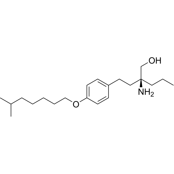
OSU-2S is a non-immunosuppressive derivative of Fingolimod hydrochloride (FTY720) (HY-12005). It is an orally active, blood-brain barrier-permeable allosteric activator of PP2A that also activates PKCδ; it restores PP2A activity by displacing the inhibitory protein SET, thereby inducing ROS-PKCδ-caspase-3-dependent apoptosis, c-Myc degradation, cell cycle arrest, and differentiation. OSU-2S is used in research on diseases such as leukemia, liver cancer, lymphoma, and lung cancer.
Nos produits utilisent uniquement pour la recherche. Nous ne vendons pas aux patients.
- CAS No.: 1351056-65-9
- Formule: C21H37NO2
- Masse moléculaire:335.52
-
Stockage:
Please store the product under the recommended conditions in the Certificate of Analysis.

To place orders, for customer services and technical support, please contact: MedChemExpress USA
Tel: 609-228-6898 E-mail: [email protected] [email protected]


-
 Activité biologique
-
Chemical Information
- Protocole
- Pureté et documentation
- Références
-
 Help & FAQs


Publications Citing Use of MedChemExpress (MCE) OSU-2S
More

Activité biologique
Description
|
PKCδ |
Cellular Effect
|
Cell Line

|
Type | Value | Description | References |
|---|---|---|---|---|
| A549 | IC50 |
4.2 μM
Compound: OSU-2S
|
Cytotoxicity against human A549 cells after 48 hrs by MTT assay
Cytotoxicity against human A549 cells after 48 hrs by MTT assay
|
[PMID: 28865276] |
| Hep 3B2 | IC50 |
2.8 μM
Compound: OSU-2S
|
Cytotoxicity against human Hep3B cells after 48 hrs by MTT assay
Cytotoxicity against human Hep3B cells after 48 hrs by MTT assay
|
[PMID: 28865276] |
| HepG2 | IC50 |
2.3 μM
Compound: OSU-2S
|
Cytotoxicity against human HepG2 cells after 48 hrs by MTT assay
Cytotoxicity against human HepG2 cells after 48 hrs by MTT assay
|
[PMID: 28865276] |
| HT-29 | IC50 |
2.7 μM
Compound: OSU-2S
|
Cytotoxicity against human HT-29 cells after 48 hrs by MTT assay
Cytotoxicity against human HT-29 cells after 48 hrs by MTT assay
|
[PMID: 28865276] |
In Vitro
OSU-2S (2-6 μM; 24 h) exhibits concentration-dependent cytotoxicity against canine B-cell lymphoma cell lines (CLBL-1 and 17–71)[7].
The cytotoxicity of OSU-2S (3 μM; 24 h) in canine B-cell lymphoma cell lines is partially mediated by ROS[7].
OSU-2S (2-4 μM; 24 h) induces ROS production and mediates cytotoxicity in primary cells from canine spontaneous B-cell lymphoma[7].
OSU-2S (24 h) downregulates PGC1α and inhibits mitochondrial biogenesis via PP2A-dependent FOXO1 dephosphorylation[6].
OSU-2S reduces mitochondrial mass and ATP production, and inhibits the expression of genes associated with mitochondrial biogenesis[6].
OSU-2S (2 μM; 24 h) induces apoptosis in primary CLL cells[4].
OSU-2S activates PP2A in CLL cells[4].
OSU-2S (5 h) induces SHP1 S591 phosphorylation via a PKC-dependent mechanism[4].
OSU-2S (5-24 h)-induced pSHP1 S591 levels correlate with CLL cell death[4].
The cytotoxicity of OSU-2S (2 μM; 24 h) is partially dependent on PKC[4].
OSU-2S (8 μM; 4 h) activates PKC in CLL cells[4].
OSU-2S (8 μM; 16 h) extensively regulates gene expression in CLL cells, affecting processes such as cell growth, proliferation, death, and the cell cycle[4].
OSU-2S (8 μM; 16-24 h) downregulates TCL1A expression in CLL cells[4].
OSU-2S (5 μM; 24 h) selectively kills CLL cells without affecting normal B cells via ROR1-targeted delivery[4].
OSU-2S (2-8 μM; 24 h) exhibits potent cytotoxicity against MCL cell lines[3].
OSU-2S (2-8 μM; 24 h) induces apoptosis in MCL cell lines[3].
OSU-2S (4-5 μM; 24 h) induces CD74 expression on the surface of MCL cells[3].
OSU-2S (4 μM; 24 h) enhances the cytotoxicity of Milatuzumab (HY-P99731) against MCL cells by upregulating CD74[3].
OSU-2S (2-10 μM; 48 h) demonstrates anti-proliferative activity against various HCC cell lines[2].
OSU-2S (1.25-2.5 μM; 48 h) enhances the anti-proliferative effect of Sorafenib (HY-10201) on HCC cells, exhibiting a synergistic effect[2].
OSU-2S (1.25-2.5 μM; 48 h) enhances Sorafenib-induced apoptosis in HCC cells[2].
OSU-2S (1.25-2.5 μM; 48 h) enhances the anti-HCC effect of Sorafenib via PKCδ and ERK pathways[2].
OSU-2S (1.25-2.5 μM; 8-24 h) in combination with Sorafenib inhibits HCC cell migration and invasion[2].
The activity of OSU-2S (1.25-7.5 μM) against HCC cells is influenced by p53 status; p53 restoration enhances sensitivity[2].
OSU-2S (1-10 μM; 24 h) inhibits A549 proliferation, with an IC50 of 4.432 μM[1].
OSU-2S (1.5-6 μM; 14 days) inhibits colony formation in A549 cells[1].
OSU-2S (1.5-6 μM; 12-24 h) inhibits A549 cell migration[1].
OSU-2S (1.5-6 μM; 24 h) downregulates p-AURKA and S1PR1 protein expression in A549 cells[1].
MedChemExpress (MCE) has not independently confirmed the accuracy of these methods. They are for reference only.
-
Cell Line:Canine B-cell lymphoma cell lines CLBL-1 and 17-71
-
Concentration:2 μM; 4 μM; 6 μM
-
Incubation Time:24 h
-
Result:Promoted cytotoxicity at concentrations as low as 2 μM.
-
Cell Line:Primary cells from canine spontaneous B-cell lymphoma
-
Concentration:2 μM; 4 μM; 8 μM
-
Incubation Time:24 h
-
Result:Induced apoptosis in canine lymphoma cells.
-
Cell Line:Primary CD19+ cells from CLL patients
-
Concentration:2 μM
-
Incubation Time:24 h
-
Result:Induced apoptosis in primary CLL cells.
-
Cell Line:CLL cells
-
Concentration:8 μM
-
Incubation Time:16 h
-
Result:Reduced TCL1A mRNA levels.
-
Cell Line:CLL cells
-
Concentration:8 μM
-
Incubation Time:24 h
-
Result:Reduced TCL1A protein levels.
-
Cell Line:MCL cell line
-
Concentration:5 μM
-
Incubation Time:24 h
-
Result:Induced apoptosis in MCL cell lines.
-
Cell Line:Huh7, Hep3B, PLC5, HepG2
-
Concentration:2 μM; 4 μM; 6 μM; 8 μM; 10 μM
-
Incubation Time:48 h
-
Result:Inhibited cell proliferation, with HepG2 being the most sensitive.
-
Cell Line:A549
-
Concentration:1.5 μM; 3 μM; 6 μM
-
Incubation Time:12 h; 24 h
-
Result:Reduced the migration ability of A549 cells.
In Vivo
MedChemExpress (MCE) has not independently confirmed the accuracy of these methods. They are for reference only.
-
Animal Model:Subcutaneous inoculation of A549 cells into BALB/c nude mice[1]
-
Dosage:2.5 mg/kg, 5 mg/kg, 10 mg/kg
-
Administration:i.p.; every other day for 35 days
-
Result:Tumor volume and weight were significantly reduced.
There was no significant decrease in mouse body weight.
Immunohistochemistry showed that OSU-2S downregulated Ki-67 and VEGF.
- Detection of Apoptosis
- TUNEL staining for apoptotic DNA fragmentation
- Flow cytometric DNA-content cell-cycle staining
- Annexin V plus membrane-impermeant dye apoptosis staining
- ROS/oxidative-stress fluorescent staining
- BrdU Incorporation Assay
- Apoptosis Solutions
- Protocol for Cell Cycle
- Liver Cancer Modeling


Chemical Information
-
CAS No. 1351056-65-9
-
Masse moléculaire 335.52
-
Formule C21H37NO2
-
SMILES
CCC[C@@](CCC1=CC=C(OCCCCCC(C)C)C=C1)(N)CO
-
Livraison
Room temperature in continental US; may vary elsewhere.
-
Stockage
Please store the product under the recommended conditions in the Certificate of Analysis.


Publications (1)
-
Journal Impact Factor
-
Most Recent
-
Int Immunopharmacol
Osthole alleviates neuropathic pain by suppressing astrocytes activation and associated inflammatory responses via the PKCδ/TRPV4 signaling pathway. [Abstract]2025 Sep 10:165:115453. PMID: 40934539


Pureté et documentation
Références

Calculators
Concentration (start) × Volume (start) = Concentration (final) × Volume (final)


OSU-2S Related Classifications
HY-117720 Related Classifications
 Powered by Bioz