



RGD-4C

Based on 1 Customer Validation
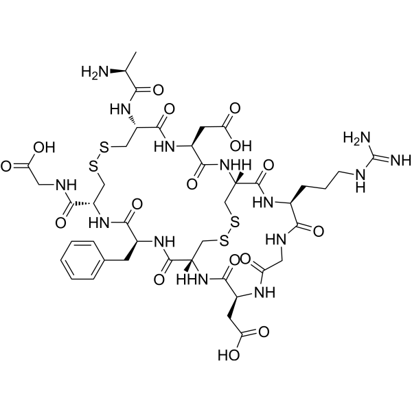
RGD-4C is a arginine-glycine-aspartic acid peptide (ACDCRGDCFC) with integrin binding activity. The Arg-Gly-Asp (RGD) sequence serves as the primary integrin recognition site in extracellular matrix proteins, and peptides containing this sequence can mimic the recognition specificity of the matrix proteins. RGD-4C is a αv-integrin ligand, can conjugate with bioactive molecule to exert antitumor effects in animal models.
Nos produits utilisent uniquement pour la recherche. Nous ne vendons pas aux patients.
- Pureté : 99.91%
- CAS No.: 332179-76-7
- Formule: C42H60N14O16S4
- Masse moléculaire:1145.27
-
Stockage:
Sealed storage, away from moisture and light.
Powder -80°C, 2 years , -20°C, 1 year* In solvent : -80°C, 6 months; -20°C, 1 month (sealed storage, away from moisture and light)

To place orders, for customer services and technical support, please contact: MedChemExpress USA
Tel: 609-228-6898 E-mail: [email protected] [email protected]


-
 Activité biologique
-
Chemical Information
-
Solvant et solubilité
- Protocole
- Pureté et documentation
- Références
-
 Help & FAQs




Activité biologique
Description
In Vitro
In Vivo
Modified RGD-4C fused with the N-terminus of plant-derived single-chain ribosome inactivating protein saporin (SAP) (called RGD-SAP), (diluted in sodium chloride, 0.9%; i.v.; 200 µL; every 5 days, for 3 times) exerts anti-tumor activity in a model of muscle invasive bladder cancer[3].
RGD-SAP in combination with mitomycin C, a chemotherapeutic drug, increases the survival of mice bearing orthotopic bladder cancer with no evidence of systemic toxicity[3].
MedChemExpress (MCE) has not independently confirmed the accuracy of these methods. They are for reference only.


Chemical Information
-
CAS No. 332179-76-7
-
Appearance Solid
-
Masse moléculaire 1145.27
-
Formule C42H60N14O16S4
-
Color White to off-white
-
Sequence Shortening
ACDCRGDCFCG(Disulfide bridge: Cys2-Cys10; Cys4-Cys8)
-
Livraison
Room temperature in continental US; may vary elsewhere.
-
Stockage
Sealed storage, away from moisture and light
Powder -80°C 2 years -20°C 1 year * In solvent : -80°C, 6 months; -20°C, 1 month (sealed storage, away from moisture and light)


Solvant et solubilité
In Vitro:
DMSO : 50 mg/mL (43.66 mM; Need ultrasonic; Hygroscopic DMSO has a significant impact on the solubility of product, please use newly opened DMSO)
Please refer to the solubility information to select the appropriate solvent. Once prepared, please aliquot and store the solution to prevent product inactivation from repeated freeze-thaw cycles.
Storage method and period of stock solution: -80°C, 6 months; -20°C, 1 month (sealed storage, away from moisture and light). When stored at -80°C, please use it within 6 months. When stored at -20°C, please use it within 1 month.
Please refer to the solubility information to select the appropriate solvent. Once prepared, please aliquot and store the solution to prevent product inactivation from repeated freeze-thaw cycles.
Storage method and period of stock solution: -80°C, 6 months; -20°C, 1 month (sealed storage, away from moisture and light). When stored at -80°C, please use it within 6 months. When stored at -20°C, please use it within 1 month.
Concentration (start) × Volume (start) = Concentration (final) × Volume (final)


Pureté et documentation
-
Fiche technique (281 KB)
-
SDS (254 KB)
- English - EN (254 KB)
- Français - FR (254 KB)
- Deutsch - DE (254 KB)
- Norwegian - NO (254 KB)
- Español - ES (254 KB)
- Swedish - SV (254 KB)
- Italian - IT (254 KB)
- Korean - KR (254 KB)
- Portuguese - PT (254 KB)
-
Instruction de manipulation (2659 KB)
Références
[1]. Assa-Munt N, et al. Solution structures and integrin binding activities of an RGD peptide with two isomers. Biochemistry. 2001 Feb 27;40(8):2373-8. [Content Brief]
[2]. Yin R, et al. Effect of RGD-4C position is more important than disulfide bonds on antiangiogenic activity of RGD-4C modified endostatin derived synthetic polypeptide. Bioconjug Chem. 2010 Jul 21;21(7):1142-7. [Content Brief]
[3]. Zuppone S, et al. A Novel RGD-4C-Saporin Conjugate Inhibits Tumor Growth in Mouse Models of Bladder Cancer. Front Oncol. 2022 Apr 11;12:846958. [Content Brief]


Complete Stock Solution Preparation Table
Please refer to the solubility information to select the appropriate solvent. Once prepared, please aliquot and store the solution to prevent product inactivation from repeated freeze-thaw cycles.
Storage method and period of stock solution: -80°C, 6 months; -20°C, 1 month (sealed storage, away from moisture and light). When stored at -80°C, please use it within 6 months. When stored at -20°C, please use it within 1 month.
| Optional Solvent | Concentration Solvent Mass | 1 mg | 5 mg | 10 mg | 25 mg |
|---|---|---|---|---|---|
| DMSO | 1 mM | 0.8732 mL | 4.3658 mL | 8.7316 mL | 21.8289 mL |
| 5 mM | 0.1746 mL | 0.8732 mL | 1.7463 mL | 4.3658 mL | |
| 10 mM | 0.0873 mL | 0.4366 mL | 0.8732 mL | 2.1829 mL | |
| 15 mM | 0.0582 mL | 0.2911 mL | 0.5821 mL | 1.4553 mL | |
| 20 mM | 0.0437 mL | 0.2183 mL | 0.4366 mL | 1.0914 mL | |
| 25 mM | 0.0349 mL | 0.1746 mL | 0.3493 mL | 0.8732 mL | |
| 30 mM | 0.0291 mL | 0.1455 mL | 0.2911 mL | 0.7276 mL | |
| 40 mM | 0.0218 mL | 0.1091 mL | 0.2183 mL | 0.5457 mL |


RGD-4C Related Classifications
HY-P3732 Related Classifications
 Powered by Bioz