



CCR4-351 hydrochloride

Based on 1 Customer Validation
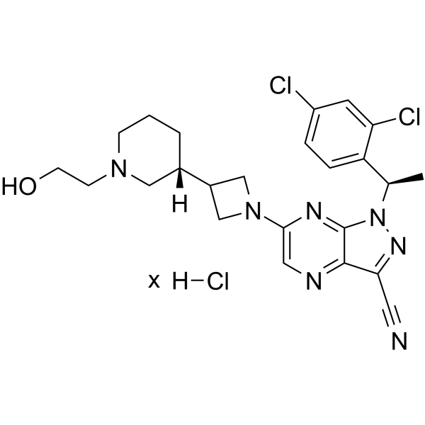
CCR4-351 hydrochloride is an orally active, potent and selective CCR4 antagonist. CCR4-351 hydrochloride, featuring a novel piperidinyl-azetidine motif, has IC50s of 22 nM and 50 nM in the calcium flux and CTX assay. CCR4-351 hydrochloride has antitumor activity.
For research use only. We do not sell to patients.
- Purity : 98.14%
- CAS No.: 2174938-71-5
- Formula: C24H28Cl3N7O
-
Storage:
4°C, sealed storage, away from moisture
* In solvent : -80°C, 6 months; -20°C, 1 month (sealed storage, away from moisture)

To place orders, for customer services and technical support, please contact: MedChemExpress USA
Tel: 609-228-6898 E-mail: [email protected] [email protected]


-
 Biological Activity
-
Chemical Information
-
Solvent & Solubility
- Protocols
- Purity & Documentation
- References
-
 Help & FAQs


-
HY-103364A
C-021 dihydrochlorideCCR4 Antagonist -
HY-148494
TivumecirnonCCR4 Antagonist -
HY-148074
ZelnecirnonCCR4 Inhibitor -
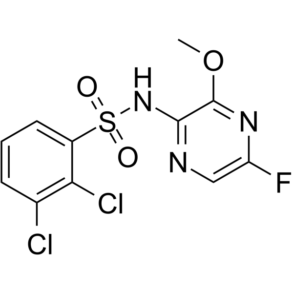
HY-109511
AZD-1678CCR4 Receptor Antagonist -
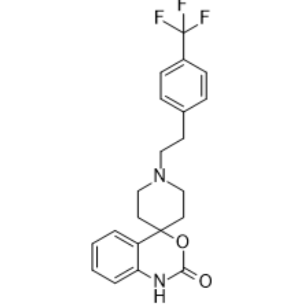
HY-18611A
AbaucinCCR Antagonist -
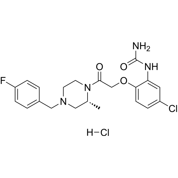
HY-12080A
BX471 hydrochlorideCCR Antagonist -
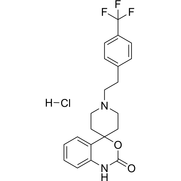
HY-18611
Abaucin hydrochlorideCCR2antagonist -
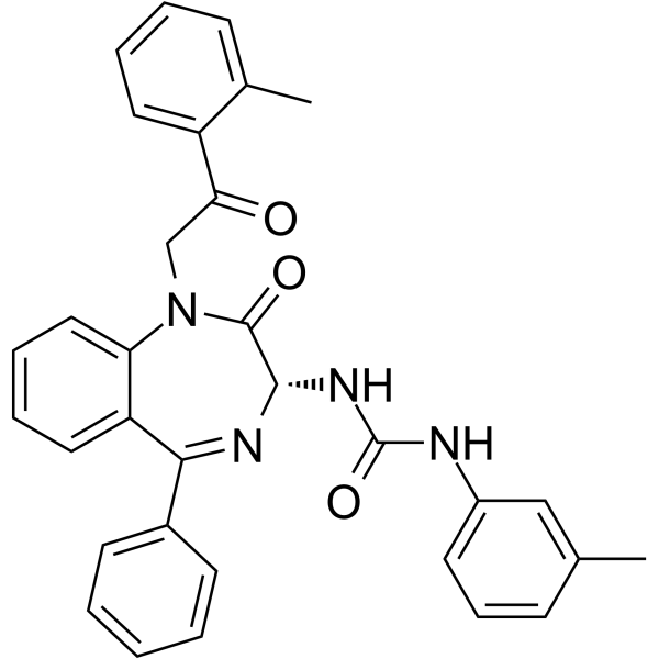
HY-103355
YM022CCK-BR Antagonist -
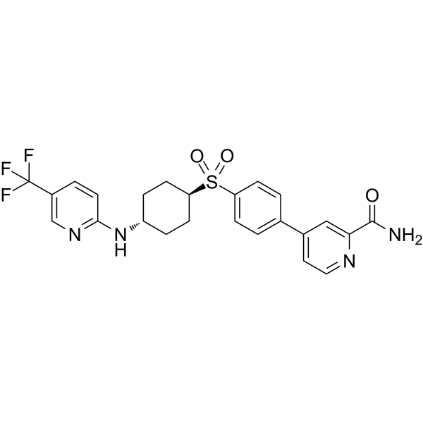
HY-112701
CCR6 inhibitor 1CCR Inhibitor -
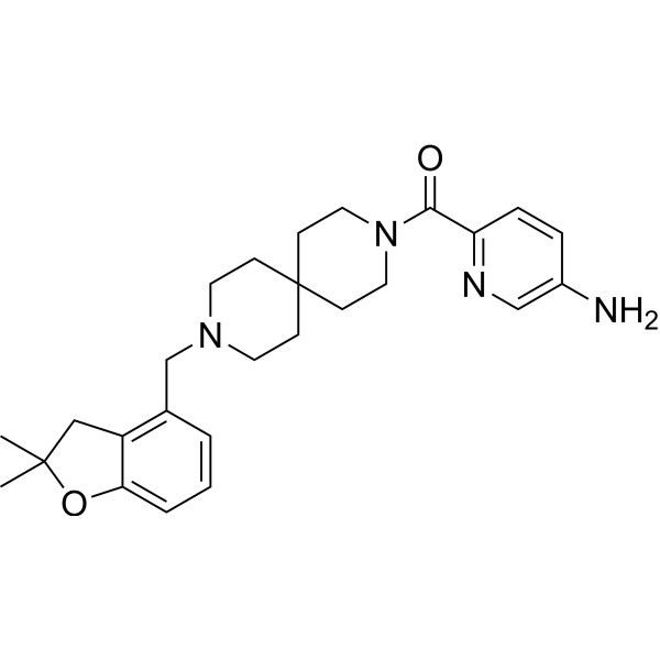
HY-119217
AZ084CCR8 Allosteric Antagonist -
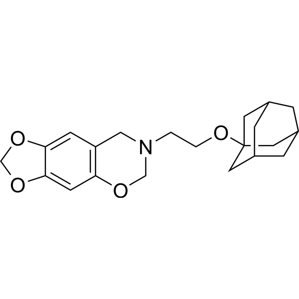
HY-122219
R243CCR8 Antagonist -
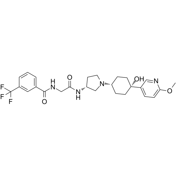
HY-15450A
INCB 3284CCR Antagonist


Biological Activity
Description
IC50 & Target
|
CCR4 |
In Vitro
CCR4-351 (compound 38) hydrochloride shows no activity in a CYP450 induction assay[1].
CCR4-351 hydrochloride inhibits the migration of mouse iTreg cells with an IC50 of 39 nM, while the IC50 in human iTreg cells is 33 nM[1].
MedChemExpress (MCE) has not independently confirmed the accuracy of these methods. They are for reference only. Further protocols information, click here.
In Vivo
CCR4-351 (0.5 mg/kg; IV) hydrochloride has low clearance (CL=10.2 mL/min/kg), medium volume of distribution (Vss=5.2 L/kg), a T1/2 of 6.9 h, and good bioavailability (%F = 29) of oral dosing in mouse[1].
CCR4-351 hydrochloride has low clearance (CL=7.3 mL/min/kg), a half-life of 12.7 hr, and is 44% bioavailable in dog. CCR4-351 hydrochloride has low clearance (CL=3.7 mL/min/kg), a long terminal half-life (10.7 hr), and good bioavailability (%F = 41) in cynomolgus monkey[1].
MedChemExpress (MCE) has not independently confirmed the accuracy of these methods. They are for reference only.
-
Animal Model:Six-to eight-week-old, female C57BL/6 mice with Pan02-OVA tumor[1]
-
Dosage:50 mg/kg
-
Administration:PO; daily; for 40 days
-
Result:Significantly reduced the tumor growth.
-
Animal Model:Rat and mouse[1]
-
Dosage:0.5 mg/kg of IV; 2 mg/kg of PO
-
Administration:IV or PO
-
Result:Possessed medium clearance (CL=47.6 mL/min/kg) and was 49% bioavailable upon oral dosing in rat.
Had low clearance (CL=10.2 mL/min/kg), medium volume of distribution (Vss=5.2 L/kg), a T1/2 of 6.9 h, and good bioavailability (%F = 29) of oral dosing in mouse.


Chemical Information
-
CAS No. 2174938-71-5
-
Appearance Solid
-
Formula C24H28Cl3N7O
-
Color White to off-white
-
SMILES
[H][C@@]1(CCCN(C1)CCO)C2CN(C2)C3=CN=C(C4=N3)C(C#N)=NN4[C@H](C)C5=CC=C(C=C5Cl)Cl.[H]Cl.[x]
-
Shipping
Room temperature in continental US; may vary elsewhere.
-
Storage
4°C, sealed storage, away from moisture
* In solvent : -80°C, 6 months; -20°C, 1 month (sealed storage, away from moisture)


Solvent & Solubility
In Vitro:
DMSO : 170 mg/mL (Need ultrasonic; Hygroscopic DMSO has a significant impact on the solubility of product, please use newly opened DMSO)
In Vivo:
Select the appropriate dissolution method based on your experimental animal and administration route.
- For the following dissolution methods, please ensure to first prepare a clear stock solution using an In Vitro approach and then sequentially add co-solvents:
- To ensure reliable experimental results, the clarified stock solution can be appropriately stored based on storage conditions. As for the working solution for In Vivo experiments, it is recommended to prepare freshly and use it on the same day.
- The percentages shown for the solvents indicate their volumetric ratio in the final prepared solution. If precipitation or phase separation occurs during preparation, heat and/or sonication can be used to aid dissolution.
Add each solvent one by one: 10% DMSO 40% PEG300 5% Tween-80 45% Saline
Solubility: ≥ 4.25 mg/mL; Clear solution
This protocol yields a clear solution of ≥ 4.25 mg/mL (saturation unknown).
Taking 1 mL working solution as an example, add 100 μL DMSO stock solution (42.5 mg/mL) to 400 μL PEG300, and mix evenly; then add 50 μL Tween-80 and mix evenly; then add 450 μL Saline to adjust the volume to 1 mL.
Preparation of Saline: Dissolve 0.9 g sodium chloride in ddH₂O and dilute to 100 mL to obtain a clear Saline solution.
In Vivo Dissolution Calculator
Please enter the basic information of animal experiments:
-
-
-
-
Recommended: Prepare an additional quantity of animals to account for potential losses during experiments.
Please enter your animal formula composition:
-
%DMSO +
Recommended: Keep the proportion of DMSO in working solution below 2% if your animal is weak.
-
%+
-
+%Tween-80 + +
-
%Saline +
The co-solvents required include: DMSO, . All of co-solvents are available by MedChemExpress (MCE). , Tween 80. All of co-solvents are available by MedChemExpress (MCE).
Working solution concentration: 0.22 mg/mL
Method for preparing stock solution: mg drug dissolved in μL DMSO. Stock solution concentration: mg/mL. * In solvent : -80°C, 6 months; -20°C, 1 month (sealed storage, away from moisture)
1. Take μL DMSO stock solution;
2. Add μL .
μL , mix evenly;
3. Then add μL Tween 80, mix evenly;
4. Then add μL
Please ensure that the stock solution in the first step is dissolved to a clear state, and add co-solvents in sequence. You can use ultrasonic heating (ultrasonic cleaner, recommended frequency 20-40 kHz), vortexing, etc. to assist dissolution.

Protocols
-
Ca2+ Staining Technique
Ca2+ staining is an experimental technique that utilizes specific fluorescent probes (such as Fluo-4 AM, Fura-2, etc.) to qualitatively or quantitatively detect dynamic changes in intracellular Ca2+ concentrations; this is achieved by monitoring the changes in fluorescent signals generated when these probes bind to free intracellular calcium ions. The underlying principle relies primarily on the presence of chelating groups within the probe's molecular structure that possess high affinity for calcium ions.
-
Subcutaneous Cell-Line-Derived Xenograft
Subcutaneous cell-line-derived xenograft (CDX) models are established by implanting cultured human cancer cell lines into immunodeficient mice, where the injected cells form localized tumors that can be monitored in vivo as a measure of tumorigenic potential, growth kinetics, and treatment response. These models are widely used in oncology research because they allow reproducible tumor formation and enable comparative assessment of tumor growth between different cell lines or genetic manipulations in a controlled in vivo microenvironment. Subcutaneous implantation of cancer cells in immunodeficient mice is a standard approach for evaluating tumor growth behavior and therapeutic response across multiple cancer types, including prostate, esophageal, pancreatic, and colon cancer models.


Purity & Documentation
-
Data Sheet (274 KB)
-
SDS (252 KB)
- English - EN (252 KB)
- Français - FR (252 KB)
- Deutsch - DE (252 KB)
- Norwegian - NO (252 KB)
- Español - ES (252 KB)
- Swedish - SV (252 KB)
- Italian - IT (252 KB)
- Korean - KR (252 KB)
- Portuguese - PT (252 KB)
-
Handling Instructions (2659 KB)
References

Calculators
Concentration (start) × Volume (start) = Concentration (final) × Volume (final)


CCR4-351 hydrochloride Related Classifications
HY-131349A Related Classifications
 Powered by Bioz