



ML281
Based on 2 publication(s) in Google Scholar
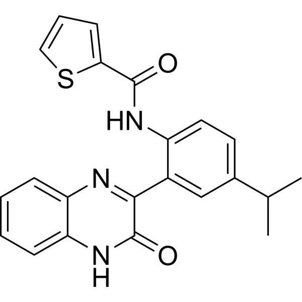
ML281 is a highly selective inhibitor of serine/threonine kinase 33 (STK33) with an IC50 value of 14 nM. ML281 shows 700-fold selectivity over PKA and 550-fold over AurB. ML281 exerts core mechanism by inhibiting STK33: in small cell lung cancer, ML281 downregulates RPS6/BAD signaling phosphorylation, induces apoptosis, and suppresses proliferation, invasion. ML281 reduces STK33-mediated 4-hydroxyphenylpyruvate dioxygenase (HPD) phosphorylation in tyrosinemia . ML281 is suitable for research on STK33 function, KRAS mutation-related cancers (pancreatic cancer, colon cancer, lung adenocarcinoma, etc.), small cell lung cancer, and tyrosinemia-related damage
Nos produits utilisent uniquement pour la recherche. Nous ne vendons pas aux patients.
- Pureté : 98.04%
- CAS No.: 1404437-62-2
- Formule: C22H19N3O2S
- Masse moléculaire:389.47
-
Stockage:Powder -20°C, 3 years , 4°C, 2 years ; In solvent -80°C, 2 years , -20°C, 1 year

To place orders, for customer services and technical support, please contact: MedChemExpress USA
Tel: 609-228-6898 E-mail: [email protected] [email protected]


-
 Activité biologique
-
Chemical Information
-
Solvant et solubilité
- Protocole
- Pureté et documentation
- Références
-
 Help & FAQs


-
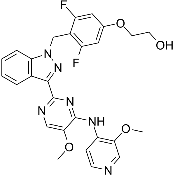
HY-103020
BAY-1816032BUB1 Kinase Inhibitor -
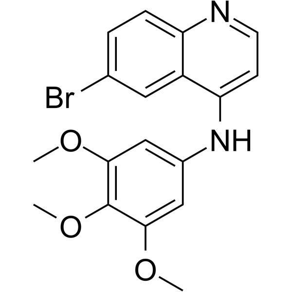
HY-122186
SGC-GAK-1GAK Inhibitor -
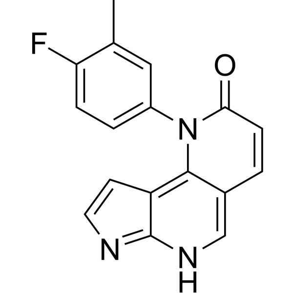
HY-101270
STK16-IN-1STK16 Inhibitor -

HY-101270R
STK16-IN-1 (Standard)STK16 Inhibitor -

HY-103020R
BAY-1816032 (Standard)Reference Standard -
HY-118134
Hepln-13Hepsin Inhibitor -
HY-W010128
6-(Dimethylamino)purineSerine Threonine Protein kinase Inhibitor -
HY-100877
GCN2-IN-1GCN2 Inhibitor -

HY-W010128R
6-(Dimethylamino)purine (Standard)Serine Threonine Protein kinase Inhibitor
Publications Citing Use of MedChemExpress (MCE) ML281
More Customer Validation & Images

-
WB
Voir tous les produits spécifiques à Isoform Aurora Kinase
More

Activité biologique
Description
IC50 & Target
[1]|
PKA |
Aurora B |
bad |
In Vitro
ML281 (10 μM; 72 hours) suppresses cell viability of NCI-H446 cells[3].
ML281 exhibits potent inhibitory activity against purified recombinant serine/threonine kinase 33 (STK33) (IC50 = 14 nM) [1].
ML281 (0.01-10 μM) has no significant effect on the viability of KRAS-dependent (NOMO-1, SKM-1) and KRAS-independent (THP-1, U937) acute myeloid leukemia-derived cell lines, ML281 does not exhibit selective cytotoxicity against KRAS-dependent cancer cells.[1].
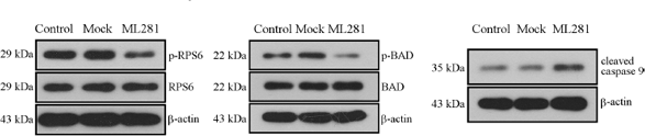
ML281 (10 μM; 72 h) suppresses RPS6/BAD signaling pathway in human small cell lung cancer NCI-H446 cells[1].
ML281 (2.5-12.5 μM; 48 h) decreases T382 site phosphorylation of 4-hydroxyphenylpyruvate dioxygenase (HPD) and increases HPD protein expression in a dosage-dependent manner in human fetal hepatocyte LO2 cells[4].
ML281 (10 μM; 48 h) abrogates enhanced HPD T382 phosphorylation and HPD degradation in Ttc36-deficient mouse primary hepatocytes[4].
MedChemExpress (MCE) has not independently confirmed the accuracy of these methods. They are for reference only.
-
Cell Line:NCI-H446 cells
-
Concentration:10 μM
-
Incubation Time:72 hours
-
Result:Suppressed cell viability of NCI-H446 cells.
-
Cell Line:LO2 cells
-
Concentration:2.5 μM, 5 μM, 7.5 μM, 10 μM, 12.5 μM
-
Incubation Time:48 h
-
Result:Decreased HPD T382 phosphorylation, increased HPD protein expression in a dosage-dependent manner; no effect on HPD T382A mutant phosphorylation.
-
Cell Line:Ttc36-/- mouse primary hepatocytes
-
Concentration:10 μM
-
Incubation Time:48 h
-
Result:Abrogated enhanced HPD T382 phosphorylation and HPD degradation induced by TTC36 deficiency.
-
Cell Line:NCI-H446 cells
-
Concentration:10 μM
-
Incubation Time:72 hours
-
Result:Decreased phosphorylation of RPS6 and BAD, increased expression of cleaved caspase 9.
- Kinase activity and phosphorylation assays
- Western Blot
- Detection of Apoptosis
- TUNEL staining for apoptotic DNA fragmentation
- Annexin V plus membrane-impermeant dye apoptosis staining
- Apoptosis Solutions
- Cell invasion
- Protocol for Kinase activity and phosphorylation assays
- Genotoxicity/Mutagenicity Study
- Cell Viability Determination by MTT Colorimetric Assay


Chemical Information
-
CAS No. 1404437-62-2
-
Appearance Solid
-
Masse moléculaire 389.47
-
Formule C22H19N3O2S
-
Color White to off-white
-
SMILES
O=C1NC2=CC=CC=C2N=C1C3=CC(C(C)C)=CC=C3NC(C4=CC=CS4)=O
-
Livraison
Room temperature in continental US; may vary elsewhere.
-
Stockage
Powder -20°C 3 years 4°C 2 years In solvent -80°C 2 years -20°C 1 year


Publications (2)
-
Journal Impact Factor
-
Most Recent
-
Nat Commun
HPD degradation regulated by the TTC36-STK33-PELI1 signaling axis induces tyrosinemia and neurological damage. [Abstract]2019 Sep 19;10(1):4266. PMID: 31537781 -
Neoplasma
Knockdown of human serine/threonine kinase 33 suppresses human small cell lung carcinoma by blocking RPS6/BAD signaling transduction. [Abstract]2017;64(6):869-879. PMID: 28895411
ML281 purchased from MedChemExpress. Usage Cited in: Neoplasma. 2017;64(6):869-879. [Abstract]
Representative images and analysis results of western blotting validation after incubation with ML281.


Solvant et solubilité
In Vitro:
DMSO : 100 mg/mL (256.76 mM; Need ultrasonic; Hygroscopic DMSO has a significant impact on the solubility of product, please use newly opened DMSO)
Please refer to the solubility information to select the appropriate solvent. Once prepared, please aliquot and store the solution to prevent product inactivation from repeated freeze-thaw cycles.
Storage method and period of stock solution: -80°C, 2 years; -20°C, 1 year. When stored at -80°C, please use it within 2 years. When stored at -20°C, please use it within 1 year.
Please refer to the solubility information to select the appropriate solvent. Once prepared, please aliquot and store the solution to prevent product inactivation from repeated freeze-thaw cycles.
Storage method and period of stock solution: -80°C, 2 years; -20°C, 1 year. When stored at -80°C, please use it within 2 years. When stored at -20°C, please use it within 1 year.
Concentration (start) × Volume (start) = Concentration (final) × Volume (final)


Pureté et documentation
-
Fiche technique (285 KB)
-
SDS (396 KB)
- English - EN (396 KB)
- Français - FR (396 KB)
- Deutsch - DE (396 KB)
- Norwegian - NO (396 KB)
- Español - ES (396 KB)
- Swedish - SV (396 KB)
- Italian - IT (396 KB)
- Korean - KR (396 KB)
- Portuguese - PT (396 KB)
-
Instruction de manipulation (2659 KB)
Références


Complete Stock Solution Preparation Table
Please refer to the solubility information to select the appropriate solvent. Once prepared, please aliquot and store the solution to prevent product inactivation from repeated freeze-thaw cycles.
Storage method and period of stock solution: -80°C, 2 years; -20°C, 1 year. When stored at -80°C, please use it within 2 years. When stored at -20°C, please use it within 1 year.
| Optional Solvent | Concentration Solvent Mass | 1 mg | 5 mg | 10 mg | 25 mg |
|---|---|---|---|---|---|
| DMSO | 1 mM | 2.5676 mL | 12.8380 mL | 25.6759 mL | 64.1898 mL |
| 5 mM | 0.5135 mL | 2.5676 mL | 5.1352 mL | 12.8380 mL | |
| 10 mM | 0.2568 mL | 1.2838 mL | 2.5676 mL | 6.4190 mL | |
| 15 mM | 0.1712 mL | 0.8559 mL | 1.7117 mL | 4.2793 mL | |
| 20 mM | 0.1284 mL | 0.6419 mL | 1.2838 mL | 3.2095 mL | |
| 25 mM | 0.1027 mL | 0.5135 mL | 1.0270 mL | 2.5676 mL | |
| 30 mM | 0.0856 mL | 0.4279 mL | 0.8559 mL | 2.1397 mL | |
| 40 mM | 0.0642 mL | 0.3209 mL | 0.6419 mL | 1.6047 mL | |
| 50 mM | 0.0514 mL | 0.2568 mL | 0.5135 mL | 1.2838 mL | |
| 60 mM | 0.0428 mL | 0.2140 mL | 0.4279 mL | 1.0698 mL | |
| 80 mM | 0.0321 mL | 0.1605 mL | 0.3209 mL | 0.8024 mL | |
| 100 mM | 0.0257 mL | 0.1284 mL | 0.2568 mL | 0.6419 mL |


ML281 Related Classifications
HY-13495 Related Classifications
 Powered by Bioz
Keywords
- ML281
- 1404437-62-2
- ML 281
- ML-281
- STK33
- PKA
- Aurora Kinase
- Bcl-2 Family
- Apoptosis
- STK33 inhibitor
- NCI-H446 cells
- DMS153 cells
- LO2 cells
- NOMO-1 cells
- SKM-1 cells
- THP-1 cells
- U937 cells
- BALB/c-nu mice
- small cell lung cancer (SCLC)
- tyrosinemia type III
- KRAS-mutant cancers
- pancreatic cancer
- colon cancer
- lung adenocarcinoma
- neurological damage
- downregulated RPS6/BAD signaling pathway
- stabilize 4-hydroxyphenylpyruvate dioxygenase (HPD) expression
- apoptosis
- suppressed tumor cell proliferation
- inhibited tumor cell invasion
- suppressed xenograft tumor growth
- Inhibitor
- inhibitor
- inhibit