



Coenzyme Q2

Based on 1 Customer Validation
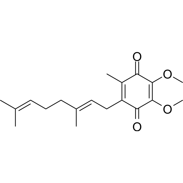
Coenzyme Q2 is a benzoquinone electron carrier in the mitochondrial electron transport chain and a p53-dependent apoptosis inducer. Coenzyme Q2 induces p53 phosphorylation at Ser15, promoting p53 accumulation and functional activation. Coenzyme Q2 induces ROS generation, caspase-3 activation, DNA fragmentation, phosphatidylserine externalization, mitochondrial permeability transition pore opening, and oxidative phosphorylation uncoupling. Coenzyme Q2 inhibits Complex I, Complex III, and Complex IV activities, disrupting electron transport and membrane potential generation. Coenzyme Q2 induces excessive mitochondrial proton leak in forebrain mitochondria. Coenzyme Q2 causes loss of righting reflex in mice, accompanied by slow-wave delta EEG activity and reversible loss of wakefulness. Coenzyme Q2 inhibits lipid peroxidation and scavenges superoxide radicals. Coenzyme Q2 is used in research on leukemia, myocardial ischemia-reperfusion injury, and mitochondrial encephalomyopathy.
For research use only. We do not sell to patients.
- CAS No.: 606-06-4
- Formula: C19H26O4
- Molecular Weight:318.41
-
Storage:
Please store the product under the recommended conditions in the Certificate of Analysis.

To place orders, for customer services and technical support, please contact: MedChemExpress USA
Tel: 609-228-6898 E-mail: [email protected] [email protected]


-
 Biological Activity
-
Chemical Information
- Protocols
- Purity & Documentation
- References
-
 Help & FAQs


-
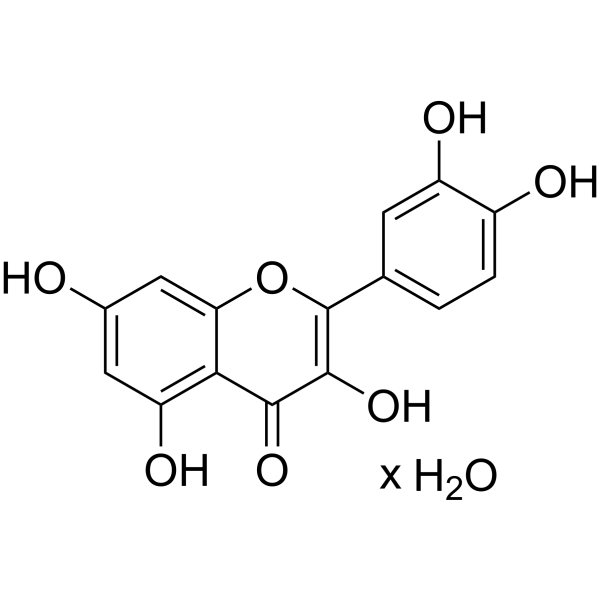
HY-18085A
Quercetin hydrateFlavonoid Antioxidant -
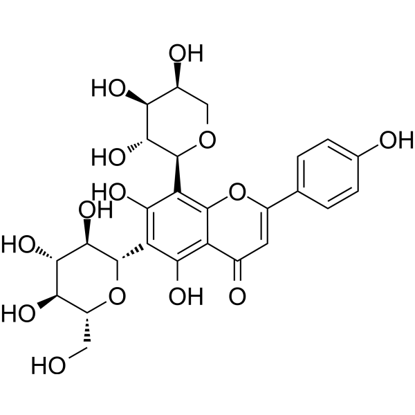
HY-N0703
SchaftosideFlavonoid -
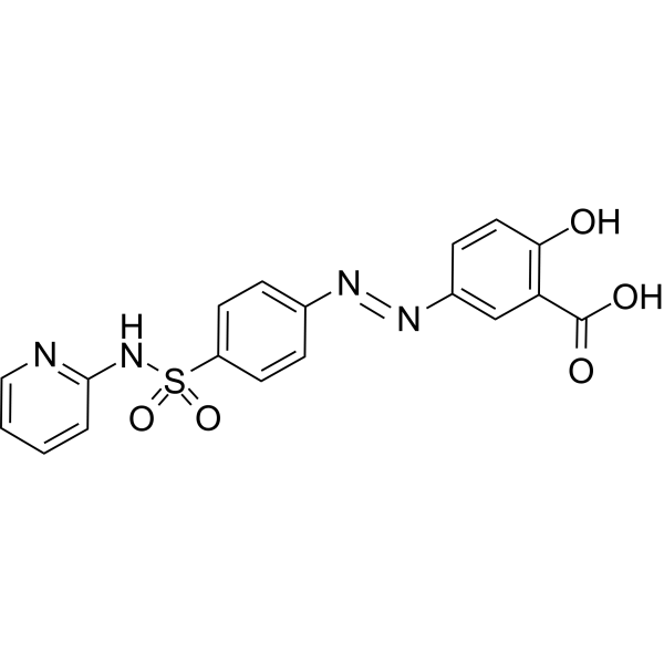
HY-14655
SulfasalazineAnti-rheumatic Agent -
HY-N0091S5
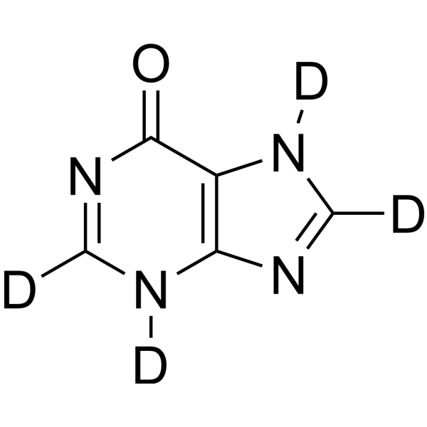
Hypoxanthine-d4Stable Isotope -

HY-14655R
Sulfasalazine (Standard)Anti-rheumatic Agent -

HY-W165608
5-(3'-Hydroxyphenyl)-γ-valerolactoneMetabolitec -

HY-135115
Oleuropein AglyconeSecoiridoid -

HY-N0703R
Schaftoside (Standard)Flavonoid -
HY-B0124S1
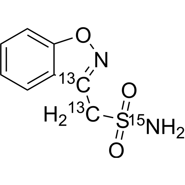
Zonisamide-13C2,15NStable Isotope -
HY-132389S
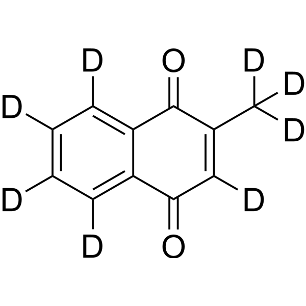
Vitamin K3-d8Stable Isotope -
HY-10208S
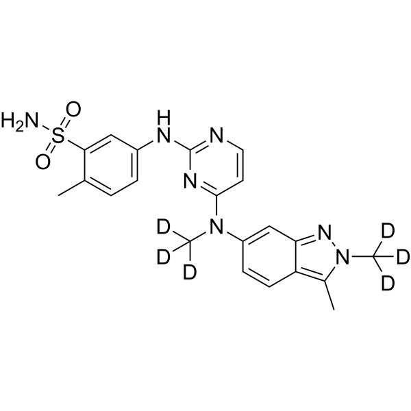
Pazopanib-d6Stable Isotope -

HY-169225
PDIC-NNSTING Activator
All Caspase Isoforms
More

Biological Activity
Description
IC50 & Target
[1]|
Caspase-3 |
Cellular Effect
|
Cell Line

|
Type | Value | Description | References |
|---|---|---|---|---|
| BALL-1 | IC50 |
20 μM
|
Inhibition of human leukemia BALL-1 cell growth assessed as reduction in cell viability incubated for 24 hrs by MTT assay.
Inhibition of human leukemia BALL-1 cell growth assessed as reduction in cell viability incubated for 24 hrs by MTT assay.
|
15905035 |
In Vitro
Coenzyme Q2 (60 μM; 24 h) induces p53-dependent apoptosis in BALL-1 cells, but not in MOLT-4F or HL-60 cells[1].
Coenzyme Q2 (20 μM; 6 h)-induced cell death depends on caspase-3 in BALL-1 cells[1].
Coenzyme Q2 (60 μM; 2-6 h) induces p53 phosphorylation at Ser15 in BALL-1 cells[1].
Coenzyme Q2 (20-100 μM; 6-24 h) inhibits BALL-1 cell growth with an IC50 of 20 μM, induces DNA fragmentation, and induces phosphatidylserine externalization[1].
Coenzyme Q2 (60 μM; 30 min) induces ROS generation in BALL-1 cells[1].
Coenzyme Q2 (23 μM) uncouples oxidative phosphorylation in isolated rabbit heart and rat liver mitochondria by increasing state 4 respiration and decreasing the respiratory control index[3].
Coenzyme Q2 (5-46 μM) decreases calcium retention capacity in isolated rabbit heart mitochondria, thereby favoring mPTP opening[3].
Coenzyme Q2 (23 μM) increases calcium retention capacity in isolated rat liver mitochondria, thereby inhibiting mPTP opening[3].
Coenzyme Q2 (23 μM) significantly decreases NADH DUb-reductase activity in isolated rabbit heart and rat liver mitochondria[3].
Coenzyme Q2 (23 μM) antagonizes the inhibition of complex I respiration by Rotenone (HY-B1756) in isolated rat liver mitochondria, but only slightly affects its action at low concentrations of Rotenone in isolated rabbit heart mitochondria[3].
Coenzyme Q2 (23 μM) increases H2O2 production in isolated rabbit heart mitochondria, but does not significantly alter H2O2 production in isolated rat liver mitochondria under basal conditions[3].
MedChemExpress (MCE) has not independently confirmed the accuracy of these methods. They are for reference only. Further protocols information, click here.
-
Cell Line:BALL-1, MOLT-4F, HL-60
-
Concentration:60 μM
-
Incubation Time:24 h
-
Result:Killed BALL-1 cells at a high rate (approximately 92% cell death), but barely killed MOLT-4F (approximately 13% cell death) or HL-60 (approximately 10% cell death) cells.
Increased caspase-3 activity approximately 30-fold in BALL-1 cells, while MOLT-4F and HL-60 cells showed little caspase-3 activation.
-
Cell Line:HepG2, HuH-7, Hep3B
-
Concentration:100 μM
-
Incubation Time:24 h
-
Result:Induced cell death strongly in HuH-7 cells (approximately 40% cell death) and induced a lower level of cell death in HepG2 cells (approximately 28% cell death).
Hep3B cells were not affected by CoQ2 (approximately 0% cell death).
Increased caspase-3 activity approximately 2-fold in HuH-7 cells and approximately 1-fold in HepG2 cells, while Hep3B showed no increase.
-
Cell Line:BALL-1
-
Concentration:0, 20, 40, 60, 80, 100 μM
-
Incubation Time:24 h
-
Result:Decreased cell viability dose-dependently with an IC50 of 20 μM.
-
Cell Line:BALL-1
-
Concentration:20 μM
-
Incubation Time:6 h
-
Result:Induced the externalization of phosphatidylserine in a time-dependent manner.\nCaspase-3 activation in BALL-1 cells was observed.
A caspase-3 specific inhibitor, Ac-DEVD-CHO, dramatically blocked CoQ2-induced cell death.
Caspase-8 and -9 were little activated by CoQ2.
-
Cell Line:BALL-1
-
Concentration:60 μM
-
Incubation Time:2, 4, or 6 h
-
Result:Phosphorylated the Ser15 site of p53, but not the Ser9, Ser20, Ser46, or Ser392 site.
-
Cell Line:BALL-1
-
Concentration:60 μM
-
Incubation Time:4 h
-
Result:Induced cell death (approximately 67% viability), caspase-3 activation (approximately 22-fold increase), and p53 Ser15 phosphorylation, which were suppressed by treatment with L-ascorbic acid (approximately 98% viability, approximately 1-fold caspase-3 activity).
In Vivo
MedChemExpress (MCE) has not independently confirmed the accuracy of these methods. They are for reference only.
-
Animal Model:C57Bl/6 N (male, 6-8 weeks old, 20-25 grams)[2]
-
Dosage:20 mg/mL (various doses); 200 mg/kg (EEG assessment)
-
Administration:i.v.; single injection
-
Result:Induced short-lived loss of righting reflex (LORR) across a range of doses.
Mice regained righting reflex within approximately 8 minutes.
Latency to return of righting reflex (RORR) showed a significant positive correlation with dose.
Mean latency to LORR was 95.0 s (95% CI: 23.3-166.7).
ED50 was approximately 100 mg/kg.


Chemical Information
-
CAS No. 606-06-4
-
Appearance Liquid
-
Molecular Weight 318.41
-
Formula C19H26O4
-
Color Light yellow to yellow
-
SMILES
O=C(C(C)=C(C1=O)C/C=C(C)/CC/C=C(C)\C)C(OC)=C1OC
-
Shipping
Room temperature in continental US; may vary elsewhere.
-
Storage
Please store the product under the recommended conditions in the Certificate of Analysis.

Protocols
-
Kinase activity and phosphorylation assays
Kinase activity assays measure the ability of kinases to transfer phosphate groups from ATP to specific substrates, while phosphorylation assays detect the presence and levels of phosphorylated proteins. Common methods include radiolabeled ATP incorporation (e. g. ,), ADP release detection via bioluminescence (e. g. ,[3]), enzyme-linked immunosorbent assays (ELISA) for phospho-specific epitopes (e. g. ,[6]), and microtiter-based formats for high-throughput screening (e. g. ,[8]). The ADP-Glo assay quantifies kinase activity by measuring ADP produced during phosphorylation using a luciferase-based system. Radiometric assays involve autoradiography or scintillation counting after incorporation of 32P-labeled ATP into substrate proteins. ELISA-based approaches rely on phospho-specific antibodies to detect activated kinases in cell lysates or purified samples.
-
Western Blot
Western blotting (WB) is a commonly used experimental method in molecular biology, biochemistry, and immunogenetics for identifying and quantifying target proteins. It combines gel electrophoresis with immunoassay, enabling researchers to analyze protein expression, post-translational modifications, and molecular weight.
-
Apoptosis
Apoptosis, also called programmed cell death, is generally characterized by distinct morphological characteristics.
-
TUNEL staining for apoptotic DNA fragmentation
TUNEL staining detects DNA strand breaks by using terminal deoxynucleotidyl transferase to add labeled nucleotides to exposed 3′-OH DNA termini, generating either microscopic staining in fixed cells or tissue sections, or fluorescence/cytometric signal in cell suspensions. TUNEL positivity reflects DNA fragmentation but should not be interpreted alone as definitive apoptosis, because TUNEL can also label necrotic, autolytic, mechanically damaged, or DNA-repair-associated DNA breaks.
-
Annexin V plus membrane-impermeant dye apoptosis staining
Annexin V-based apoptosis assays rely on the detection of phosphatidylserine (PS) externalization from the inner leaflet of the plasma membrane to the outer leaflet, an early biochemical hallmark of apoptosis. Fluorescently labeled Annexin V binds PS in a calcium-dependent manner, enabling identification of early apoptotic cells by flow cytometry or fluorescence microscopy. When combined with a membrane-impermeant DNA-binding dye (e. g. , propidium iodide), this approach allows discrimination between viable (Annexin V−/dye−), early apoptotic (Annexin V+/dye−), and late apoptotic or necrotic (Annexin V+/dye+) cell populations by assessing membrane integrity and PS exposure.
-
ROS/oxidative-stress fluorescent staining
ROS/oxidative-stress fluorescent staining uses cell-permeant fluorogenic probes that become fluorescent after oxidation inside cells or tissues; commonly used examples include DCFH-DA/DCFDA for broad cellular oxidant detection, DHE for superoxide-related signal detection, MitoSOX for mitochondrial superoxide-related signal detection, and CellROX probes for oxidative-stress-associated fluorescence readouts. The assay detects probe oxidation rather than a single ROS species unless the probe and analysis method have been chemically validated for that species. DCFH-DA enters cells, is deacetylated by intracellular esterases to DCFH, and produces fluorescent DCF after oxidation, so the readout is used as an operational measure of total cellular oxidative stress rather than a species-specific ROS measurement. DHE and MitoSOX can report superoxide-related oxidation, but red fluorescence alone can include non-specific ethidium-like oxidation products; HPLC or optimized spectral approaches are
-
Fluorescent plasma-membrane potential dye assay
Fluorescent plasma-membrane potential dye assays measure changes in cell membrane potential using voltage-sensitive dyes whose fluorescence changes when cells depolarize or hyperpolarize. Anionic bis-oxonol dyes such as DiBAC4(3) enter depolarized cells more readily and show increased fluorescence after intracellular binding, while hyperpolarization reduces dye accumulation and fluorescence. FMP/FLIPR membrane-potential dyes are used for faster, homogeneous microplate assays of ion-channel or receptor-mediated membrane-potential changes.
-
Apoptosis Solutions
Apoptosis is a regulated, generally non-lytic cell-death pathway that removes unwanted, damaged, infected, or abnormal cells through coordinated morphological changes, caspase activation, DNA fragmentation, and membrane remodeling. The intrinsic apoptosis pathway is controlled mainly by mitochondrial outer membrane permeabilization, BCL-2 family proteins, cytochrome c release, apoptosome formation, caspase-9 activation, and downstream executioner caspase-3/7 activation. The extrinsic apoptosis pathway is initiated by death receptors such as Fas, TNFR, and TRAIL receptors, which recruit adaptor proteins and activate caspase-8 before engaging executioner caspases or mitochondrial amplification through BID cleavage. Apoptosis is linked to many phenotypes, including cancer cell killing, tissue homeostasis, immune regulation, neurodegeneration, infection response, and treatment-induced cytotoxicity; unresolved questions include how apoptosis interacts with necroptosis, pyroptosis, ferroptos
-
Transepithelial/transendothelial electrical resistance assay
TEER measures electrical resistance across epithelial or endothelial monolayers cultured on permeable supports, and the readout reflects ionic conductance through the cell barrier, especially the paracellular pathway regulated by junctional integrity. TEER can be measured without destroying the monolayer and is commonly used before or during transport, permeability, barrier-disruption, and barrier-maturation experiments. TEER values are influenced by biological maturation and technical conditions; reported factors include temperature, medium formulation, passage number, electrode geometry, membrane properties, and junctional length during early monolayer maturation. Therefore, TEER should be interpreted with blank-insert subtraction, area normalization, repeated readings, and, when possible, orthogonal barrier readouts such as FITC-dextran flux or tight-junction staining.
-
Protocol for Kinase activity and phosphorylation assays
Kinase activity assays measure transfer of phosphate from ATP to a protein or peptide substrate, generating phosphorylated substrate, ADP, or incorporated radiolabeled phosphate as the readout; phosphorylation assays measure site-specific phosphorylation in cells or tissues as a proxy for kinase-pathway activation, inhibition, or substrate regulation. Phosphorylation can be detected by phospho-specific Western blot, immunoprecipitation kinase assay, phospho-immunofluorescence, phospho-flow cytometry, luminescent ADP detection, radiolabeled ATP incorporation, or reporter-based pathway assays, and these readouts can be applied to cancer cells, primary neurons, mouse tumors, organoids, inflammatory macrophages, ferroptosis studies, and mitophagy studies when the kinase target is biologically relevant.
-
Ferroptosis Solutions
Ferroptosis is an iron-dependent, non-apoptotic form of regulated cell death characterized by lethal lipid peroxidation and sensitivity to suppression by iron chelators or lipophilic radical-trapping antioxidants. The core pathway links cystine uptake through system Xc−, glutathione availability, GPX4-dependent detoxification of phospholipid hydroperoxides, iron-dependent oxidative reactions, and polyunsaturated-phospholipid metabolism into a cell-death program that is biochemically and morphologically distinct from apoptosis, necrosis, and autophagy. The ferroptosis pathway is experimentally linked to phenotype through chemical and genetic perturbation. Erastin induces ferroptosis by inhibiting cystine uptake through system Xc− and weakening antioxidant defenses, while GPX4 inhibition or depletion causes lipid peroxide accumulation and ferroptotic cancer-cell death. ACSL4 and oxidizable arachidonoyl- or adrenoyl-containing phosphatidylethanolamines shape ferroptosis sensitivity by con


Purity & Documentation
-
Data Sheet (284 KB)
-
SDS (393 KB)
- English - EN (393 KB)
- Français - FR (393 KB)
- Deutsch - DE (393 KB)
- Norwegian - NO (393 KB)
- Español - ES (393 KB)
- Swedish - SV (393 KB)
- Italian - IT (393 KB)
- Korean - KR (393 KB)
- Portuguese - PT (393 KB)
-
Handling Instructions (2659 KB)
References

Calculators
Concentration (start) × Volume (start) = Concentration (final) × Volume (final)


Coenzyme Q2 Related Classifications
HY-118075 Related Classifications
 Powered by Bioz
Keywords
- Coenzyme Q2
- 606-06-4
- Coenzyme Q 2
- Coenzyme Q-2
- MDM-2/p53
- Apoptosis
- Reactive Oxygen Species (ROS)
- Caspase
- Oxidative Phosphorylation
- p53-dependent apoptosis
- caspase-3 activation
- ROS generation
- mitochondrial permeability transition pore opening
- BALL-1 cells
- p53 phosphorylation at Ser15
- HuH-7 cells
- HL-60 cells
- human hepatoma cell lines
- MOLT-4F
- Inhibitor
- inhibitor
- inhibit