



SF1126

Based on 1 Customer Validation
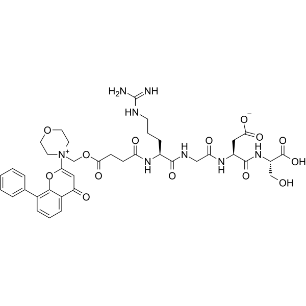
SF1126 is a relevant pan and dual first-in-class PI3K/BRD4 inhibitor, has antitumor and anti-angiogenic activity. SF1126 is an RGDS-conjugated LY294002 proagent, which is designed to exhibit increased solubility and bind to specific integrins within the tumor compartment. SF1126 induces cell apoptosis.
Para uso exclusivo en investigación. No vendemos a pacientes.
- No. CAS: 936487-67-1
- Fòrmula: C39H48N8O14
- Peso molecular:852.84
-
Almacenamiento:
-20°C, sealed storage, away from moisture and light
* In solvent : -80°C, 6 months; -20°C, 1 month (sealed storage, away from moisture and light)

To place orders, for customer services and technical support, please contact: MedChemExpress USA
Tel: 609-228-6898 E-mail: [email protected] [email protected]


-
 Actividad biológica
-
Chemical Information
- Protocolo
- Pureza y Documentación
- Referencias
-
 Help & FAQs




Actividad biológica
Descripciòn
IC50 & Target
PI3K/BRD4[1]
In Vitro
SF1126 (0-6 μM; 48 hours) inhibits Hep3B, HepG2, SK-Hep1, and Huh7 cells proliferation with IC50s of 5.05, 6.89, 3.14, and 2.14 μM, respectively[1]. SF1126 (1-10 μM; 24 hours) results in cell-cycle arrest with a proportional increase in G0-G1 and a decrease in the number of cells in the S-phase in Hep 3B, Hep G2, SK-Hep1, and Huh7 cells[1]. SF1126 (0.5-2.5 μM; pre-30 minutes) and sorafenib suggests that combined treatment of SF1126 and sorafenib blocks multiple key enzymes in PI3K/AKT/mTOR and Ras/Raf/MAPK pathway[1].
MedChemExpress (MCE) has not independently confirmed the accuracy of these methods. They are for reference only. Further protocols information, click here.
-
Cell Line:Hep3B, HepG2, SK-Hep1, and Huh7 cells
-
Concentration:0 µM; 1 µM; 2 µM; 3 µM; 4 µM; 5 µM; 6 µM
-
Incubation Time:48 hours
-
Result:Resulted in an increased inhibition of HCC proliferation.
-
Cell Line:Hep3B, HepG2, SK-Hep1, and Huh7 cells
-
Concentration:1 µM; 5 µM; 10 µM
-
Incubation Time:24 hours
-
Result:Induced cell-cycle arrest.
-
Cell Line:Hep3B, HepG2, SK-Hep1, and Huh7 cells
-
Concentration:0.5 µM and 2.5 µM
-
Incubation Time:Pre-30 mins
-
Result:Suppressed phosphorylation of AKT, p70S6K, 4EBP1, and ERK in all the cell lines together with sorafenib.
Ensayo clínico
| NCT Number | Sponsor | Condition | Start Date |
Phase

|
|---|---|---|---|---|
| NCT01329991 | Plexxikon| | 2011-05 | PHASE1 |


Chemical Information
-
No. CAS 936487-67-1
-
Appearance Solid
-
Peso molecular 852.84
-
Fòrmula C39H48N8O14
-
Color White to off-white
-
SMILES
N=C(N)NCCC[C@H](NC(CCC(OC[N+]1(CCOCC1)C(OC(C2=CC=C3)=C3C4=CC=CC=C4)=CC2=O)=O)=O)C(NCC(N[C@@H](CC([O-])=O)C(N[C@@H](CO)C(O)=O)=O)=O)=O
-
Envío
Room temperature in continental US; may vary elsewhere.
-
Almacenamiento
-20°C, sealed storage, away from moisture and light
* In solvent : -80°C, 6 months; -20°C, 1 month (sealed storage, away from moisture and light)

Protocolo
-
Detection of Apoptosis
Apoptosis, also called programmed cell death, is generally characterized by distinct morphological characteristics.
-
TUNEL staining for apoptotic DNA fragmentation
TUNEL staining detects DNA strand breaks by using terminal deoxynucleotidyl transferase to add labeled nucleotides to exposed 3′-OH DNA termini, generating either microscopic staining in fixed cells or tissue sections, or fluorescence/cytometric signal in cell suspensions. TUNEL positivity reflects DNA fragmentation but should not be interpreted alone as definitive apoptosis, because TUNEL can also label necrotic, autolytic, mechanically damaged, or DNA-repair-associated DNA breaks.
-
Annexin V plus membrane-impermeant dye apoptosis staining
Annexin V-based apoptosis assays rely on the detection of phosphatidylserine (PS) externalization from the inner leaflet of the plasma membrane to the outer leaflet, an early biochemical hallmark of apoptosis. Fluorescently labeled Annexin V binds PS in a calcium-dependent manner, enabling identification of early apoptotic cells by flow cytometry or fluorescence microscopy. When combined with a membrane-impermeant DNA-binding dye (e. g. , propidium iodide), this approach allows discrimination between viable (Annexin V−/dye−), early apoptotic (Annexin V+/dye−), and late apoptotic or necrotic (Annexin V+/dye+) cell populations by assessing membrane integrity and PS exposure.
-
Endothelial Tube Formation Assay
Endothelial tube formation assay evaluates the ability of endothelial cells to attach, migrate, align, and organize into capillary-like networks when cultured on gelled basement membrane extract or Matrigel; the readout is the morphology and quantity of tube-like networks, which reflects an in vitro endothelial morphogenesis step related to angiogenesis. Basement membrane extract/Matrigel provides laminin-rich extracellular matrix cues that support endothelial differentiation into capillary-like structures, but it can contain biologically active growth factors, so growth-factor-reduced matrix is preferred when testing defined angiogenic stimulators or inhibitors.
-
Apoptosis Solutions
Apoptosis is a regulated, generally non-lytic cell-death pathway that removes unwanted, damaged, infected, or abnormal cells through coordinated morphological changes, caspase activation, DNA fragmentation, and membrane remodeling. The intrinsic apoptosis pathway is controlled mainly by mitochondrial outer membrane permeabilization, BCL-2 family proteins, cytochrome c release, apoptosome formation, caspase-9 activation, and downstream executioner caspase-3/7 activation. The extrinsic apoptosis pathway is initiated by death receptors such as Fas, TNFR, and TRAIL receptors, which recruit adaptor proteins and activate caspase-8 before engaging executioner caspases or mitochondrial amplification through BID cleavage. Apoptosis is linked to many phenotypes, including cancer cell killing, tissue homeostasis, immune regulation, neurodegeneration, infection response, and treatment-induced cytotoxicity; unresolved questions include how apoptosis interacts with necroptosis, pyroptosis, ferroptos


Pureza y Documentación
-
Ficha de datos (272 KB)
-
SDS (252 KB)
- Français - FR (252 KB)
- Deutsch - DE (252 KB)
- Norwegian - NO (252 KB)
- Español - ES (252 KB)
- Swedish - SV (252 KB)
- Italian - IT (252 KB)
- Korean - KR (252 KB)
- Portuguese - PT (252 KB)
-
Instrucciones de manejo (2659 KB)
Referencias

Calculators
Concentration (start) × Volume (start) = Concentration (final) × Volume (final)


SF1126 Related Classifications
HY-10220 Related Classifications
 Powered by Bioz