



Phleomycin
Based on 3 publication(s) in Google Scholar
Phleomycin is a copper-dependent DNA damaging agent and antibiotic with antitumor activity. Phleomycin binds to DNA and produces ROS in the presence of reducing agents (such as dithiothreitol and glutathione), inducing single-strand and double-strand breaks in DNA. Phleomycin can induce cell apoptosis or mutation and is widely used in cancer inhibition, microbial genetic transformation (as a screening marker to improve fungal transformation efficiency) and DNA repair mechanism research.
For research use only. We do not sell to patients.
- Purity : 95.0%
- CAS No.: 11006-33-0
-
Storage:
-20°C, stored under nitrogen
* In solvent : -80°C, 6 months; -20°C, 1 month (stored under nitrogen)


To place orders, for customer services and technical support, please contact: MedChemExpress USA
Tel: 609-228-6898 E-mail: [email protected] [email protected]


-
 Biological Activity
-
Chemical Information
-
Solvent & Solubility
- Protocols
- Purity & Documentation
- References
-
 Help & FAQs


-
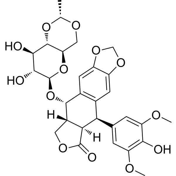
HY-13629
EtoposideTopoisomerase II Inhibitor -
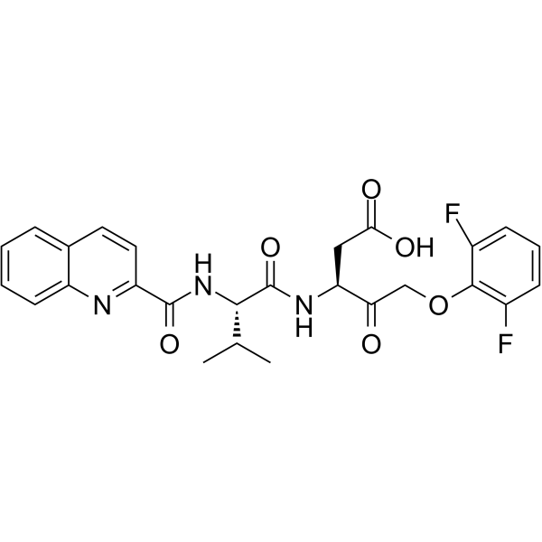
HY-12305
Q-VD-OPhPan-caspase Inhibitor -
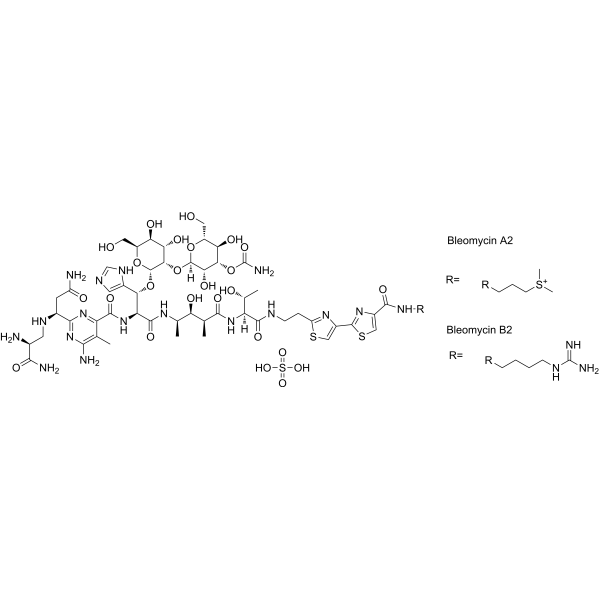
HY-17565
Bleomycin sulfateDNA Synthesis Inhibitor -
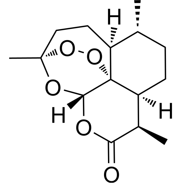
HY-B0094
ArtemisininParasite Inhibitor -
HY-N0281
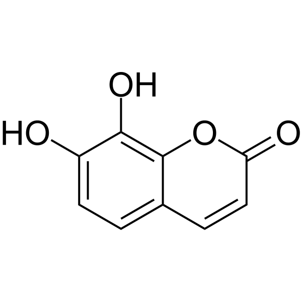
DaphnetinProtein Kinase Inhibitor -

HY-B0094R
Artemisinin (Standard)Parasite Inhibitor -

HY-N0281R
Daphnetin (Standard)Protein Kinase Inhibitor
Publications Citing Use of MedChemExpress (MCE) Phleomycin
MoreAll Antibiotic Isoforms
MoreAll DNA/RNA Synthesis Isoforms
More

Biological Activity
Description
IC50 & Target
|
Glycopeptide |
In Vitro
1. Phleomycin (1-25 μg/mL; overnight) can increase the transformation efficiency by more than 10 times in the protoplast transformation experiment of Schizophyllum commune, promote single copy integration, and upregulate the expression of DNA repair genes KU70 and RAD52[2].
2. Phleomycin (2 μg/mL, 10 μg/mL; 90 min-4 h) selectively inhibits DNA synthesis in Escherichia coli (E. coli) and HeLa cells and reduces 3H-thymidine incorporation into DNA. At 20 μg/mL, the DNA degradation activity is significantly lower than that of Mitomycin C (HY-13316)[3].
MedChemExpress (MCE) has not independently confirmed the accuracy of these methods. They are for reference only.


Chemical Information
-
CAS No. 11006-33-0
-
Appearance Solid
-
Color Light blue to blue
-
SMILES
[Phleomycin]
-
Structure Classification
-
Initial Source
Streptoalloteichus hindustanus
-
Shipping
Room temperature in continental US; may vary elsewhere.
-
Storage
-20°C, stored under nitrogen
* In solvent : -80°C, 6 months; -20°C, 1 month (stored under nitrogen)


Publications (3)
-
Journal Impact Factor
-
Most Recent
-
FEMS Yeast Res
Characterization of oncohistone H2B variants in Schizosaccharomyces pombe reveals a key role of H2B monoubiquitination deficiency in genomic instability by altering gene expression. [Abstract]2025 Jan 30:25:foaf027. PMID: 40402811 -
-
Genes Cells
SRPKs Homolog Dsk1 Regulates Homologous Recombination Repair in Schizosaccharomyces pombe. [Abstract]2025 Jan;30(1):e13192. PMID: 39789818


Solvent & Solubility
In Vitro:
H2O : 100 mg/mL (Need ultrasonic)


Purity & Documentation
-
Data Sheet (270 KB)
-
SDS (393 KB)
- English - EN (393 KB)
- Français - FR (393 KB)
- Deutsch - DE (393 KB)
- Norwegian - NO (393 KB)
- Español - ES (393 KB)
- Swedish - SV (393 KB)
- Italian - IT (393 KB)
- Korean - KR (393 KB)
- Portuguese - PT (393 KB)
-
Handling Instructions (2659 KB)
References
[1]. Kamila Stokowa-Sołtys, et al. Phleomycin complex-Coordination mode and in vitro cleavage of DNA. J Inorg Biochem. 2019 Jun;195:71-82. [Content Brief]
[2]. van Peer AF, et al. Phleomycin increases transformation efficiency and promotes single integrations in Schizophyllum commune. Appl Environ Microbiol. 2009 Mar;75(5):1243-7. [Content Brief]
[3]. TANAKA N, et al. Mechanism of action of phleomycin, a tumor-inhibitory antibiotic. Biochem Biophys Res Commun. 1963 Jan 31;10:171-4. [Content Brief]

Calculators
Concentration (start) × Volume (start) = Concentration (final) × Volume (final)


Phleomycin Related Classifications
HY-126490 Related Classifications
 Powered by Bioz