



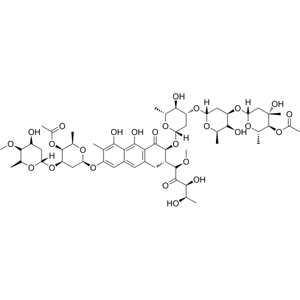
Chromomycin A3
Based on 1 publication(s) in Google Scholar
Chromomycin A3 is an inhibitor that selectively binds to GC-rich DNA sequences. Chromomycin A3 targets the DNA minor groove after forming a dimer with Mg2+. Chromomycin A3 inhibits DNA replication and transcription, blocks the binding of Sp1 transcription factor to target gene promoters, downregulates the expression of anti-apoptotic proteins such as FLIP, Mcl-1, and XIAP, and induces S-phase cycle arrest and caspase-dependent apoptosis in tumor cells. Chromomycin A3 can antagonize oxidative stress induced by glutathione depletion and neuronal apoptosis induced by Camptothecin (HY-15660). Chromomycin A3 can be used in basic research on malignant tumors such as cholangiocarcinoma, and is a potential chemosensitizer and GC-rich region probe.
For research use only. We do not sell to patients.
- Purity : 99.48%
- CAS No.: 7059-24-7
- Formula: C57H82O26
- Molecular Weight:1183.25
-
Storage:Powder -20°C, 3 years ; In solvent -80°C, 6 months , -20°C, 1 month

To place orders, for customer services and technical support, please contact: MedChemExpress USA
Tel: 609-228-6898 E-mail: [email protected] [email protected]


-
 Biological Activity
-
Chemical Information
-
Solvent & Solubility
- Protocols
- Purity & Documentation
- References
-
 Help & FAQs


-
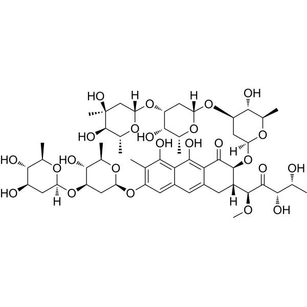
HY-A0122
PlicamycinSp1 Inhibitor -
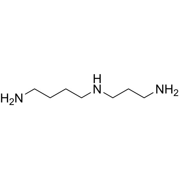
HY-B1776
SpermidinePolyamine Compound -
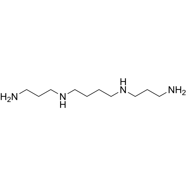
HY-B1777
SpermineEndogenous Metabolite -
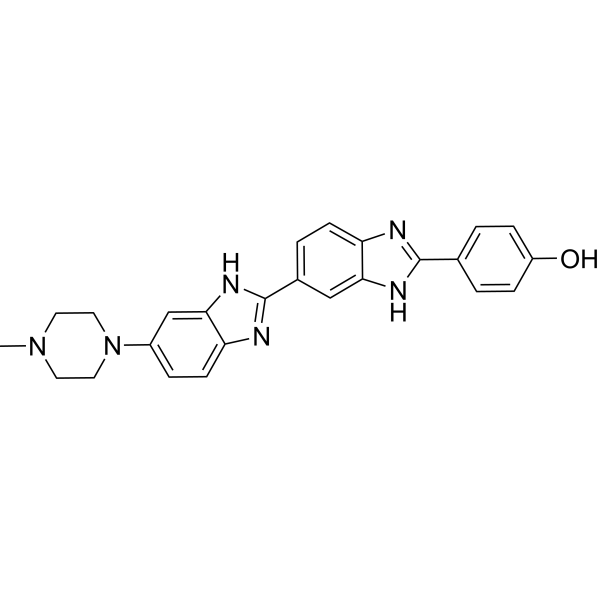
HY-15558
Hoechst 33258DNA Minor Groove Stain -

HY-W040129R
Chromomycin A3 (Standard) -
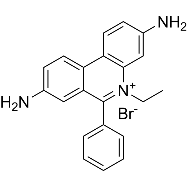
HY-D0021
EthD bromideFluorescent Tag -

HY-DY1050
Ethidium bromide (solution)Fluorescent Tag -
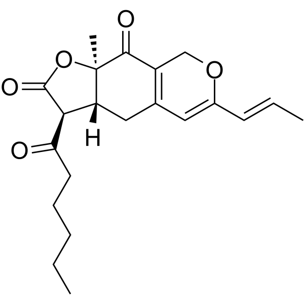
HY-N6641
MonascinAzaphilonoid Pigment -

HY-N6641R
Monascin (Standard)Azaphilonoid Pigment
Publications Citing Use of MedChemExpress (MCE) Chromomycin A3
MoreAll DNA/RNA Synthesis Isoforms
MoreAll Caspase Isoforms
More

Biological Activity
Description
Cellular Effect
|
Cell Line

|
Type | Value | Description | References |
|---|---|---|---|---|
| A2780 | IC50 |
0.04 μM
Compound: 2
|
Cytotoxicity against human A2780 cells
Cytotoxicity against human A2780 cells
|
[PMID: 22647722] |
| A549 | IC50 |
0.042 μM
Compound: 1
|
Cytotoxicity against human A549 cells after 96 hrs by Cell Titer Glo assay
Cytotoxicity against human A549 cells after 96 hrs by Cell Titer Glo assay
|
[PMID: 21807523] |
| A549 | IC50 |
0.1 μM
Compound: 2
|
Cytotoxicity against human A549 cells
Cytotoxicity against human A549 cells
|
[PMID: 22647722] |
| BXPC-3 | IC50 |
0.13 μM
Compound: 2
|
Cytotoxicity against human BxPC3 cells
Cytotoxicity against human BxPC3 cells
|
[PMID: 22647722] |
| COLO 205 | IC50 |
0.063 μM
Compound: 2
|
Cytotoxicity against human COLO205 cells
Cytotoxicity against human COLO205 cells
|
[PMID: 22647722] |
| HCC44 | IC50 |
0.001 μM
Compound: 1
|
Cytotoxicity against human HCC44 cells after 96 hrs by Cell Titer Glo assay
Cytotoxicity against human HCC44 cells after 96 hrs by Cell Titer Glo assay
|
[PMID: 21807523] |
| NIH3T3 | IC50 |
1 μM
Compound: 2
|
Cytotoxicity against mouse NIH/3T3 cells
Cytotoxicity against mouse NIH/3T3 cells
|
[PMID: 22647722] |
In Vitro
Chromomycin A3 (2.5-160 nM; 24 h, 48 h) inhibits the proliferation of cholangiocarcinoma cells KKU-213, KKU-055, and KKU-100 in a dose- and time-dependent manner, with IC50s ??of 22.48 nM, 21.14 nM, and 30.52 nM at 24 h, respectively.
Chromomycin A3 (2.5-10 nM; 24 h) induces S phase arrest of KKU-213 and KKU-055 cells at low concentrations. At high concentrations, Chromomycin A3 (10-40 nM; 24 h) induces dose-dependent apoptosis by activating the caspase-8/-9/-3 pathway, and the proportion of early and late apoptotic cells increased significantly[1].
MedChemExpress (MCE) has not independently confirmed the accuracy of these methods. They are for reference only.
-
Cell Line:KKU-213, KKU-055, KKU-100 cholangiocarcinoma cells
-
Concentration:0, 2.5, 5, 10, 20, 40, 80, 160 nM (CMA3); 0, 12.5, 25, 50, 100, 200 nM (MTA)
-
Incubation Time:24 h and 48 h
-
Result:Reduced cell viability in a dose- and time-dependent manner, with IC50 values of 22.48±4.08 nM (KKU-213), 21.14±2.24 nM (KKU-055), and 30.52±2.91 nM (KKU-100) at 24 h; with IC50 values of 9.79±1.15 nM, 13.34±1.28 nM, and 14.74±1.34 nM at 48 h, for the respective cell lines.
In Vivo
MedChemExpress (MCE) has not independently confirmed the accuracy of these methods. They are for reference only.
-
Animal Model:Rag-2/Jak3 double-deficient Balb/c mouse (male; 22-25 g, 6-8 weeks old) cholangiocarcinoma xenograft model[1]
-
Dosage:0.5 mg/kg
-
Administration:Intravenous injection via tail vein, once weekly, for 3 weeks
-
Result:Significantly reduced tumor growth compared to the DMSO control group, decreased tumor weight.
Resulted a significant reduction in Ki-67-positive proliferating cells, and increased expression of pro-apoptotic proteins caspase-9 and Bax in tumor tissues.
No significant toxicity was observed, as body weight and general health remained stable throughout the experiment.
- Nuclear Protein Extraction (High-Salt/Hypotonic Fractionation)
- RNA extraction experimental
- Detection of Apoptosis
- TUNEL staining for apoptotic DNA fragmentation
- PCNA Immunodetection Proliferation Assay
- Annexin V plus membrane-impermeant dye apoptosis staining
- ROS/oxidative-stress fluorescent staining
- Apoptosis Solutions


Chemical Information
-
CAS No. 7059-24-7
-
Appearance Solid
-
Molecular Weight 1183.25
-
Formula C57H82O26
-
Color Light yellow to yellow
-
SMILES
O=C1C2=C(C[C@H]([C@@H]1O[C@](C[C@@H](O[C@](C[C@@H](O[C@@]3([H])C[C@](O)([C@H]([C@@H](O3)C)OC(C)=O)C)[C@@H]4O)([H])O[C@@H]4C)[C@@H]5O)([H])O[C@@H]5C)[C@@H](C([C@H]([C@@H](C)O)O)=O)OC)C=C(C=C6O[C@H]7C[C@H]([C@H]([C@H](O7)C)OC(C)=O)O[C@]8([H])C[C@H]([C@H]([C@H](O8)C)OC)O)C(C(O)=C6C)=C2O
-
Structure Classification
-
Initial Source
Streptomyces griseu
-
Shipping
Room temperature in continental US; may vary elsewhere.
-
Storage
Powder -20°C 3 years In solvent -80°C 6 months -20°C 1 month


Publications (1)
-
Journal Impact Factor
-
Most Recent
-
J Asian Nat Prod Res
Chromomycins from soil-derived Streptomyces sp. inhibit the growth of human non-small cell lung cancer cells by targeting c-FLIP. [Abstract]2024 Jul 8:1-16. PMID: 38975979


Solvent & Solubility
In Vitro:
DMSO : ≥ 100 mg/mL (84.51 mM; Hygroscopic DMSO has a significant impact on the solubility of product, please use newly opened DMSO)
* "≥" means soluble, but saturation unknown.
Please refer to the solubility information to select the appropriate solvent. Once prepared, please aliquot and store the solution to prevent product inactivation from repeated freeze-thaw cycles.
Storage method and period of stock solution: -80°C, 6 months; -20°C, 1 month. When stored at -80°C, please use it within 6 months. When stored at -20°C, please use it within 1 month.
Please refer to the solubility information to select the appropriate solvent. Once prepared, please aliquot and store the solution to prevent product inactivation from repeated freeze-thaw cycles.
Storage method and period of stock solution: -80°C, 6 months; -20°C, 1 month. When stored at -80°C, please use it within 6 months. When stored at -20°C, please use it within 1 month.
Concentration (start) × Volume (start) = Concentration (final) × Volume (final)
In Vivo:
Select the appropriate dissolution method based on your experimental animal and administration route.
- For the following dissolution methods, please ensure to first prepare a clear stock solution using an In Vitro approach and then sequentially add co-solvents:
- To ensure reliable experimental results, the clarified stock solution can be appropriately stored based on storage conditions. As for the working solution for In Vivo experiments, it is recommended to prepare freshly and use it on the same day.
- The percentages shown for the solvents indicate their volumetric ratio in the final prepared solution. If precipitation or phase separation occurs during preparation, heat and/or sonication can be used to aid dissolution.
Add each solvent one by one: 10% DMSO 40% PEG300 5% Tween-80 45% Saline
Solubility: 2.5 mg/mL (2.11 mM); Suspended solution; Need ultrasonic
This protocol yields a suspended solution of 2.5 mg/mL. Suspended solution can be used for oral and intraperitoneal injection.
Taking 1 mL working solution as an example, add 100 μL DMSO stock solution (25.0 mg/mL) to 400 μL PEG300, and mix evenly; then add 50 μL Tween-80 and mix evenly; then add 450 μL Saline to adjust the volume to 1 mL.
Preparation of Saline: Dissolve 0.9 g sodium chloride in ddH₂O and dilute to 100 mL to obtain a clear Saline solution.
In Vivo Dissolution Calculator
Please enter the basic information of animal experiments:
-
-
-
-
Recommended: Prepare an additional quantity of animals to account for potential losses during experiments.
Please enter your animal formula composition:
-
%DMSO +
Recommended: Keep the proportion of DMSO in working solution below 2% if your animal is weak.
-
%+
-
+%Tween-80 + +
-
%Saline +
The co-solvents required include: DMSO, . All of co-solvents are available by MedChemExpress (MCE). , Tween 80. All of co-solvents are available by MedChemExpress (MCE).
Working solution concentration: 0.22 mg/mL
Method for preparing stock solution: mg drug dissolved in μL DMSO. Stock solution concentration: mg/mL.
1. Take μL DMSO stock solution;
2. Add μL .
μL , mix evenly;
3. Then add μL Tween 80, mix evenly;
4. Then add μL
Please ensure that the stock solution in the first step is dissolved to a clear state, and add co-solvents in sequence. You can use ultrasonic heating (ultrasonic cleaner, recommended frequency 20-40 kHz), vortexing, etc. to assist dissolution.


Purity & Documentation
-
Data Sheet (289 KB)
-
SDS (644 KB)
- English - EN (644 KB)
- Français - FR (644 KB)
- Deutsch - DE (644 KB)
- Norwegian - NO (644 KB)
- Español - ES (644 KB)
- Swedish - SV (644 KB)
- Italian - IT (644 KB)
- Korean - KR (644 KB)
- Portuguese - PT (644 KB)
-
Handling Instructions (2659 KB)
References
[1]. Saranaruk P, et al. Chromomycin A3 suppresses cholangiocarcinoma growth by induction of S phase cell cycle arrest and suppression of Sp1‑related anti‑apoptotic proteins. Int J Mol Med. 2020 Apr;45(4):1005-1016. [Content Brief]
[2]. Murase H, et al. Evaluation of simultaneous binding of Chromomycin A3 to the multiple sites of DNA by the new restriction enzyme assay. Bioorg Med Chem Lett. 2018 Jun 1;28(10):1832-1835. [Content Brief]
[3]. Chatterjee S, et al. Sequence-selective DNA binding drugs mithramycin A and chromomycin A3 are potent inhibitors of neuronal apoptosis induced by oxidative stress and DNA damage in cortical neurons. Ann Neurol. 2001 Mar;49(3):345-54. [Content Brief]


Complete Stock Solution Preparation Table
Please refer to the solubility information to select the appropriate solvent. Once prepared, please aliquot and store the solution to prevent product inactivation from repeated freeze-thaw cycles.
Storage method and period of stock solution: -80°C, 6 months; -20°C, 1 month. When stored at -80°C, please use it within 6 months. When stored at -20°C, please use it within 1 month.
| Optional Solvent | Concentration Solvent Mass | 1 mg | 5 mg | 10 mg | 25 mg |
|---|---|---|---|---|---|
| DMSO | 1 mM | 0.8451 mL | 4.2256 mL | 8.4513 mL | 21.1282 mL |
| 5 mM | 0.1690 mL | 0.8451 mL | 1.6903 mL | 4.2256 mL | |
| 10 mM | 0.0845 mL | 0.4226 mL | 0.8451 mL | 2.1128 mL | |
| 15 mM | 0.0563 mL | 0.2817 mL | 0.5634 mL | 1.4085 mL | |
| 20 mM | 0.0423 mL | 0.2113 mL | 0.4226 mL | 1.0564 mL | |
| 25 mM | 0.0338 mL | 0.1690 mL | 0.3381 mL | 0.8451 mL | |
| 30 mM | 0.0282 mL | 0.1409 mL | 0.2817 mL | 0.7043 mL | |
| 40 mM | 0.0211 mL | 0.1056 mL | 0.2113 mL | 0.5282 mL | |
| 50 mM | 0.0169 mL | 0.0845 mL | 0.1690 mL | 0.4226 mL | |
| 60 mM | 0.0141 mL | 0.0704 mL | 0.1409 mL | 0.3521 mL | |
| 80 mM | 0.0106 mL | 0.0528 mL | 0.1056 mL | 0.2641 mL |


Chromomycin A3 Related Classifications
HY-W040129 Related Classifications
 Powered by Bioz