



Roridin E

Based on 1 Customer Validation
Roridin E is a glucose-6-phosphatase (G6Pase) inhibitor and antibiotic, and is a metabolic byproduct of Roridin A (HY-N9599). Roridin E induces significant oxidative stress, characterized by depletion of glutathione in vivo, induction of hepatic lipid peroxidation, and inhibition of renal superoxide dismutase activity. Roridin E reduces blood glucose levels in rats, but exhibits acute toxicity (which is enhanced when co-administered with linoleic acid (HY-N0729)) and causes hepatotoxicity in male albino mice. Roridin E induces a decrease in total blood protein and increases in the levels of total lipids, γ-glutamyltransferase, alkaline phosphatase, and 5'-nucleotidase. Roridin E can be isolated from molds, and possesses cytostatic and antifungal activities similar to those of Verrucarin A (HY-107426) and Roridin A. Roridin E exhibits in vivo activity in rodents and is commonly used in hepatotoxicity-related studies.
商品は「研究用試薬」です。人や動物の医療用・臨床診断用・食品用の製品ではありません。
研究用途以外に使用した場合、当社は一切の責任を負いかねます。
- 純度 : 99.89%
- CAS 番号: 16891-85-3
- 分子式: C29H38O8
- 分子量:514.61
-
保管条件:Powder -20°C, 3 years ; In solvent -80°C, 6 months , -20°C, 1 month


To place orders, for customer services and technical support, please contact: MedChemExpress USA
Tel: 609-228-6898 E-mail: [email protected] [email protected]


-
 生物活性
生物活性
-
化学情報
- プロトコル
- 純度とドキュメンテーション
- 参考文献
-
 Help & FAQs


-
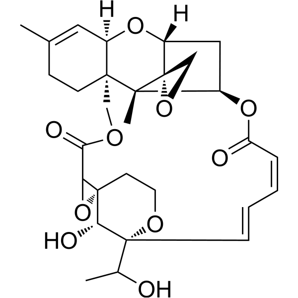
HY-130476
Satratoxin GLarge Cyclic Mucor Toxin. -
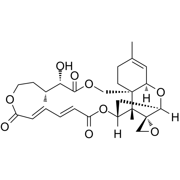
HY-107426
Verrucarin AProtein Synthesis Inhibitor -
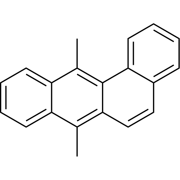
HY-W011845
DMBA -
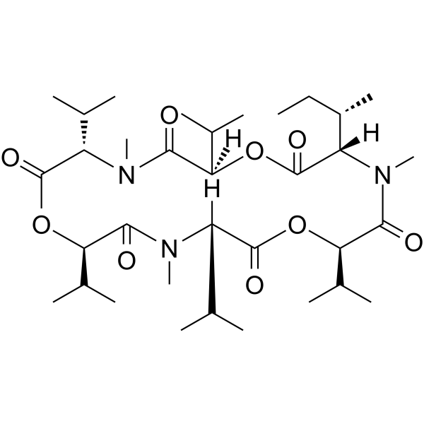
HY-N3807
Enniatin B1ACAT Inhibitor -
HY-123647
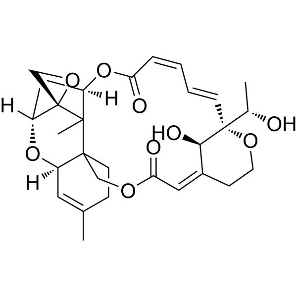
Satratoxin HRibosome Binding Agent\nApoptosis Inducer\nGenotoxin -
HY-124923
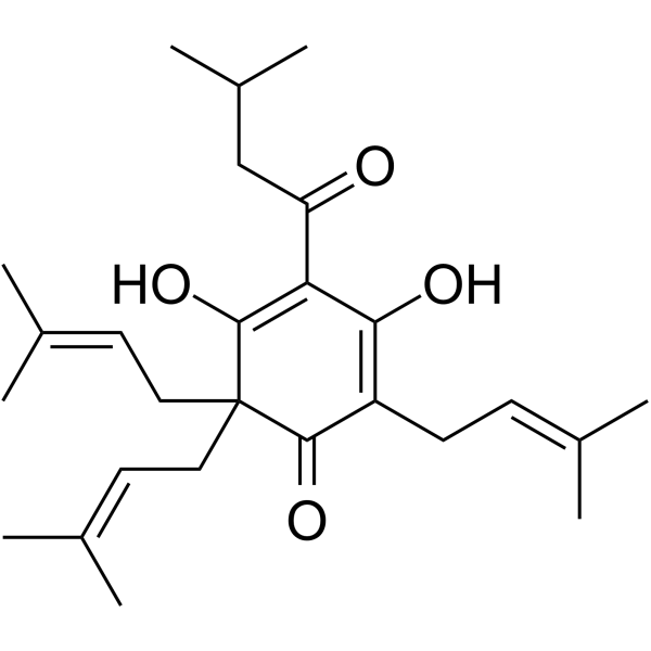
LupuloneBacterial Inhibitor -
HY-W013863
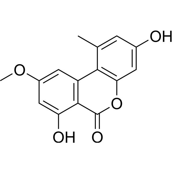
Djalonensone -

HY-W749510
Spiroxamine-d4Stable Isotope -

HY-W011845R
DMBA (Standard) -
HY-108766
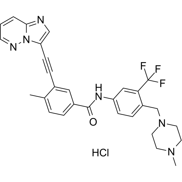
Ponatinib hydrochlorideMulti-targeted Kinase Inhibitor -
HY-116909
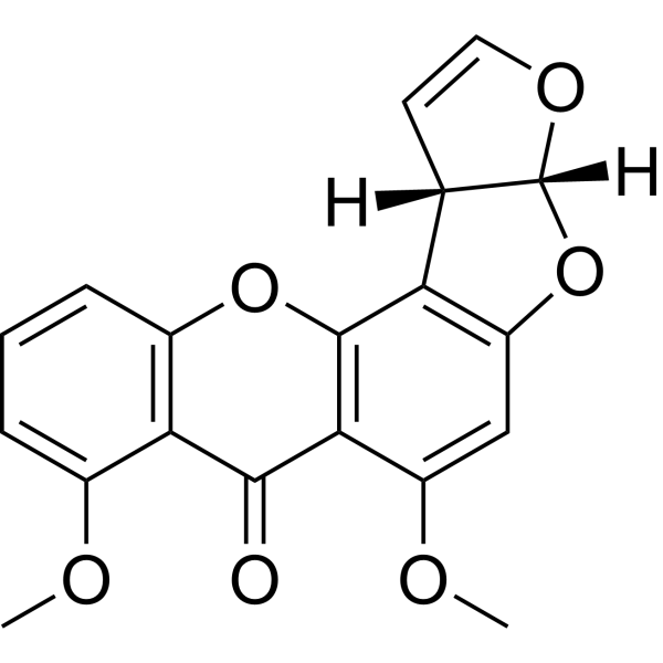
O-MethylsterigmatocystinFungal Metabolites
Antibiotic アイソフォーム固有の製品をすべて表示
More

生物活性
製品説明
Cellular Effect
|
Cell Line

|
Type | Value | Description | References |
|---|---|---|---|---|
| A549 | IC50 |
<9.7 μM
Compound: Roridin E
|
Cytotoxicity against human A549 cells after 72 hrs
Cytotoxicity against human A549 cells after 72 hrs
|
[PMID: 21907583] |
| BT-549 | IC50 |
uM μM
Compound: 4
|
Cytotoxicity against human BT549 cells assessed as growth inhibition after 72 hrs by SRB assay
Cytotoxicity against human BT549 cells assessed as growth inhibition after 72 hrs by SRB assay
|
[PMID: 30457333] |
| HCC70 | IC50 |
uM μM
Compound: 4
|
Cytotoxicity against human HCC70 cells assessed as growth inhibition after 72 hrs by SRB assay
Cytotoxicity against human HCC70 cells assessed as growth inhibition after 72 hrs by SRB assay
|
[PMID: 30457333] |
| HL-60 | IC50 |
0.3 ng/mL
Compound: 2
|
Cytotoxicity against human HL60 cells after 72 hrs by WST8 assay
Cytotoxicity against human HL60 cells after 72 hrs by WST8 assay
|
[PMID: 11277768] |
| K562 | IC50 |
0.54 μM
Compound: 11
|
Cytotoxicity against human K562 cells by disk diffusion soft agar colony formation assay
Cytotoxicity against human K562 cells by disk diffusion soft agar colony formation assay
|
[PMID: 31117520] |
| KB | EC50 |
0.5 ng/mL
Compound: 4
|
Cytotoxicity against human KB cells
Cytotoxicity against human KB cells
|
[PMID: 10075777] |
| L1210 | IC50 |
0.2 ng/mL
Compound: 2
|
Cytotoxicity against mouse L1210 cells after 72 hrs by WST8 assay
Cytotoxicity against mouse L1210 cells after 72 hrs by WST8 assay
|
[PMID: 11277768] |
| MDA-MB-231 | IC50 |
uM μM
Compound: 4
|
Cytotoxicity against human MDA-MB-231 cells assessed as growth inhibition after 72 hrs by SRB assay
Cytotoxicity against human MDA-MB-231 cells assessed as growth inhibition after 72 hrs by SRB assay
|
[PMID: 30457333] |
| MDA-MB-468 | IC50 |
uM μM
Compound: 4
|
Cytotoxicity against human MDA-MB-468 cells assessed as growth inhibition after 72 hrs by SRB assay
Cytotoxicity against human MDA-MB-468 cells assessed as growth inhibition after 72 hrs by SRB assay
|
[PMID: 30457333] |
| SW 1116 | IC50 |
0.039 μM
Compound: 11
|
Cytotoxicity against human SW1116 cells by disk diffusion soft agar colony formation assay
Cytotoxicity against human SW1116 cells by disk diffusion soft agar colony formation assay
|
[PMID: 31117520] |
| Vero | EC50 |
0.4 ng/mL
Compound: 4
|
Cytotoxicity against african green monkey Vero cells
Cytotoxicity against african green monkey Vero cells
|
[PMID: 10075777] |
体内実験
Roridin E (2 mg/kg; i.p.; single dose) induces significant hepatotoxicity in male albino mice, as evidenced by increased liver oxidative stress markers and altered serum liver function parameters[2].
MedChemExpress (MCE) has not independently confirmed the accuracy of these methods. They are for reference only.
-
Animal Model:Sprague-Dawley (male, 150-170 g)[1]
-
Dosage:2 mg/kg
-
Administration:s.c.; single dose
-
Result:Showed no significant changes in levels of TBARS, total lipids, glucose, glutathione, creatinine, zinc, or calcium in serum compared to controls.
Increased liver glucose-6-phosphatase activity to 10.6 nmol/min/mg proteins, with no significant changes in TBARS, total lipid, glutathione, 5-nucleotidase, or superoxide dismutase levels in liver compared to controls.
Increased kidney glutathione level to 6.76 μg/mg proteins, with no significant changes in TBARS, total lipid, 5-nucleotidase, glucose-6-phosphatase, or superoxide dismutase levels in kidney compared to controls.
-
Animal Model:albino mice (male, 6 weeks old, 20-25 g)[2]
-
Dosage:2 mg/kg
-
Administration:i.p.; single dose
-
Result:Increased liver tissue TBARs to 2.3 mmol/mL.
Increased liver tissue SOD to 1.9 ng/mL.
Increased liver tissue GSH to 2.9 mmol/mL.
Decreased blood serum total protein to 4.4 g/dl.
Increased blood serum total lipids to 5.8 mg/mL.
Increased blood serum GGT to 32.1 u/mL.
Increased blood serum alkaline phosphatase to 116 u/L.
Increased blood serum 5'-nucleotidase to 1.8 u/mL.


化学情報
-
CAS 番号 16891-85-3
-
性状 Solid
-
分子量 514.61
-
分子式 C29H38O8
-
Color White to off-white
-
SMILES
C[C@@]12[C@]3(CO3)[C@@]4([H])C[C@@]1([H])OC(/C=C\C=C\[C@@]([C@H](O)C)([H])OCC/C(C)=C/C(OC[C@]25[C@](C=C(C)CC5)([H])O4)=O)=O
-
Structure Classification
-
Initial Source
Myrothecium verrucaria
-
輸送条件
Room temperature in continental US; may vary elsewhere.
-
保管条件
Powder -20°C 3 years In solvent -80°C 6 months -20°C 1 month


純度とドキュメンテーション
-
データシート (277 KB)
-
SDS (393 KB)
- English - EN (393 KB)
- Français - FR (393 KB)
- Deutsch - DE (393 KB)
- Norwegian - NO (393 KB)
- Español - ES (393 KB)
- Swedish - SV (393 KB)
- Italian - IT (393 KB)
- Korean - KR (393 KB)
- Portuguese - PT (393 KB)
-
取扱説明書 (2659 KB)
参考文献

Calculators
濃度 (開始) × 体積 (開始) = 濃度 (終了) × 体積 (終了)


Roridin E 関連分類
HY-125539 関連分類
 Powered by Bioz