



Olprinone
Based on 1 publication(s) in Google Scholar
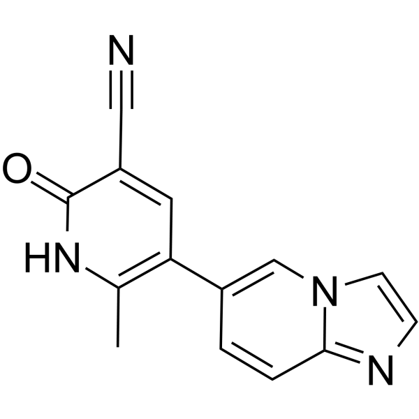
Olprinone (Loprinone) is a potent and selective phosphodiesterase 3 (PDE3) inhibitor, with IC50 values of 150, 100, 0.35, and 14 μM against PDE1, PDE2, PDE3, and PDE4, respectively. Olprinone exerts comprehensive protective effects including anti-inflammatory, antioxidant, and anti-apoptotic activities by elevating intracellular cAMP levels and inhibiting the NF-κB and MAPK signaling pathways. Olprinone can be used in studies related to lung injury, spinal cord injury, heart failure, myocardial ischemia-reperfusion injury, and cerebral ischemic injury.
연구목적의 판매만을 진행합니다. 환자를 대상으로 한 판매는 하지 않습니다.
- Purity : 99.58%
- CAS No.: 106730-54-5
- 화학식: C14H10N4O
- 분자량:250.26
-
보관:
4°C, protect from light
* In solvent : -80°C, 6 months; -20°C, 1 month (protect from light)

To place orders, for customer services and technical support, please contact: MedChemExpress USA
Tel: 609-228-6898 E-mail: [email protected] [email protected]


-
 Biological Activity
-
Chemical Information
-
용액&용해도
- Protocol
- 순도&문서
- References
-
 Help & FAQs


-
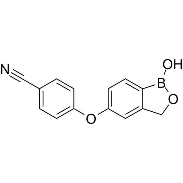
HY-10978
CrisaborolePDE Inhibitor -
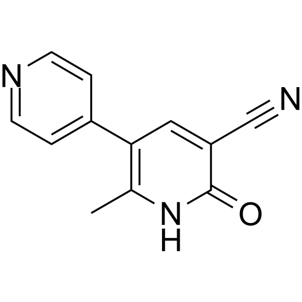
HY-14252
Milrinone -
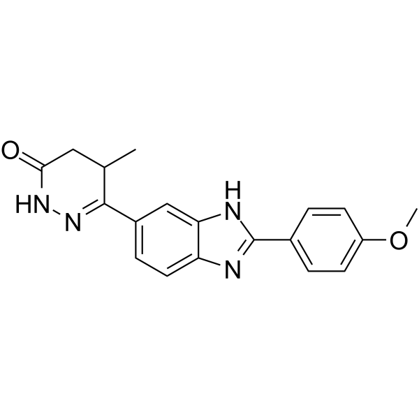
HY-B0204
PimobendanPDE Inhibitor -
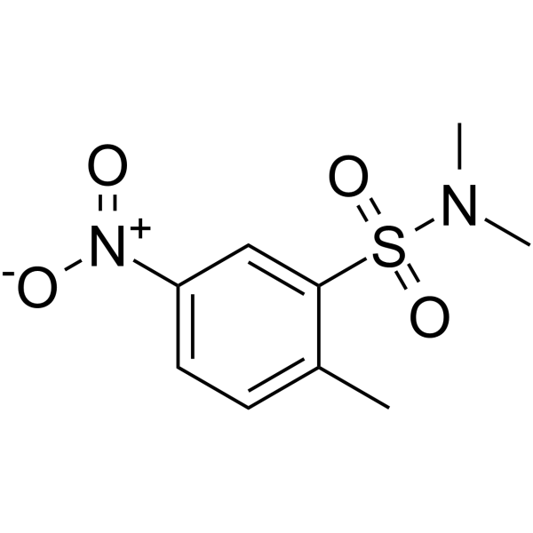
HY-109586
BRL-50481PDE Inhibitor -
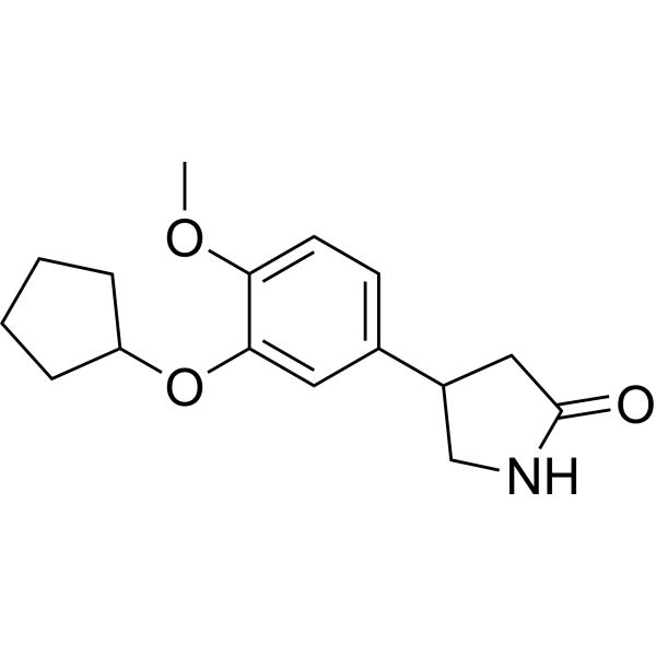
HY-16900
RolipramPDE4 Inhibitor -
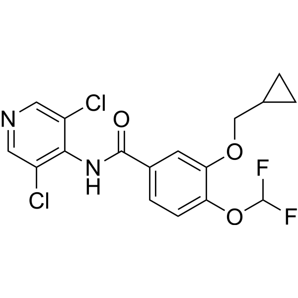
HY-15455
RoflumilastPDE4 Inhibitor -
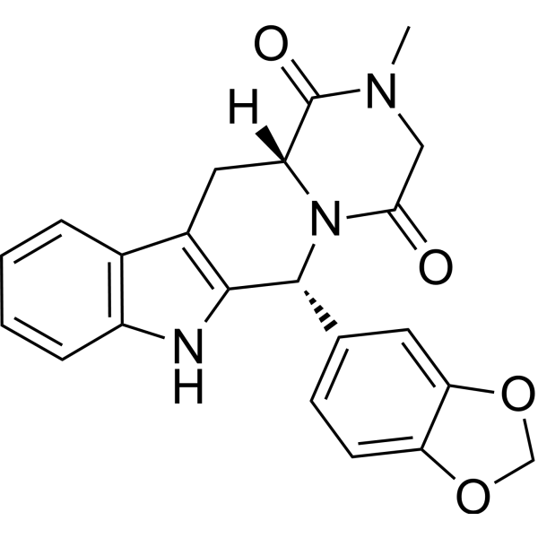
HY-90009A
TadalafilPDE5 Inhibitor -
HY-12085
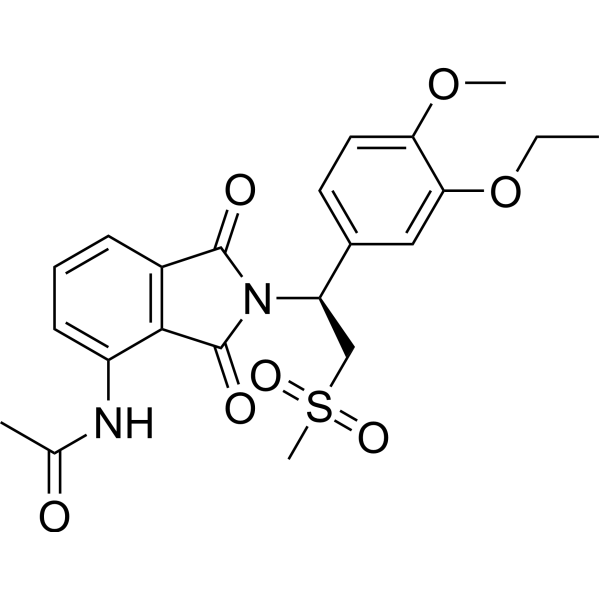
ApremilastPDE-4 Inhibitor -
HY-B0442
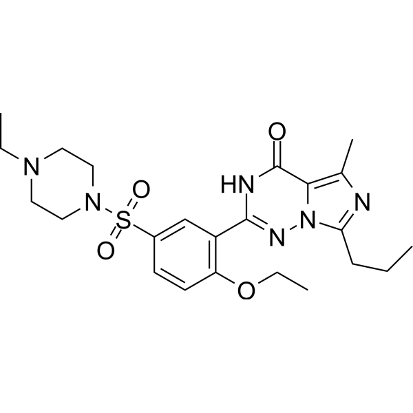
VardenafilPDE5 Inhibitor -
HY-10790
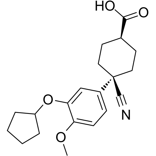
CilomilastPDE4 Inhibitor -
HY-B0140
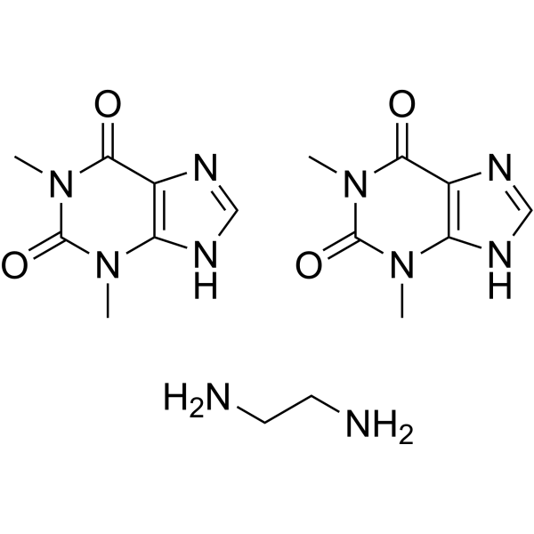
AminophyllinePDE3/4 Inhibitor/Adenosine Receptor Antagonist -
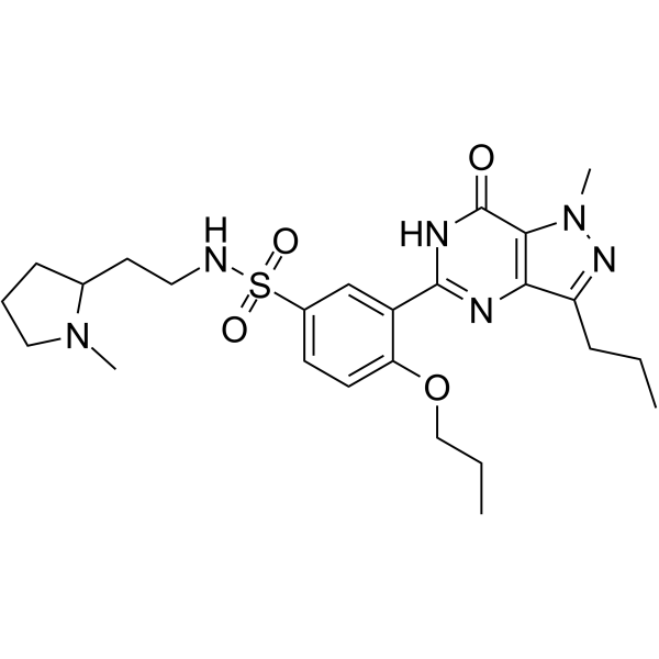
HY-18253
UdenafilPDE Inhibitor
Publications Citing Use of MedChemExpress (MCE) Olprinone
More

Biological Activity
제품 설명
IC50 & Target
[1]|
PDE1 150 μM (IC50) |
PDE2 100 μM (IC50) |
PDE3 0.35 μM (IC50) |
PDE4 14 μM (IC50) |
In Vitro
Olprinone (Loprinone) hydrochloride (10 mM; 48-72 h) hydrate inhibits LPS (HY-D1056)-induced production of TNF-α and IL-6, and promotes the production of IL-10, in isolated rat alveolar macrophages[2].
MedChemExpress (MCE) has not independently confirmed the accuracy of these methods. They are for reference only.
-
Cell Line:Primary rat alveolar macrophages
-
Concentration:10 mM
-
Incubation Time:48, 72 h
-
Result:Significantly suppressed the levels of TNF-α and IL-6 at 48 h and 72 h.
Augmented the production of IL-10 at 48 h and 72 h.
In Vivo
Olprinone (0.2 mg/kg; i.p.; administered at 1 h and 6 h post-injury, followed by daily administration until day 9 post-injury; observation for 10 days) hydrochloride hydrate exerts effects of alleviating spinal cord inflammation, reducing cell apoptosis and improving hindlimb motor function in a mouse model of spinal cord injury[3].
Olprinone (0.2 mg/kg; i.v.; single administration; observation for 5 h) hydrochloride hydrate exerts effects of alleviating pulmonary oxidative stress, reducing pulmonary edema and inflammatory cell infiltration in a rabbit model of acute lung injury induced by meconium aspiration syndrome[4].
Olprinone (0.2 mg/kg; i.p.; single administration) hydrochloride hydrate exerts effects of reducing myocardial infarction size, anti-inflammation and anti-apoptosis in a rat model of myocardial ischemic injury[5].
Olprinone (0.2 mg/kg; i.p.; single administration; 5 minutes before reperfusion) hydrochloride hydrate exerts effects of reducing cerebral infarction volume, improving neurological deficits, anti-inflammation and anti-apoptosis in a rat model of cerebral ischemia/reperfusion injury[6].
MedChemExpress (MCE) has not independently confirmed the accuracy of these methods. They are for reference only.
-
Animal Model:Wistar rats (male, 180-220 g, acute lung injury model via intravenous Escherichia coli serotype 055:B5 lipopolysaccharide injection at 5 mg/kg)[2]
-
Dosage:0.2 mg/kg
-
Administration:i.p.; single dose (30 minutes prior to LPS exposure); 6 h
-
Result:Significantly inhibited the LPS-induced neutrophil influx into the lungs.
Suppressed inflammatory cytokines TNF-α and IL-6 in the serum.
Augmented the production of the anti-inflammatory cytokine IL-10 in the serum.
-
Animal Model:CD1 mice (male adult, 25-30 g, spinal cord injury induced by extradural compression of T5-T8 spinal cord with 24 g closing force for 1 min after four-level T5-T8 laminectomy)[3]
-
Dosage:0.2 mg/kg
-
Administration:i.p.; 1 h and 6 h post-injury, then daily until day 9 post-injury (motor function assessment); 10 days
-
Result:Reduced the degree of spinal cord inflammation and tissue injury.
Attenuated neutrophil infiltration (reduced myeloperoxidase activity) and nitrotyrosine formation.
Decreased the expression of pro-inflammatory cytokines (TNF-α, IL-1β) and adhesion molecules (ICAM-1, P-selectin).
Inhibited NF-κB expression, p-ERK1/2, and p-p38 MAP kinase activation.
Decreased apoptosis, evident by reduced TUNEL staining, Fas ligand, and Bax expression, alongside preserved Bcl-2 expression.
Ameliorated the recovery of hind-limb function (BMS score).
-
Animal Model:Chinchilla rabbit (adult, 2.7 kg; meconium aspiration syndrome model via intratracheal meconium instillation)[4]
-
Dosage:0.2 mg/kg
-
Administration:i.v.; single dose; 5 h
-
Result:Reduced the numbers of neutrophils and eosinophils in the BAL fluid.
Decreased the formation of oxidation markers (conjugated dienes, TBARS, dityrosine, and lysine-lipid peroxidation products) in lung mitochondria.
Reduced lung edema (decreased wet/dry weight ratio) and prevented a decrease in total antioxidant status (TAS) in the lung homogenate and plasma.
Decreased TBARS levels in blood plasma and preserved cytochrome coxidase (COX) activity in the lung.
-
Animal Model:Wistar (male, 270-290 g, transient right hemisphere middle cerebral artery occlusion for 2 hours followed by 22 hours of reperfusion)[6]
-
Dosage:0.2 mg/kg
-
Administration:i.p.; single dose; 5 minutes before reperfusion
-
Result:Reduced the infarct volume and improved the neurological deficit score.
Blocked the acute turning behavior significantly.
Suppressed the formation of nitrotyrosine and the expression of iNOS, IL-1β, and ICAM-1 in ischemic tissues.
Reduced levels of apoptosis (decreased TUNEL-positive cells and Bax expression, and maintained Bcl-2 expression).


Chemical Information
-
CAS No. 106730-54-5
-
Appearance Solid
-
분자량 250.26
-
화학식 C14H10N4O
-
Color White to off-white
-
SMILES
CC(N1)=C(C=C(C#N)C1=O)C2=CN3C(C=C2)=NC=C3
-
Synonyms
Loprinone
-
선적
Room temperature in continental US; may vary elsewhere.
-
보관
4°C, protect from light
* In solvent : -80°C, 6 months; -20°C, 1 month (protect from light)


Publications (1)
-
Journal Impact Factor
-
Most Recent


용액&용해도
In Vitro:
DMSO : 20.83 mg/mL (83.23 mM; ultrasonic and warming and heat to 60°C; Hygroscopic DMSO has a significant impact on the solubility of product, please use newly opened DMSO)
Please refer to the solubility information to select the appropriate solvent. Once prepared, please aliquot and store the solution to prevent product inactivation from repeated freeze-thaw cycles.
Storage method and period of stock solution: -80°C, 6 months; -20°C, 1 month (protect from light). When stored at -80°C, please use it within 6 months. When stored at -20°C, please use it within 1 month.
Please refer to the solubility information to select the appropriate solvent. Once prepared, please aliquot and store the solution to prevent product inactivation from repeated freeze-thaw cycles.
Storage method and period of stock solution: -80°C, 6 months; -20°C, 1 month (protect from light). When stored at -80°C, please use it within 6 months. When stored at -20°C, please use it within 1 month.
Concentration (start) × Volume (start) = Concentration (final) × Volume (final)


순도&문서
-
Data Sheet (300 KB)
-
SDS (394 KB)
- English - EN (394 KB)
- Français - FR (394 KB)
- Deutsch - DE (394 KB)
- Norwegian - NO (394 KB)
- Español - ES (394 KB)
- Swedish - SV (394 KB)
- Italian - IT (394 KB)
- Korean - KR (394 KB)
- Portuguese - PT (394 KB)
-
Handling Instructions (2659 KB)
References
[1].
Sugioka M, et al. Identification and characterization of isoenzymes of cyclic nucleotide phosphodiesterase in human kidney and heart, and the effects of new cardiotonic agents on these isoenzymes. Naunyn Schmiedebergs Arch Pharmacol. 1994 Sep;350(3):284-93.
[Content Brief]
[2]. Koike T, et al. Pretreatment with olprinone hydrochloride, a phosphodiesterase III inhibitor, attenuates lipopolysaccharide-induced lung injury via an anti-inflammatory effect. Pulmonary pharmacology & therapeutics. 2008;21(1):166-71. [Content Brief]
[3]. Esposito E, et al. Olprinone attenuates the acute inflammatory response and apoptosis after spinal cord trauma in mice. PloS one. 2010 Sep 07;5(9):e12170. [Content Brief]
[4]. Mokra D, et al. Selective phosphodiesterase 3 inhibitor olprinone attenuates meconium-induced oxidative lung injury. Pulmonary pharmacology & therapeutics. 2012 Jun;25(3):216-22. [Content Brief]
[5]. Di Paola R, et al. Olprinone, a PDE3 inhibitor, modulates the inflammation associated with myocardial ischemia-reperfusion injury in rats. European journal of pharmacology. 2011 Jan 15;650(2-3):612-20. [Content Brief]
[6]. Genovese T, et al. Neuroprotective effects of olprinone after cerebral ischemia/reperfusion injury in rats. Neuroscience letters. 2011 Oct 03;503(2):93-9. [Content Brief]


Complete Stock Solution Preparation Table
Please refer to the solubility information to select the appropriate solvent. Once prepared, please aliquot and store the solution to prevent product inactivation from repeated freeze-thaw cycles.
Storage method and period of stock solution: -80°C, 6 months; -20°C, 1 month (protect from light). When stored at -80°C, please use it within 6 months. When stored at -20°C, please use it within 1 month.
| Optional Solvent | Concentration Solvent Mass | 1 mg | 5 mg | 10 mg | 25 mg |
|---|---|---|---|---|---|
| DMSO | 1 mM | 3.9958 mL | 19.9792 mL | 39.9584 mL | 99.8961 mL |
| 5 mM | 0.7992 mL | 3.9958 mL | 7.9917 mL | 19.9792 mL | |
| 10 mM | 0.3996 mL | 1.9979 mL | 3.9958 mL | 9.9896 mL | |
| 15 mM | 0.2664 mL | 1.3319 mL | 2.6639 mL | 6.6597 mL | |
| 20 mM | 0.1998 mL | 0.9990 mL | 1.9979 mL | 4.9948 mL | |
| 25 mM | 0.1598 mL | 0.7992 mL | 1.5983 mL | 3.9958 mL | |
| 30 mM | 0.1332 mL | 0.6660 mL | 1.3319 mL | 3.3299 mL | |
| 40 mM | 0.0999 mL | 0.4995 mL | 0.9990 mL | 2.4974 mL | |
| 50 mM | 0.0799 mL | 0.3996 mL | 0.7992 mL | 1.9979 mL | |
| 60 mM | 0.0666 mL | 0.3330 mL | 0.6660 mL | 1.6649 mL | |
| 80 mM | 0.0499 mL | 0.2497 mL | 0.4995 mL | 1.2487 mL |


Olprinone Related Classifications
HY-14254A Related Classifications
 Powered by Bioz
Keywords