



Aurein 2.6
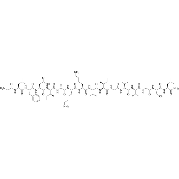
Aurein 2.6 is an antibiotic antimicrobial peptide. Aurein 2.6 is active against Gram-positive bacterial (MIC: 25, 25, 30, 25, 30 μM for M. luteus, S. aureus, S. epidermis, S. mutans, B. subtilis).
연구목적의 판매만을 진행합니다. 환자를 대상으로 한 판매는 하지 않습니다.
- CAS No.: 302343-10-8
- 화학식: C77H133N19O19
- 분자량:1629.00
-
보관:
Please store the product under the recommended conditions in the Certificate of Analysis.

To place orders, for customer services and technical support, please contact: MedChemExpress USA
Tel: 609-228-6898 E-mail: [email protected] [email protected]


-
 Biological Activity
-
Chemical Information
- Protocol
- 순도&문서
- References
-
 Help & FAQs


All Antibiotic Isoforms
More

Biological Activity
제품 설명


Chemical Information
-
CAS No. 302343-10-8
-
분자량 1629.00
-
화학식 C77H133N19O19
-
Sequence
Gly-Leu-Phe-Asp-Ile-Ala-Lys-Lys-Val-Ile-Gly-Val-Ile-Gly-Ser-Leu-NH2
-
Sequence Shortening
GLFDIAKKVIGVIGSL-NH2
-
선적
Room temperature in continental US; may vary elsewhere.
-
보관
Please store the product under the recommended conditions in the Certificate of Analysis.


순도&문서
References
[1]. Rozek T, et al. The antibiotic and anticancer active aurein peptides from the Australian Bell Frogs Litoria aurea and Litoria raniformis the solution structure of aurein 1.2. Eur J Biochem. 2000 Sep;267(17):5330-41. [Content Brief]
[2]. Dennison SR, et al. PEGylation enhances the antibacterial and therapeutic potential of amphibian host defence peptides. Biochim Biophys Acta Biomembr. 2022 Feb 1;1864(1):183806. [Content Brief]

Calculators
Concentration (start) × Volume (start) = Concentration (final) × Volume (final)


Aurein 2.6 Related Classifications
HY-P5573 Related Classifications
 Powered by Bioz