
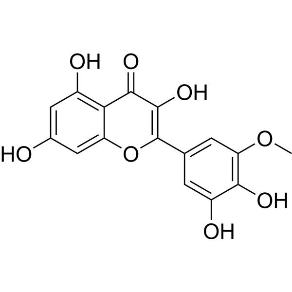
Laricitrin
Based on 1 Customer Validation
Laricitrin is a natural product with multiple biological activities. Laricitrin inhibits the phosphorylation and DNA-binding activity of STAT3, and downregulates the expression of IL-10. Laricitrin acts as an immunomodulator to regulate the differentiation, maturation and function of dendritic cells (DCs). Laricitrin inhibits the migration and invasion of lung cancer cells, and ameliorates lung cancer progression. Laricitrin can be used in research related to hypertension and lung cancer.
For research use only. We do not sell to patients.
- Purity : 99.36%
- CAS No.: 53472-37-0
- Formula: C16H12O8
- Molecular Weight:332.26
-
Storage:
4°C, sealed storage, away from moisture and light
* In solvent : -80°C, 6 months; -20°C, 1 month (sealed storage, away from moisture and light)
Biological Activity
Description
IC50 & Target
[1]|
STAT3 |
IL-10 |
IL-12 |
In Vitro
Laricitrin binds to regions adjacent to the ACE active site, active site residues of renin, and core active site residues of ATR; it inhibits LPS-induced phosphorylation and DNA-binding activity of STAT3 in human CD14+ monocytes, and downregulates the expression of IL-10[1].
Laricitrin (2 μM; 5 days) restores the differentiation, maturation and cytokine balance (increased IL-12, decreased IL-10) of human CD14+ monocyte-derived dendritic cells impaired by the conditioned medium of A549 or CL1-5 lung cancer cells[1].
Laricitrin (2 μM) restores the T cell activation function of dendritic cells treated with conditioned medium from human A549 or CL1-5 lung cancer cells, promotes the proliferation of naive CD4+ T cells, and shifts the cytokine secretion profile from a Th2-type to a Th1-type pattern[1].
Laricitrin (2 µM; 1 h pretreatment of dendritic cells followed by 24-48 h chemoattractant exposure) inhibits the migration of human lung adenocarcinoma H1395 and HCC2935 cells induced by H1395-TADC-CM, BaP-H1395-TADC-CM, HCC2935-TADC-CM, and BaP-HCC2935-TADC-CM[2].
MedChemExpress (MCE) has not independently confirmed the accuracy of these methods. They are for reference only. Further protocols information, click here.
-
Cell Line:human lung adenocarcinoma H1395 cells, human lung adenocarcinoma HCC2935 cells
-
Concentration:2 µM (pretreated on dendritic cells)
-
Incubation Time:1 h (dendritic cell pretreatment); 24, 48 h (conditioned media incubation as chemoattractant)
-
Result:Inhibited the migration of human lung adenocarcinoma H1395 and HCC2935 cells induced by H1395-TADC-CM, BaP-H1395-TADC-CM, HCC2935-TADC-CM, and BaP-HCC2935-TADC-CM.
In Vivo
Laricitrin (30 mg/kg; i.p.; 3 times over 5 days) potentiates the anticancer activity of Cisplatin (HY-17394) (2.5 mg/kg; i.p.; weekly) in LLC-bearing mice, resulting in fewer lung tumor nodules than Cisplatin treatment alone[1].
MedChemExpress (MCE) has not independently confirmed the accuracy of these methods. They are for reference only.
Chemical Information
-
CAS No. 53472-37-0
-
Appearance Solid
-
Molecular Weight 332.26
-
Formula C16H12O8
-
Color Light yellow to yellow
-
SMILES
O=C1C(O)=C(C2=CC(OC)=C(O)C(O)=C2)OC3=CC(O)=CC(O)=C13
-
Structure Classification
-
Initial Source
-
Shipping
Room temperature in continental US; may vary elsewhere.
-
Storage
4°C, sealed storage, away from moisture and light
* In solvent : -80°C, 6 months; -20°C, 1 month (sealed storage, away from moisture and light)
Protocols
-
Kinase activity and phosphorylation assays
Kinase activity assays measure the ability of kinases to transfer phosphate groups from ATP to specific substrates, while phosphorylation assays detect the presence and levels of phosphorylated proteins. Common methods include radiolabeled ATP incorporation (e. g. ,), ADP release detection via bioluminescence (e. g. ,[3]), enzyme-linked immunosorbent assays (ELISA) for phospho-specific epitopes (e. g. ,[6]), and microtiter-based formats for high-throughput screening (e. g. ,[8]). The ADP-Glo assay quantifies kinase activity by measuring ADP produced during phosphorylation using a luciferase-based system. Radiometric assays involve autoradiography or scintillation counting after incorporation of 32P-labeled ATP into substrate proteins. ELISA-based approaches rely on phospho-specific antibodies to detect activated kinases in cell lysates or purified samples.
-
Western Blot
Western blotting (WB) is a commonly used experimental method in molecular biology, biochemistry, and immunogenetics for identifying and quantifying target proteins. It combines gel electrophoresis with immunoassay, enabling researchers to analyze protein expression, post-translational modifications, and molecular weight.
-
RNA extraction experimental
By lysing cells, releasing RNA, and removing impurities such as proteins and DNA, high-purity RNA products are finally obtained. The commonly used traditional method is the guanidine isothiocyanate/phenol/chloroform method (Trizol), which is suitable for a variety of animal materials including animal tissues, microorganisms, cultured cells, etc., and most plant materials.
-
Cell migration
Cell migration is a method that plays an important role in wound healing, cell differentiation, embryonic development, etc.
-
Cell invasion
Cell invasion is the ability of cells to migrate from one area to another via the extracellular matrix. Cell invasion is the response of normal and cancer cells to chemical and mechanical stimuli. Before migrating to a new region, the extracellular matrix is degraded by proteases within the cell. Cell invasion often occurs during wound repair, vascularization and inflammation, abnormal tissue invasion, and tumor cell metastasis.
-
Research Protocol for Cardiovascular Diseases
Cardiovascular disease can be modeled as maladaptive cardiac remodeling, where ischemic injury or pressure overload activates inflammatory signaling, fibroblast activation, extracellular-matrix deposition, cardiomyocyte hypertrophy, vascular remodeling, and progressive ventricular dysfunction. The TGF-β/SMAD axis is a central profibrotic pathway after myocardial injury and pressure overload, while innate immune and cytokine pathways regulate leukocyte recruitment, scar formation, and adverse remodeling. Key unresolved questions include which inflammatory signals are reparative versus harmful, when fibrosis is protective versus maladaptive, and whether pathway inhibition improves function without weakening necessary infarct healing or compensatory remodeling.
-
Protocol for Kinase activity and phosphorylation assays
Kinase activity assays measure transfer of phosphate from ATP to a protein or peptide substrate, generating phosphorylated substrate, ADP, or incorporated radiolabeled phosphate as the readout; phosphorylation assays measure site-specific phosphorylation in cells or tissues as a proxy for kinase-pathway activation, inhibition, or substrate regulation. Phosphorylation can be detected by phospho-specific Western blot, immunoprecipitation kinase assay, phospho-immunofluorescence, phospho-flow cytometry, luminescent ADP detection, radiolabeled ATP incorporation, or reporter-based pathway assays, and these readouts can be applied to cancer cells, primary neurons, mouse tumors, organoids, inflammatory macrophages, ferroptosis studies, and mitophagy studies when the kinase target is biologically relevant.
-
Monocyte-derived dendritic cell differentiation
Human monocyte-derived dendritic cells are generated by isolating PBMC-derived monocytes and culturing them with GM-CSF plus IL-4, which produces cells with dendritic-cell antigen-presenting properties, reduced monocyte phenotype, and increased dendritic-cell functional readouts such as antigen uptake, allogeneic T-cell stimulation, and expression of markers including HLA-DR, CD80, CD86, CD83, CD1a, or CD209 depending on protocol and maturation state. The main readout is phenotypic and functional differentiation: immature MoDCs are commonly evaluated by loss or reduction of CD14 with acquisition of dendritic-cell markers and antigen uptake capacity, whereas mature MoDCs are evaluated by increased CD83, CD80, CD86, HLA-DR, and T-cell stimulatory function after exposure to maturation stimuli such as TNF-α or a cytokine/PGE2 cocktail.
Purity & Documentation
-
Data Sheet (284 KB)
-
SDS (252 KB)
- English - EN (252 KB)
- Français - FR (252 KB)
- Deutsch - DE (252 KB)
- Norwegian - NO (252 KB)
- Español - ES (252 KB)
- Swedish - SV (252 KB)
- Italian - IT (252 KB)
- Korean - KR (252 KB)
- Portuguese - PT (252 KB)
-
Handling Instructions (2659 KB)
References
[1]. Chang WA, et al. Laricitrin ameliorates lung cancer-mediated dendritic cell suppression by inhibiting signal transducer and activator of transcription 3. Oncotarget. 2016 Dec 20;7(51):85220-85234. [Content Brief]
[2]. Chang WA, et al. Laricitrin suppresses increased benzo(a)pyrene-induced lung tumor-associated monocyte-derived dendritic cell cancer progression. Oncology letters. 2016 Mar;11(3):1783-1790. [Content Brief]
Calculators
Concentration (start) × Volume (start) = Concentration (final) × Volume (final)

