



Mavodelpar

Based on 1 Customer Validation
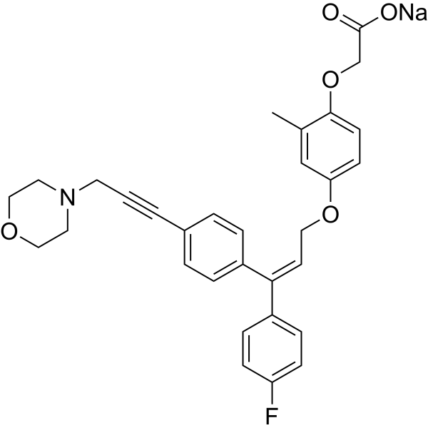
Mavodelpar (REN001) is a selective PPARδ agonist. Mavodelpar suppresses glomerular injury and renal fibrosis. Mavodelpar can be used for the research of primary mitochondrial myopathies (PMM) and long-chain fatty acid oxidation disorders (LC-FAOD). Mavodelpar is a click chemistry reagent, it contains an Alkyne group and can undergo copper-catalyzed azide-alkyne cycloaddition (CuAAc) with molecules containing Azide groups.
연구목적의 판매만을 진행합니다. 환자를 대상으로 한 판매는 하지 않습니다.
- Purity : 99.93%
- CAS No.: 1604815-32-8
- 화학식: C31H29FNNaO5
- 분자량:537.55
-
보관:
4°C, sealed storage, away from moisture
* In solvent : -80°C, 6 months; -20°C, 1 month (sealed storage, away from moisture)

To place orders, for customer services and technical support, please contact: MedChemExpress USA
Tel: 609-228-6898 E-mail: [email protected] [email protected]


-
 Biological Activity
-
Chemical Information
-
용액&용해도
- Protocol
- 순도&문서
- References
-
 Help & FAQs




Biological Activity
제품 설명
In Vivo
MedChemExpress (MCE) has not independently confirmed the accuracy of these methods. They are for reference only.
-
Animal Model:Male and female B6129SF1-Col4a3-/- mice[1]
-
Dosage:10 mg/kg
-
Administration:Intraperitoneal injection; 10 mg/kg, once daily, from 6 to 17 weeks of age
-
Result:Suppressed proteinuria and blood urea nitrogen (BUN) levels. Reduced glomerular injury, renal fibrosis, phosho-Stat3 and connective tissue growth factor (CTGF) levels. Decreased the expression level of the activated fibroblast marker alpha-SMA and Collagen I and IV.


Chemical Information
-
CAS No. 1604815-32-8
-
Appearance Solid
-
분자량 537.55
-
화학식 C31H29FNNaO5
-
Color White to off-white
-
SMILES
O=C(O[Na])COC1=CC=C(OC/C=C(C2=CC=C(F)C=C2)\C3=CC=C(C#CCN4CCOCC4)C=C3)C=C1C
-
Synonyms
REN001; HPP593
-
선적
Room temperature in continental US; may vary elsewhere.
-
보관
4°C, sealed storage, away from moisture
* In solvent : -80°C, 6 months; -20°C, 1 month (sealed storage, away from moisture)


용액&용해도
In Vitro:
DMSO : 100 mg/mL (186.03 mM; Need ultrasonic; Hygroscopic DMSO has a significant impact on the solubility of product, please use newly opened DMSO)
Please refer to the solubility information to select the appropriate solvent. Once prepared, please aliquot and store the solution to prevent product inactivation from repeated freeze-thaw cycles.
Storage method and period of stock solution: -80°C, 6 months; -20°C, 1 month (sealed storage, away from moisture). When stored at -80°C, please use it within 6 months. When stored at -20°C, please use it within 1 month.
Please refer to the solubility information to select the appropriate solvent. Once prepared, please aliquot and store the solution to prevent product inactivation from repeated freeze-thaw cycles.
Storage method and period of stock solution: -80°C, 6 months; -20°C, 1 month (sealed storage, away from moisture). When stored at -80°C, please use it within 6 months. When stored at -20°C, please use it within 1 month.
Concentration (start) × Volume (start) = Concentration (final) × Volume (final)
In Vivo:
Select the appropriate dissolution method based on your experimental animal and administration route.
- For the following dissolution methods, please ensure to first prepare a clear stock solution using an In Vitro approach and then sequentially add co-solvents:
- To ensure reliable experimental results, the clarified stock solution can be appropriately stored based on storage conditions. As for the working solution for In Vivo experiments, it is recommended to prepare freshly and use it on the same day.
- The percentages shown for the solvents indicate their volumetric ratio in the final prepared solution. If precipitation or phase separation occurs during preparation, heat and/or sonication can be used to aid dissolution.
Add each solvent one by one: 10% DMSO 40% PEG300 5% Tween-80 45% Saline
Solubility: ≥ 2.5 mg/mL (4.65 mM); Clear solution
This protocol yields a clear solution of ≥ 2.5 mg/mL (saturation unknown).
Taking 1 mL working solution as an example, add 100 μL DMSO stock solution (25.0 mg/mL) to 400 μL PEG300, and mix evenly; then add 50 μL Tween-80 and mix evenly; then add 450 μL Saline to adjust the volume to 1 mL.
Preparation of Saline: Dissolve 0.9 g sodium chloride in ddH₂O and dilute to 100 mL to obtain a clear Saline solution.
Add each solvent one by one: 10% DMSO 90% (20% SBE-β-CD in Saline)
Solubility: ≥ 2.5 mg/mL (4.65 mM); Clear solution
This protocol yields a clear solution of ≥ 2.5 mg/mL (saturation unknown).
Taking 1 mL working solution as an example, add 100 μL DMSO stock solution (25.0 mg/mL) to 900 μL 20% SBE-β-CD in Saline, and mix evenly.
Preparation of 20% SBE-β-CD in Saline (4°C, storage for one week): 2 g SBE-β-CD powder is dissolved in 10 mL Saline, completely dissolve until clear.
In Vivo Dissolution Calculator
Please enter the basic information of animal experiments:
-
-
-
-
Recommended: Prepare an additional quantity of animals to account for potential losses during experiments.
Please enter your animal formula composition:
-
%DMSO +
Recommended: Keep the proportion of DMSO in working solution below 2% if your animal is weak.
-
%+
-
+%Tween-80 + +
-
%Saline +
The co-solvents required include: DMSO, . All of co-solvents are available by MedChemExpress (MCE). , Tween 80. All of co-solvents are available by MedChemExpress (MCE).
Working solution concentration: 0.22 mg/mL
Method for preparing stock solution: mg drug dissolved in μL DMSO. Stock solution concentration: mg/mL. * In solvent : -80°C, 6 months; -20°C, 1 month (sealed storage, away from moisture)
1. Take μL DMSO stock solution;
2. Add μL .
μL , mix evenly;
3. Then add μL Tween 80, mix evenly;
4. Then add μL
Please ensure that the stock solution in the first step is dissolved to a clear state, and add co-solvents in sequence. You can use ultrasonic heating (ultrasonic cleaner, recommended frequency 20-40 kHz), vortexing, etc. to assist dissolution.


순도&문서
-
Data Sheet (274 KB)
-
SDS (252 KB)
- English - EN (252 KB)
- Français - FR (252 KB)
- Deutsch - DE (252 KB)
- Norwegian - NO (252 KB)
- Español - ES (252 KB)
- Swedish - SV (252 KB)
- Italian - IT (252 KB)
- Korean - KR (252 KB)
- Portuguese - PT (252 KB)
-
Handling Instructions (2659 KB)
References


Complete Stock Solution Preparation Table
Please refer to the solubility information to select the appropriate solvent. Once prepared, please aliquot and store the solution to prevent product inactivation from repeated freeze-thaw cycles.
Storage method and period of stock solution: -80°C, 6 months; -20°C, 1 month (sealed storage, away from moisture). When stored at -80°C, please use it within 6 months. When stored at -20°C, please use it within 1 month.
| Optional Solvent | Concentration Solvent Mass | 1 mg | 5 mg | 10 mg | 25 mg |
|---|---|---|---|---|---|
| DMSO | 1 mM | 1.8603 mL | 9.3015 mL | 18.6029 mL | 46.5073 mL |
| 5 mM | 0.3721 mL | 1.8603 mL | 3.7206 mL | 9.3015 mL | |
| 10 mM | 0.1860 mL | 0.9301 mL | 1.8603 mL | 4.6507 mL | |
| 15 mM | 0.1240 mL | 0.6201 mL | 1.2402 mL | 3.1005 mL | |
| 20 mM | 0.0930 mL | 0.4651 mL | 0.9301 mL | 2.3254 mL | |
| 25 mM | 0.0744 mL | 0.3721 mL | 0.7441 mL | 1.8603 mL | |
| 30 mM | 0.0620 mL | 0.3100 mL | 0.6201 mL | 1.5502 mL | |
| 40 mM | 0.0465 mL | 0.2325 mL | 0.4651 mL | 1.1627 mL | |
| 50 mM | 0.0372 mL | 0.1860 mL | 0.3721 mL | 0.9301 mL | |
| 60 mM | 0.0310 mL | 0.1550 mL | 0.3100 mL | 0.7751 mL | |
| 80 mM | 0.0233 mL | 0.1163 mL | 0.2325 mL | 0.5813 mL | |
| 100 mM | 0.0186 mL | 0.0930 mL | 0.1860 mL | 0.4651 mL |


Mavodelpar Related Classifications
HY-112597A Related Classifications
 Powered by Bioz