



Methionine-d3

Based on 1 Customer Validation
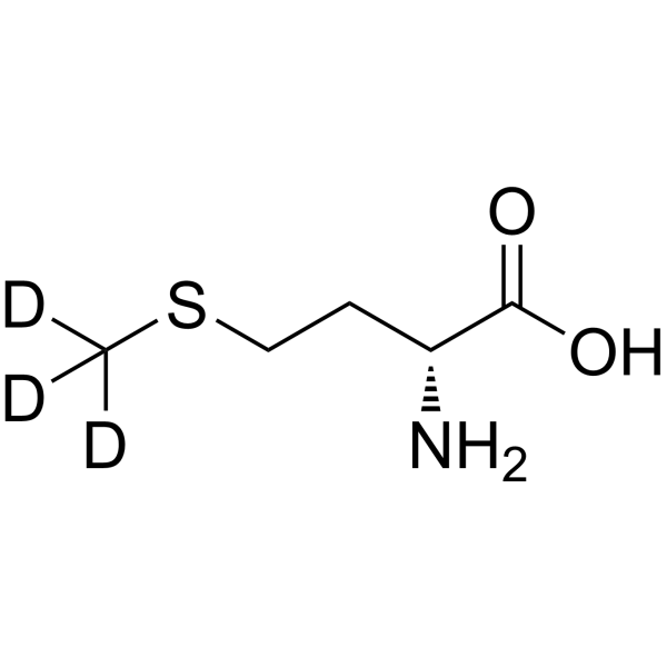
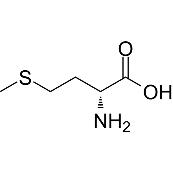
Methionine-d3 is the deuterium labeled Methionine. Methionine (MRX-1024; D-Methionine) is an effective chemoprotective agent which can also inhibit the neuronal activity through GABAA receptor activation.
For research use only. We do not sell to patients.
- Purity : 99.22%
- CAS No.: 284665-18-5
- Formula: C5H8D3NO2S
- Molecular Weight:152.23
-
Storage:Powder -20°C, 3 years , 4°C, 2 years ; In solvent -80°C, 6 months , -20°C, 1 month

To place orders, for customer services and technical support, please contact: MedChemExpress USA
Tel: 609-228-6898 E-mail: [email protected] [email protected]


-
 Biological Activity
-
Chemical Information
- Protocols
- Purity & Documentation
- References
-
 Help & FAQs


All Endogenous Metabolite Isoforms
More

Biological Activity
Description
In Vitro
Stable heavy isotopes of hydrogen, carbon, and other elements have been incorporated into drug molecules, largely as tracers for quantitation during the drug development process. Deuteration has gained attention because of its potential to affect the pharmacokinetic and metabolic profiles of drugs[1].
MedChemExpress (MCE) has not independently confirmed the accuracy of these methods. They are for reference only. Further protocols information, click here.
Application
1. This compound can be used as a tracer
2. This compound can be used as an internal standard for quantitative analysis by NMR, GC-MS, or LC-MS.


Chemical Information
-
CAS No. 284665-18-5
-
Unlabeled CAS 348-67-4
-
Appearance Solid
-
Molecular Weight 152.23
-
Formula C5H8D3NO2S
-
Color White to off-white
-
SMILES
OC([C@H](N)CCSC([2H])([2H])[2H])=O
-
Synonyms
MRX-1024-d3; D-Methionine-d3
-
Shipping
Room temperature in continental US; may vary elsewhere.
-
Storage
Powder -20°C 3 years 4°C 2 years In solvent -80°C 6 months -20°C 1 month

Protocols
-
Neuronal voltage-sensitive dye imaging
Neuronal voltage-sensitive dye imaging detects membrane-potential-dependent optical changes from dyes associated with neuronal membranes, enabling optical recording of electrical activity from single neurons, dendrites, axons, spines, or neuronal populations in brain slices and cultured neurons. VSD signals are typically reported as fractional fluorescence or absorbance changes over baseline, such as ΔF/F or ΔI/I, and published protocols use high-speed cameras or photodiode arrays because neuronal voltage signals occur on millisecond time scales. Fast VSD imaging can be applied at two common scales: bulk staining of brain slices to measure circuit-level spatiotemporal activity, and single-cell loading or biolistic delivery to record membrane-potential transients from individual neuronal compartments. Optical signals should be interpreted as membrane-potential-related readouts, and validation by simultaneous electrophysiology or pharmacological controls is recommended when the experimen


Purity & Documentation
-
Data Sheet (267 KB)
-
SDS (394 KB)
- English - EN (394 KB)
- Français - FR (394 KB)
- Deutsch - DE (394 KB)
- Norwegian - NO (394 KB)
- Español - ES (394 KB)
- Swedish - SV (394 KB)
- Italian - IT (394 KB)
- Korean - KR (394 KB)
- Portuguese - PT (394 KB)
-
Handling Instructions (2659 KB)
References
[1]. Russak EM, et al. Impact of Deuterium Substitution on the Pharmacokinetics of Pharmaceuticals. Ann Pharmacother. 2019;53(2):211-216. [Content Brief]
[2]. Wu C, et al. Antioxidants L-carnitine and D-methionine modulate neuronal activity through GABAergic inhibition. J Neural Transm (Vienna). 2014 Jul;121(7):683-93. [Content Brief]
[3]. Sooriyaarachchi M, et al. Chemoprotection by D-methionine against cisplatin-induced side-effects: insight from in vitrostudies using human plasma. Metallomics. 2014 Mar;6(3):532-41. [Content Brief]
[4]. Hinduja S, et al. D-methionine protects against cisplatin-induced neurotoxicity in the hippocampus of the adult rat. Neurotox Res. 2015 Apr;27(3):199-204. [Content Brief]

Calculators
Concentration (start) × Volume (start) = Concentration (final) × Volume (final)


Methionine-d3 Related Classifications
HY-13694S Related Classifications
 Powered by Bioz