



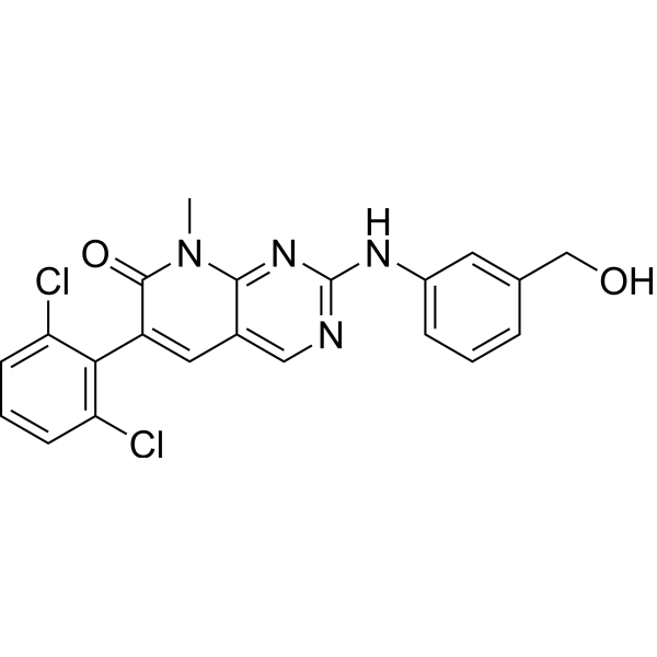
PD166326

Based on 1 Customer Validation
PD166326 is an orally active tyrosine kinase inhibitor with a IC50 of 8 nM against abl tyrosine kinase and a IC50 of 6 nM against src tyrosine kinase. PD166326 blocks Bcr/Abl kinase activity. PD166326 inhibits Bcr/Abl-dependent proliferation and cell cycle progression. PD166326 reduces peripheral blood granulocytosis, alleviates splenomegaly and prolongs survival in a mouse model of chronic myeloid leukemia. PD166326 can be used in research related to chronic myeloid leukemia.
For research use only. We do not sell to patients.
- Purity : 99.99%
- CAS No.: 185039-91-2
- Formula: C21H16Cl2N4O2
- Molecular Weight:427.28
-
Storage:
4°C, sealed storage, away from moisture and light
* In solvent : -80°C, 6 months; -20°C, 1 month (sealed storage, away from moisture and light)

To place orders, for customer services and technical support, please contact: MedChemExpress USA
Tel: 609-228-6898 E-mail: [email protected] [email protected]


-
 Biological Activity
-
Chemical Information
-
Solvent & Solubility
- Protocols
- Purity & Documentation
- References
-
 Help & FAQs


-
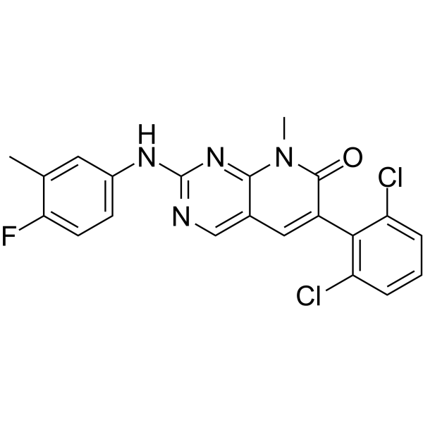
HY-103274
PD180970p210Bcr-Abl Kinase Inhibitor -
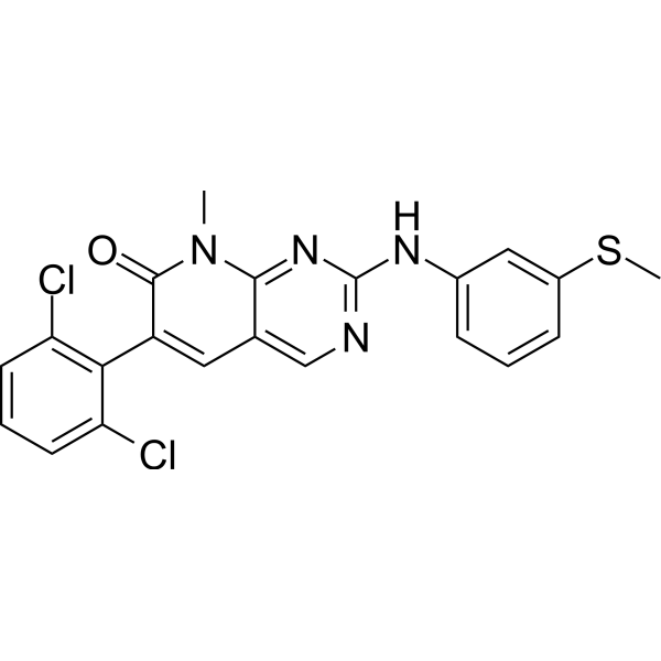
HY-10395
PD173955Src Inhibitor -
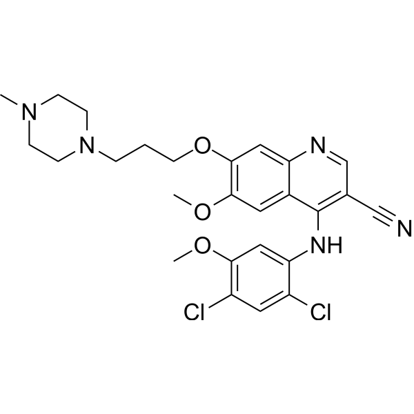
HY-10158
BosutinibSrc/Abl Inhibitor -
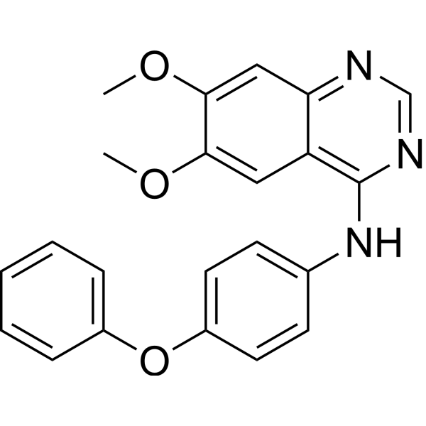
HY-101053
Src Inhibitor 1Src Inhibitor -
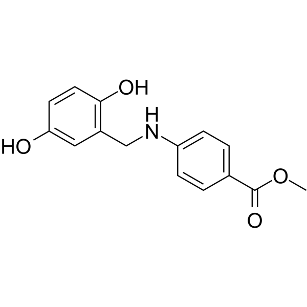
HY-117718
AG957Bcr/Abl Inhibitor -
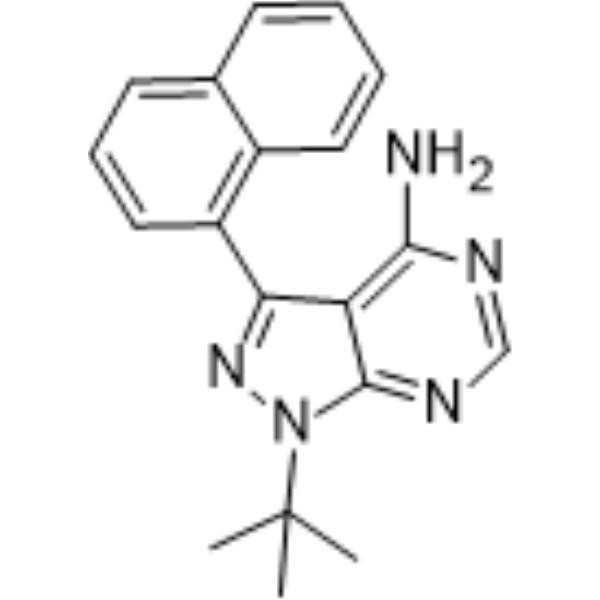
HY-13941
1-Naphthyl PP1Src Inhibitor -
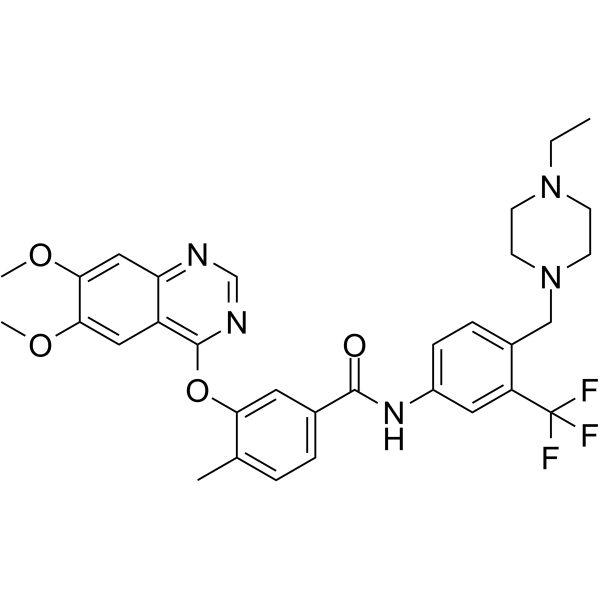
HY-112852
TL02-59Fgr Inhibitor -
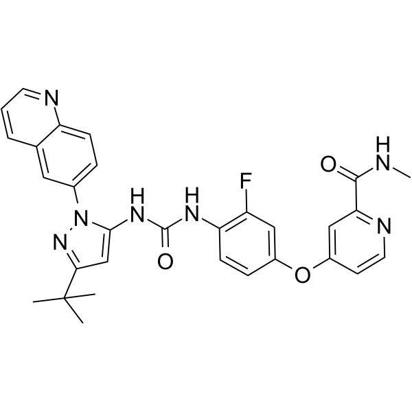
HY-13024
RebastinibKinase Inhibitor -
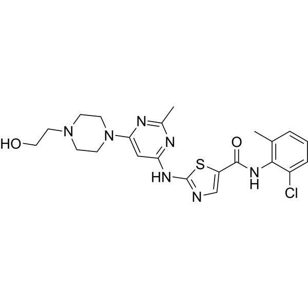
HY-10181
DasatinibSrc/Bcr-Abl Inhibitor -
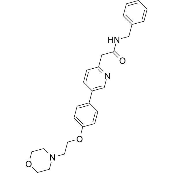
HY-10340
TirbanibulinSrc Inhibitor -
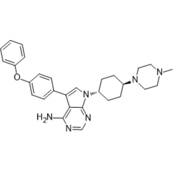
HY-15764
A 419259Src Inhibitor -
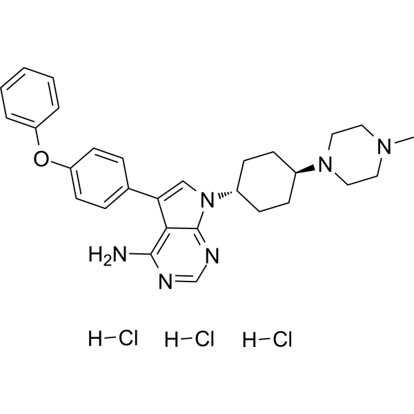
HY-15764A
A 419259 trihydrochlorideSrc Inhibitor


Biological Activity
Description
IC50 & Target
[1]|
Abl 8 nM (IC50) |
Cellular Effect
|
Cell Line

|
Type | Value | Description | References |
|---|---|---|---|---|
| HCT-8 | IC50 |
<5 μM
Compound: PD-0166326
|
Inhibition of HCT-8 clonogenic growth in soft agar with pretreatment
Inhibition of HCT-8 clonogenic growth in soft agar with pretreatment
|
[PMID: 10974196] |
| HCT-8 | IC50 |
<5 μM
Compound: PD-0166326
|
Inhibition of HCT-8 clonogenic growth in soft agar without pretreatment
Inhibition of HCT-8 clonogenic growth in soft agar without pretreatment
|
[PMID: 10974196] |
| HCT-8 | IC50 |
1.11 μM
Compound: PD-0166326
|
Inhibition of HCT-8 cell proliferation
Inhibition of HCT-8 cell proliferation
|
[PMID: 10974196] |
| HT-29 | IC50 |
0.526 μM
Compound: PD-0166326
|
Inhibition of HT-29 cell proliferation
Inhibition of HT-29 cell proliferation
|
[PMID: 10974196] |
| HT-29 | IC50 |
10.3 μM
Compound: PD-0166326
|
Inhibition of HT-29 clonogenic growth in soft agar without pretreatment
Inhibition of HT-29 clonogenic growth in soft agar without pretreatment
|
[PMID: 10974196] |
| HT-29 | IC50 |
7.96 μM
Compound: PD-0166326
|
Inhibition of HT-29 clonogenic growth in soft agar with pretreatment
Inhibition of HT-29 clonogenic growth in soft agar with pretreatment
|
[PMID: 10974196] |
| Jurkat | IC50 |
2100 nM
Compound: 1, PD-166326
|
Cytotoxicity against Philadelphia chromosome negative human Jurkat cells by Alamar Blue fluorescent assay
Cytotoxicity against Philadelphia chromosome negative human Jurkat cells by Alamar Blue fluorescent assay
|
[PMID: 19889540] |
| K562 | IC50 |
<10 nM
Compound: 1, PD-166326
|
Cytotoxicity against Philadelphia chromosome positive human K562 cells by Alamar Blue fluorescent assay
Cytotoxicity against Philadelphia chromosome positive human K562 cells by Alamar Blue fluorescent assay
|
[PMID: 19889540] |
| KU812 cell line | IC50 |
<10 nM
Compound: 1, PD-166326
|
Cytotoxicity against Philadelphia chromosome positive human KU812 cells by Alamar Blue fluorescent assay
Cytotoxicity against Philadelphia chromosome positive human KU812 cells by Alamar Blue fluorescent assay
|
[PMID: 19889540] |
| Macrophage | CC50 |
5 μM
Compound: 66; PD 166326
|
Cytotoxicity against macrophage (unknown origin) cells
Cytotoxicity against macrophage (unknown origin) cells
|
[PMID: 33539089] |
| MEG-01 | IC50 |
<10 nM
Compound: 1, PD-166326
|
Cytotoxicity against Philadelphia chromosome positive human MEG01 cells by Alamar Blue fluorescent assay
Cytotoxicity against Philadelphia chromosome positive human MEG01 cells by Alamar Blue fluorescent assay
|
[PMID: 19889540] |
| SW-620 | IC50 |
<5 μM
Compound: PD-0166326
|
Inhibition of SW-620 clonogenic growth in soft agar without pretreatment
Inhibition of SW-620 clonogenic growth in soft agar without pretreatment
|
[PMID: 10974196] |
| SW-620 | IC50 |
1.8 μM
Compound: PD-0166326
|
Inhibition of SW-620 cell proliferation
Inhibition of SW-620 cell proliferation
|
[PMID: 10974196] |
In Vitro
PD166326 (0.1-2.5 nM; 48 h) potently inhibits Bcr/Abl-dependent proliferation of R10(-) cells, with an IC50 of 0.2 nM[1].
PD166326 (5-50 nM; 48 h) inhibits stem cell factor-dependent proliferation of parental MO7E cells, with an IC50 of 12 nM[1].
PD166326 (10 min; 30°C) potently inhibits purified recombinant c-abl tyrosine kinase in vitro with an IC50 of 8 nM[2].
PD166326 potently inhibits purified recombinant c-src tyrosine kinase in vitro with an IC50 of 6 nM[2].
PD166326 (4-7 days) potently inhibits the growth of K562 Bcr-abl-positive cells with an IC50 of 0.3 nM[2].
PD166326 (4-7 days) potently inhibits the growth of BaF3 p210Bcr-abl Bcr-abl-positive cells with an IC50 of 6.0 nM[2].
MedChemExpress (MCE) has not independently confirmed the accuracy of these methods. They are for reference only.
-
Cell Line:parental MO7E cells (stem cell factor-dependent)
-
Concentration:5 nM, 10 nM, 50 nM
-
Incubation Time:48 h
-
Result:Inhibited stem cell factor-dependent proliferation of parental MO7E cells with an IC50 of 12 nM.
Inhibited proliferation at 5 to 10 nM.
Completely suppressed growth at 50 nM.
-
Cell Line:K562 cells
-
Concentration:0.05-5 nM
-
Incubation Time:24 h
-
Result:Inhibited MAPK phosphorylation in parallel with reduced Bcr-abl autophosphorylation.
In Vivo
mesylate (HY-50946)[1].
MedChemExpress (MCE) has not independently confirmed the accuracy of these methods. They are for reference only.
-
Animal Model:Balb/c mice[1]
-
Dosage:50 mg/kg
-
Administration:p.o.; twice a day
-
Result:Ensured 100% of treated mice survived 3 to 4 weeks of treatment with no clinical signs of toxicity.
Reduced median spleen weight to 78 mg, over 8-fold less than placebo-treated animals (P < .001); two thirds of treated mice showed complete resolution of splenomegaly (spleen weights in the normal 50-100 mg range).
Lowered median peripheral white blood cell count to 15.0 × 10^9/L, almost 10-fold lower than placebo-treated animals (P < .01); 70% of treated mice achieved a WBC count less than 20.0 × 10^9/L.
Induced a marked reduction in constitutive tyrosine phosphorylation of leukemia-cell proteins, including an average phosphorylated Crk-L (p-Crk-L)/Crk-L ratio of 0.23 and a marked reduction in constitutive Lyn kinase activation, while ERK1/2 activation remained similar to placebo-treated mice.


Chemical Information
-
CAS No. 185039-91-2
-
Appearance Solid
-
Molecular Weight 427.28
-
Formula C21H16Cl2N4O2
-
Color White to off-white
-
SMILES
O=C1C(C2=C(C=CC=C2Cl)Cl)=CC3=CN=C(N=C3N1C)NC4=CC=CC(CO)=C4
-
Shipping
Room temperature in continental US; may vary elsewhere.
-
Storage
4°C, sealed storage, away from moisture and light
* In solvent : -80°C, 6 months; -20°C, 1 month (sealed storage, away from moisture and light)


Solvent & Solubility
In Vitro:
DMSO : 100 mg/mL (234.04 mM; Need ultrasonic; Hygroscopic DMSO has a significant impact on the solubility of product, please use newly opened DMSO)
Please refer to the solubility information to select the appropriate solvent. Once prepared, please aliquot and store the solution to prevent product inactivation from repeated freeze-thaw cycles.
Storage method and period of stock solution: -80°C, 6 months; -20°C, 1 month (sealed storage, away from moisture and light). When stored at -80°C, please use it within 6 months. When stored at -20°C, please use it within 1 month.
Please refer to the solubility information to select the appropriate solvent. Once prepared, please aliquot and store the solution to prevent product inactivation from repeated freeze-thaw cycles.
Storage method and period of stock solution: -80°C, 6 months; -20°C, 1 month (sealed storage, away from moisture and light). When stored at -80°C, please use it within 6 months. When stored at -20°C, please use it within 1 month.
Concentration (start) × Volume (start) = Concentration (final) × Volume (final)


Purity & Documentation
-
Data Sheet (277 KB)
-
SDS (252 KB)
- English - EN (252 KB)
- Français - FR (252 KB)
- Deutsch - DE (252 KB)
- Norwegian - NO (252 KB)
- Español - ES (252 KB)
- Swedish - SV (252 KB)
- Italian - IT (252 KB)
- Korean - KR (252 KB)
- Portuguese - PT (252 KB)
-
Handling Instructions (2659 KB)
References
[1]. Wolff NC, et al. PD166326, a novel tyrosine kinase inhibitor, has greater antileukemic activity than imatinib mesylate in a murine model of chronic myeloid leukemia. Blood. 2005;105(10):3995-4003. [Content Brief]
[2]. Huron DR, et al. A novel pyridopyrimidine inhibitor of abl kinase is a picomolar inhibitor of Bcr-abl-driven K562 cells and is effective against STI571-resistant Bcr-abl mutants. Clin Cancer Res. 2003 Apr;9(4):1267-73. [Content Brief]


Complete Stock Solution Preparation Table
Please refer to the solubility information to select the appropriate solvent. Once prepared, please aliquot and store the solution to prevent product inactivation from repeated freeze-thaw cycles.
Storage method and period of stock solution: -80°C, 6 months; -20°C, 1 month (sealed storage, away from moisture and light). When stored at -80°C, please use it within 6 months. When stored at -20°C, please use it within 1 month.
| Optional Solvent | Concentration Solvent Mass | 1 mg | 5 mg | 10 mg | 25 mg |
|---|---|---|---|---|---|
| DMSO | 1 mM | 2.3404 mL | 11.7019 mL | 23.4039 mL | 58.5096 mL |
| 5 mM | 0.4681 mL | 2.3404 mL | 4.6808 mL | 11.7019 mL | |
| 10 mM | 0.2340 mL | 1.1702 mL | 2.3404 mL | 5.8510 mL | |
| 15 mM | 0.1560 mL | 0.7801 mL | 1.5603 mL | 3.9006 mL | |
| 20 mM | 0.1170 mL | 0.5851 mL | 1.1702 mL | 2.9255 mL | |
| 25 mM | 0.0936 mL | 0.4681 mL | 0.9362 mL | 2.3404 mL | |
| 30 mM | 0.0780 mL | 0.3901 mL | 0.7801 mL | 1.9503 mL | |
| 40 mM | 0.0585 mL | 0.2925 mL | 0.5851 mL | 1.4627 mL | |
| 50 mM | 0.0468 mL | 0.2340 mL | 0.4681 mL | 1.1702 mL | |
| 60 mM | 0.0390 mL | 0.1950 mL | 0.3901 mL | 0.9752 mL | |
| 80 mM | 0.0293 mL | 0.1463 mL | 0.2925 mL | 0.7314 mL | |
| 100 mM | 0.0234 mL | 0.1170 mL | 0.2340 mL | 0.5851 mL |


PD166326 Related Classifications
HY-118144 Related Classifications
 Powered by Bioz