



Nicotinic acid mononucleotide
Based on 1 publication(s) in Google Scholar
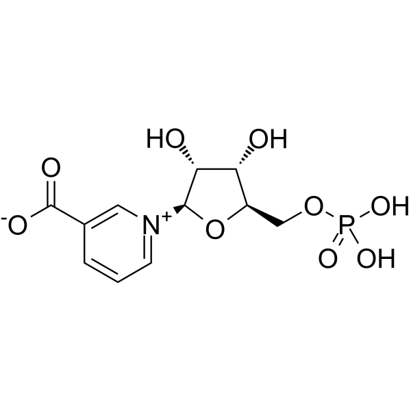
Nicotinic acid mononucleotide acts as a SARM1 inhibitor and a NAD+ biosynthesis intermediate, with an IC50 value of 93.3 μM against SARM1. Nicotinic acid mononucleotide exerts axon-protective effects, delays axonal degeneration, elevates NAD+ levels, enhances Sirt1 activity, improves myocardial capillary density and alleviates myocardial fibrosis. Nicotinic acid mononucleotide reverses diabetic cardiomyopathy in diabetic mice by increasing myocardial NAD+ levels. Nicotinic acid mononucleotide is applicable to research related to cancer, multiple sclerosis, diabetic cardiomyopathy, neurodegenerative diseases and Huntington's disease.
For research use only. We do not sell to patients.
- Purity : 98.08%
- CAS No.: 321-02-8
- Formula: C11H14NO9P
- Molecular Weight:335.20
-
Storage:Powder -20°C, 3 years , 4°C, 2 years ; In solvent -80°C, 6 months , -20°C, 1 month

To place orders, for customer services and technical support, please contact: MedChemExpress USA
Tel: 609-228-6898 E-mail: [email protected] [email protected]


-
 Biological Activity
-
Chemical Information
-
Solvent & Solubility
- Protocols
- Purity & Documentation
- References
-
 Help & FAQs


-
HY-123033A
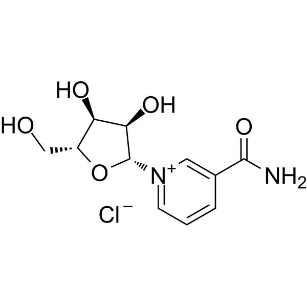
Nicotinamide riboside chlorideEndogenous Metabolite -
HY-B0445
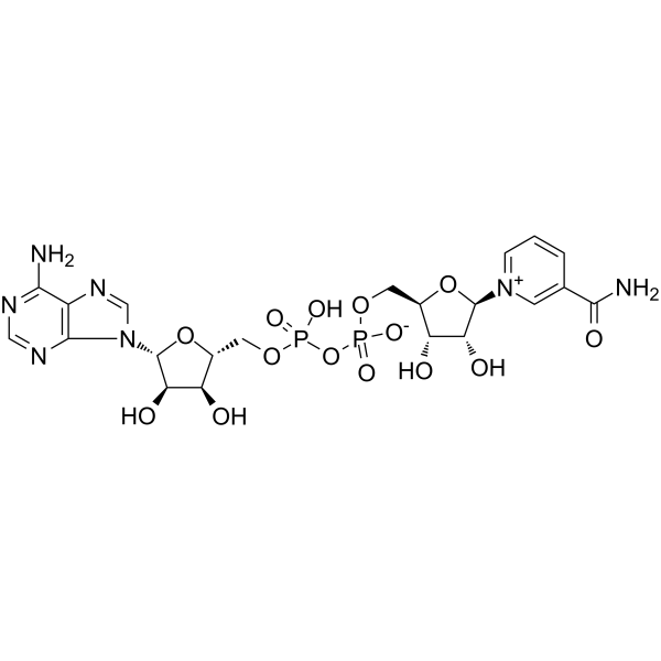
NAD+Endogenous Metabolite -
HY-W104368
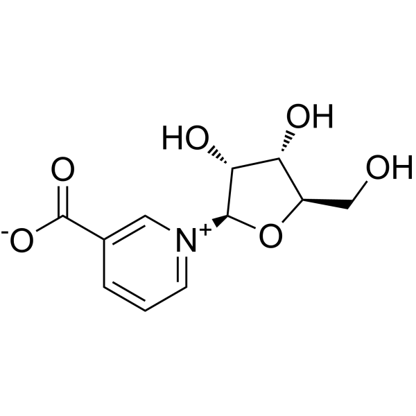
Nicotinic acid ribosideNAD+ Precursor -
HY-B2156
Menaquinone-4Endogenous Metabolite -

HY-10071
Y-27632ROCK Inhibitor -
HY-F0004
FosribnicotinamideNAD+ Intermediate -
HY-N6821
2-O-α-D-Glucopyranosyl-L-ascorbic AcidGlucoside Derivative -

HY-B2156R
Menaquinone-4 (Standard)Endogenous Metabolite -
HY-B1278
D-α-Tocopherol acetateEndogenous Metabolite -
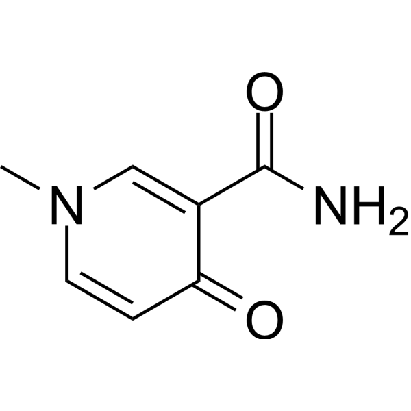
HY-113472
N-Methyl-4-pyridone-3-carboxamideNAD Metabolite -
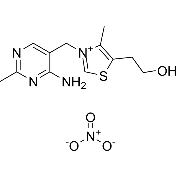
HY-B2223
Thiamine nitrateVitamin B1 Supplement -
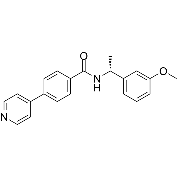
HY-119937
ROCK inhibitor-2ROCK Inhibitor
Publications Citing Use of MedChemExpress (MCE) Nicotinic acid mononucleotide
MoreAll Endogenous Metabolite Isoforms
More

Biological Activity
Description
In Vitro
Nicotinic acid mononucleotide (0-250 μM; assay duration) inhibits the NAD+ hydrolase activity of full-length human SARM1 by binding to its N-terminal ARM domain; its IC50 is 93.3 μM in the absence of NMN, while this inhibitory effect is attenuated in the presence of 5 μM NMN (IC50 = 147.2 μM)[1].
Nicotinic acid mononucleotide delays axonal degeneration of mammalian axons by restoring reduced intracellular NAD+ levels[3].
Nicotinic acid mononucleotide (NAM) increases the expression of Sirt1 and the phosphorylation level of eNOS in human umbilical vein endothelial cells (HUVECs) treated with high glucose[4].
MedChemExpress (MCE) has not independently confirmed the accuracy of these methods. They are for reference only. Further protocols information, click here.
In Vivo
MedChemExpress (MCE) has not independently confirmed the accuracy of these methods. They are for reference only.
-
Animal Model:C57BL/6J (8-week-old male; streptozotocin-induced diabetic)[4]
-
Dosage:400 mg/kg/day
-
Administration:i.p.; daily; 8 weeks
-
Result:Partially increased myocardial NAD+ levels in diabetic mice.
Partially reversed impaired left ventricular ejection fraction (LVEF) and fractional shortening (FS) relative to untreated diabetic mice.
Improved myocardial ultrastructural abnormalities (reduced Z-line misalignment, mitochondrial swelling, and myofibril disorganization).
Increased myocardial capillary density relative to untreated diabetic mice.
Reduced myocardial expression of fibrosis markers TGF-β1 and collagen I relative to untreated diabetic mice.
Restored myocardial Sirt1 mRNA and protein expression relative to untreated diabetic mice.
Had no effect on blood glucose or body weight in diabetic mice, and no significant effect on interventricular septal thickness (IVSD) or left ventricular posterior wall thickness (LVPWD).


Chemical Information
-
CAS No. 321-02-8
-
Appearance Solid
-
Molecular Weight 335.20
-
Formula C11H14NO9P
-
Color White to off-white
-
SMILES
O[C@@H]1[C@H](O)[C@@H](COP(O)(O)=O)O[C@H]1[N+]2=CC=CC(C([O-])=O)=C2
-
Structure Classification
-
Initial Source
-
Shipping
Room temperature in continental US; may vary elsewhere.
-
Storage
Powder -20°C 3 years 4°C 2 years In solvent -80°C 6 months -20°C 1 month


Publications (1)
-
Journal Impact Factor
-
Most Recent
-
Diabetes Res Clin Pract
ACMSD mediated de novo NAD+ biosynthetic impairment in cardiac endothelial cells as a potential therapeutic target for diabetic cardiomyopathy. [Abstract]2023 Dec:206:111014. PMID: 37977551


Solvent & Solubility
In Vitro:
H2O : 10 mg/mL (29.83 mM; Need ultrasonic)
Please refer to the solubility information to select the appropriate solvent. Once prepared, please aliquot and store the solution to prevent product inactivation from repeated freeze-thaw cycles.
Storage method and period of stock solution: -80°C, 6 months; -20°C, 1 month. When stored at -80°C, please use it within 6 months. When stored at -20°C, please use it within 1 month.
* Note: If you choose water as the stock solution, please dilute it to the working solution, then filter and sterilize it with a 0.22 μm filter before use.
Please refer to the solubility information to select the appropriate solvent. Once prepared, please aliquot and store the solution to prevent product inactivation from repeated freeze-thaw cycles.
Storage method and period of stock solution: -80°C, 6 months; -20°C, 1 month. When stored at -80°C, please use it within 6 months. When stored at -20°C, please use it within 1 month.
* Note: If you choose water as the stock solution, please dilute it to the working solution, then filter and sterilize it with a 0.22 μm filter before use.
Concentration (start) × Volume (start) = Concentration (final) × Volume (final)

Protocols
-
Fibrosis/Collagen Morphometry
Fibrosis and collagen morphometry is based on the quantitative visualization of fibrillar collagen deposition in tissue sections using histochemical stains such as Sirius Red (Picrosirius Red) or Masson's trichrome, followed by image-based or polarization-enhanced analysis to estimate collagen proportional area as a surrogate of extracellular matrix accumulation during fibrotic remodeling. Sirius Red combined with polarized light microscopy enhances detection of collagen fibers due to birefringence properties, enabling more specific visualization of collagen type I and III fibrils compared to conventional bright-field histology, while whole-section or region-restricted digital morphometry reduces field-selection bias in fibrosis assessment. Alternative quantitative approaches include second harmonic generation (SHG) and two-photon excited fluorescence microscopy, which enable label-free detection of fibrillar collagen and have been validated against histological staining and biochemica
-
Collagen: Sirius Red Staining
Sirius Red or picrosirius red staining is a histochemical method for visualizing collagen-rich extracellular matrix in tissue sections, and collagen fibers are detected as red-stained structures under bright-field microscopy with enhanced birefringence under polarized light. Picrosirius red is useful for assessing total collagen organization, distribution, and fibrosis burden, but polarized color should not be interpreted as a definitive collagen type I versus type III readout because color is affected by fiber orientation, thickness, and packing.
-
Cell Cytotoxicity Assay
Cytotoxicity assays are usually based on the assessment of cell membrane damage, which can also be indirectly detected by measuring cell viability. Detection methods include MTT assay, CKK-8 assay, LDH assay and ATP assay, etc.
-
Connective Tissue: Masson's Trichrome/Collagen Trichrome Staining
Masson’s Trichrome (collagen/trichrome staining) is a histological technique that differentially stains tissue compartments using sequential acidic dyes to distinguish collagen from muscle and cytoplasmic components based on dye affinity and tissue permeability differences, enabling visualization of fibrosis and connective tissue architecture in histological sections. The classical formulation typically uses Weigert's iron hematoxylin for nuclear staining, Biebrich scarlet-acid fuchsin for cytoplasm and muscle, and aniline blue (or light green variants) for collagen, producing a characteristic blue/green collagen signal contrasted against red cytoplasm and dark nuclei. The staining principle relies on selective displacement of smaller dye molecules by larger anionic dyes in collagen-rich regions under controlled acidified conditions, which enhances collagen-specific dye retention. This property makes the method widely used for fibrosis assessment in organs such as heart, liver, lung, a


Purity & Documentation
-
Data Sheet (280 KB)
-
SDS (252 KB)
- English - EN (252 KB)
- Français - FR (252 KB)
- Deutsch - DE (252 KB)
- Norwegian - NO (252 KB)
- Español - ES (252 KB)
- Swedish - SV (252 KB)
- Italian - IT (252 KB)
- Korean - KR (252 KB)
- Portuguese - PT (252 KB)
-
Handling Instructions (2659 KB)
References
[1]. Sasaki Y, et al. Nicotinic acid mononucleotide is an allosteric SARM1 inhibitor promoting axonal protection. Exp Neurol. 2021;345:113842. [Content Brief]
[2]. O'Hara JK, et al. Targeting NAD+ metabolism in the human malaria parasite Plasmodium falciparum. PLoS One. 2014;9(4):e94061. Published 2014 Apr 18. [Content Brief]
[3]. Khan JA, et al. Nicotinamide adenine dinucleotide metabolism as an attractive target for drug discovery. Expert Opin Ther Targets. 2007;11(5):695-705. [Content Brief]
[4]. Zeng F, et al. ACMSD mediated de novo NAD+ biosynthetic impairment in cardiac endothelial cells as a potential therapeutic target for diabetic cardiomyopathy. Diabetes Res Clin Pract. 2023;206:111014. [Content Brief]


Complete Stock Solution Preparation Table
Please refer to the solubility information to select the appropriate solvent. Once prepared, please aliquot and store the solution to prevent product inactivation from repeated freeze-thaw cycles.
Storage method and period of stock solution: -80°C, 6 months; -20°C, 1 month. When stored at -80°C, please use it within 6 months. When stored at -20°C, please use it within 1 month.
| Optional Solvent | Concentration Solvent Mass | 1 mg | 5 mg | 10 mg | 25 mg |
|---|---|---|---|---|---|
| H2O | 1 mM | 2.9833 mL | 14.9165 mL | 29.8329 mL | 74.5823 mL |
| 5 mM | 0.5967 mL | 2.9833 mL | 5.9666 mL | 14.9165 mL | |
| 10 mM | 0.2983 mL | 1.4916 mL | 2.9833 mL | 7.4582 mL | |
| 15 mM | 0.1989 mL | 0.9944 mL | 1.9889 mL | 4.9722 mL | |
| 20 mM | 0.1492 mL | 0.7458 mL | 1.4916 mL | 3.7291 mL | |
| 25 mM | 0.1193 mL | 0.5967 mL | 1.1933 mL | 2.9833 mL |
* Note: If you choose water as the stock solution, please dilute it to the working solution, then filter and sterilize it with a 0.22 μm filter before use.


Nicotinic acid mononucleotide Related Classifications
HY-128700 Related Classifications
 Powered by Bioz