



Pichromene

Based on 1 Customer Validation
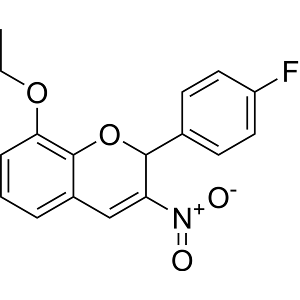
Pichromene (S14161) is an anticancer agent and weak PI3K inhibitor. Pichromene can effectively inhibit tumor growth in leukemia mouse models and can be used in cancer research.
For research use only. We do not sell to patients.
- Purity : 98.0%
- CAS No.: 883046-50-2
- Formula: C17H14FNO4
- Molecular Weight:315.30
-
Storage:
Store at room temperature 3 years.
In solvent -80°C, 2 years , -20°C, 1 year

To place orders, for customer services and technical support, please contact: MedChemExpress USA
Tel: 609-228-6898 E-mail: [email protected] [email protected]


-
 Biological Activity
-
Chemical Information
- Protocols
- Purity & Documentation
- References
-
 Help & FAQs






Biological Activity
Description
Cellular Effect
|
Cell Line

|
Type | Value | Description | References |
|---|---|---|---|---|
| B16 | GI50 |
23.28 μM
Compound: 2, S14161
|
Cytotoxicity against mouse B16 cells after 24 to 72 hrs by SRB assay
Cytotoxicity against mouse B16 cells after 24 to 72 hrs by SRB assay
|
[PMID: 23601711] |
| K562 | GI50 |
1.24 μM
Compound: 2, S14161
|
Cytotoxicity against human K562 cells after 24 to 72 hrs by SRB assay
Cytotoxicity against human K562 cells after 24 to 72 hrs by SRB assay
|
[PMID: 23601711] |
| LP-1 | GI50 |
0.57 μM
Compound: 2, S14161
|
Cytotoxicity against human LP-1 cells after 24 to 72 hrs by SRB assay
Cytotoxicity against human LP-1 cells after 24 to 72 hrs by SRB assay
|
[PMID: 23601711] |
| MDA-MB-231 | GI50 |
8.53 μM
Compound: 2, S14161
|
Cytotoxicity against human MDA-MB-231 cells after 24 to 72 hrs by SRB assay
Cytotoxicity against human MDA-MB-231 cells after 24 to 72 hrs by SRB assay
|
[PMID: 23601711] |
| Platelet | IC50 |
3.79 μM
Compound: S14161
|
Inhibition of collagen-induced platelet aggregation in human platelet rich plasma preincubated for 10 mins followed by collagen addition by aggregometric method
Inhibition of collagen-induced platelet aggregation in human platelet rich plasma preincubated for 10 mins followed by collagen addition by aggregometric method
|
[PMID: 27996269] |
| U-87MG ATCC | GI50 |
18.72 μM
Compound: 2, S14161
|
Cytotoxicity against human U87 cells after 24 to 72 hrs by SRB assay
Cytotoxicity against human U87 cells after 24 to 72 hrs by SRB assay
|
[PMID: 23601711] |


Chemical Information
-
CAS No. 883046-50-2
-
Appearance Solid
-
Molecular Weight 315.30
-
Formula C17H14FNO4
-
Color Off-white to light yellow
-
SMILES
O=[N+](C1=CC2=CC=CC(OCC)=C2OC1C3=CC=C(F)C=C3)[O-]
-
Synonyms
S14161
-
Shipping
Room temperature in continental US; may vary elsewhere.
-
Storage
Store at room temperature 3 years
In solvent -80°C 2 years -20°C 1 year

Protocols
-
Patient-Derived Xenograft (PDX)
Patient-derived xenograft (PDX) models are generated by engrafting primary human tumor tissue directly into immunodeficient mice, allowing in vivo propagation of patient tumor biology without initial in vitro adaptation. These models are used to preserve key histopathological and molecular characteristics of the original tumor and enable assessment of tumor growth dynamics and therapeutic response in a living organism. The biological readout is tumor engraftment and subsequent growth in the murine host, which reflects the ability of human tumor cells to survive, vascularize, and expand in an immunocompromised microenvironment.
-
Patient-Derived Orthotopic Xenograft (PDOX)
Patient-derived orthotopic xenograft (PDOX) modeling implants fresh patient tumor tissue or patient-derived tumor cells into the anatomically corresponding organ or tissue site of immunodeficient mice, usually by surgical orthotopic implantation, to preserve patient tumor histology, local microenvironmental context, invasion, metastatic behavior, and treatment-response features better than subcutaneous implantation. PDOX readouts include tumor engraftment, orthotopic tumor growth, local invasion, metastasis, recurrence after resection, histologic similarity to the donor tumor, biomarker retention, molecular concordance, survival, and response or resistance to therapy. PDOX models are used for preclinical drug testing and individualized therapy evaluation, but engraftment success varies by tumor type and specimen quality.
-
Subcutaneous Cell-Line-Derived Xenograft
Subcutaneous cell-line-derived xenograft (CDX) models are established by implanting cultured human cancer cell lines into immunodeficient mice, where the injected cells form localized tumors that can be monitored in vivo as a measure of tumorigenic potential, growth kinetics, and treatment response. These models are widely used in oncology research because they allow reproducible tumor formation and enable comparative assessment of tumor growth between different cell lines or genetic manipulations in a controlled in vivo microenvironment. Subcutaneous implantation of cancer cells in immunodeficient mice is a standard approach for evaluating tumor growth behavior and therapeutic response across multiple cancer types, including prostate, esophageal, pancreatic, and colon cancer models.
-
Orthotopic Cell-Line Xenograft
Orthotopic cell-line xenograft models involve implantation of human cancer cell lines into the anatomically corresponding organ of immunodeficient mice to reproduce tumor growth within a native microenvironment, enabling more clinically relevant tumor behavior compared with subcutaneous models. These models are widely used because orthotopic placement better recapitulates tumor progression, including invasion and metastatic spread, which are often underrepresented in heterotopic implantation systems. Compared with conventional xenografts, orthotopic implantation is described as more technically complex but provides improved simulation of tumor-microenvironment interactions and metastatic behavior, making it particularly valuable for translational oncology research. Surgical orthotopic implantation approaches have been emphasized as enabling faithful reproduction of clinical cancer features, including metastasis and disease progression patterns that align with the tumor’s organ of origi


Purity & Documentation
-
Data Sheet (270 KB)
-
SDS (392 KB)
- English - EN (392 KB)
- Français - FR (392 KB)
- Deutsch - DE (392 KB)
- Norwegian - NO (392 KB)
- Español - ES (392 KB)
- Swedish - SV (392 KB)
- Italian - IT (392 KB)
- Korean - KR (392 KB)
- Portuguese - PT (392 KB)
-
Handling Instructions (2659 KB)
References
[1]. Mao X, et al. A small-molecule inhibitor of D-cyclin transactivation displays preclinical efficacy in myeloma and leukemia via phosphoinositide 3-kinase pathway. Blood. 2011 Feb 10;117(6):1986-97. [Content Brief]
[2]. Fouqué A, et al. A Novel Covalent mTOR Inhibitor, DHM25, Shows in Vivo Antitumor Activity against Triple-Negative Breast Cancer Cells. J Med Chem. 2015 Aug 27;58(16):6559-73. [Content Brief]

Calculators
Concentration (start) × Volume (start) = Concentration (final) × Volume (final)


Pichromene Related Classifications
HY-111209 Related Classifications
 Powered by Bioz