



Protegrin-1
Based on 2 publication(s) in Google Scholar
Protegrin-1 is an orally active antibacterial peptide. Protegrin-1 activates ERK, COX2, NFκB, inhibits Apoptosis, and NO production. Protegrin-1 has antibacterial activity against S. aureus, E. faecium, E. coli, P. aeruginosa and K. Pneumoniae, with MBC values between 10 and 20 µM. Protegrin-1 has antiviral activity against dengue NS2B-NS3. Protegrin-1 has anti-inflammatory activity. Protegrin-1 is used in the study of inflammatory diseases and infections.
For research use only. We do not sell to patients.
- Purity : 99.96%
- CAS No.: 168831-77-4
- Formula: C88H147N37O19S4
- Molecular Weight:2155.61
-
Storage:
Sealed storage, away from moisture.
Powder -80°C, 2 years , -20°C, 1 year* In solvent : -80°C, 6 months; -20°C, 1 month (sealed storage, away from moisture)

To place orders, for customer services and technical support, please contact: MedChemExpress USA
Tel: 609-228-6898 E-mail: [email protected] [email protected]


-
 Biological Activity
-
Chemical Information
-
Solvent & Solubility
- Protocols
- Purity & Documentation
- References
-
 Help & FAQs


Publications Citing Use of MedChemExpress (MCE) Protegrin-1
More

Biological Activity
Description
In Vitro
Protegrin-1 (2.5-12.5 μM; 24-72 h) inhibits dengue NS2B-NS3 serine protease and viral replication in MK2 cells[1].
Protegrin-1 (1-9 μM; 16 h) promotes the migration of IPEC-J2 cells[2].
Protegrin-1 (>100 μM; 24 h) reduces the viability of 661W and PMN cells[3].
Protegrin-1 (0.1-10 μg/mL; 24 h) inhibits porcine ovarian granulosa cell apoptosis from Hydrogen peroxide-induced oxidative stress via the PERK/eIF2a/CHOP signaling pathway[5].
Protegrin-1 shows antimicrobial activity against S. aureus, E. faecium, E. coli, P. aeruginosa and K. Pneumoniae, with MBC values between 10 and 20 µM[6].
Protegrin-1 (1.25-20 μM) inhibits LPS-induced activation of RAW macrophages, inhibiting the NO production[6].
MedChemExpress (MCE) has not independently confirmed the accuracy of these methods. They are for reference only. Further protocols information, click here.
-
Cell Line:IPEC-J2
-
Concentration:9 μM
-
Incubation Time:3 h
-
Result:Increased CCL2, COX2, IRF7, NFκB, EGFR, and MCL-1.
In Vivo
Protegrin-1 (5 mg/kg; i.v.; single) reduces the mortality of mice inoculated intravenously with Staphylococcus aureus[7].
Protegrin-1 (10 mg/kg; p.o.) reduces Citrobacter rodentium intestinal infection in mice as evidenced by reduced histopathologic change in the colon, prevention of body weight loss, milder clinical signs of disease[8].
MedChemExpress (MCE) has not independently confirmed the accuracy of these methods. They are for reference only.
-
Animal Model:Immunocompetent mice inoculated with Pseudomonas aeruginosa or Staphylococcus aureus by intraperitoneal injection; mice inoculated with Staphylococcus aureus by intravenous injection; leukopenic mice[7]
-
Dosage:0.5 mg/kg (mice inoculated with Pseudomonas aeruginosa or Staphylococcus aureus by intraperitoneal injection); 5 mg/kg (mice inoculated with Staphylococcus aureus by intravenous injection); 2.5 mg/kg (leukopenic mice)
-
Administration:Intraperitoneal injection (i.p.) (0.5 mg/kg); Intravenous injection (i.v.) (2.5, 5 mg/kg); single
-
Result:Caused 0% to 27% mortality (0.5 mg/kg).
Caused 7% to 33% mortality (5 mg/kg).
Caused 33% mortality (2.5 mg/kg).
Clinical Trial
| NCT Number | Sponsor | Condition | Start Date |
Phase

|
|---|---|---|---|---|
| NCT01329991 | Plexxikon| | 2011-05 | PHASE1 |


Chemical Information
-
CAS No. 168831-77-4
-
Appearance Solid
-
Molecular Weight 2155.61
-
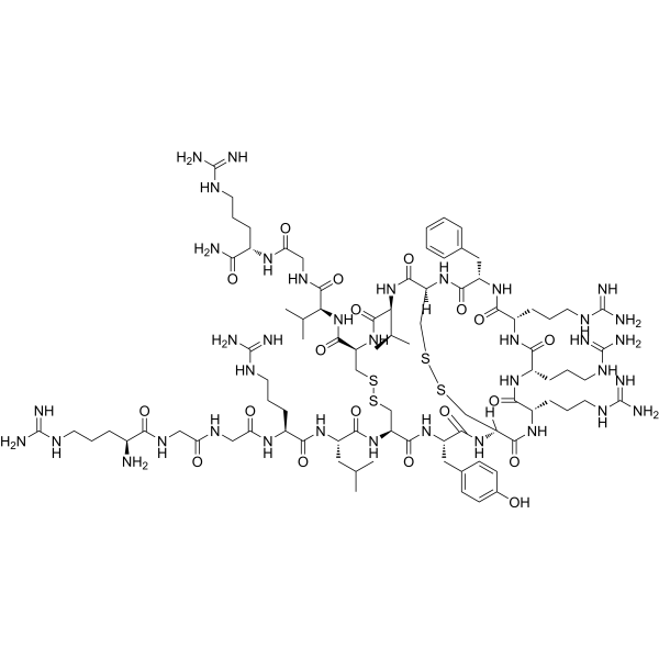
Formula C88H147N37O19S4
-
Color White to off-white
-
Synonyms
PG1
-
Sequence
Arg-Gly-Gly-Arg-Leu-Cys-Tyr-Cys-Arg-Arg-Arg-Phe-Cys-Val-Cys-Val-Gly-Arg-NH2 (Disulfide bridge:Cys6-Cys15;Cys8-Cys13)
-
Sequence Shortening
RGGRLCYCRRRFCVCVGR-NH2 (Disulfide bridge:Cys6-Cys15;Cys8-Cys13)
-
Shipping
Room temperature in continental US; may vary elsewhere.
-
Storage
Sealed storage, away from moisture
Powder -80°C 2 years -20°C 1 year * In solvent : -80°C, 6 months; -20°C, 1 month (sealed storage, away from moisture)


Publications (2)
-
Journal Impact Factor
-
Most Recent
-
Int J Pharm
Combined antimicrobial and pro-healing effects of cathelicidins in a polysaccharide-based hydrogel scaffold: treatment of carbapenemase-resistant Pseudomonas aeruginosa wound infections in a porcine model. [Abstract]2026 Jun 25:699:126992. PMID: 42150643 -
Folia Microbiol (Praha)
EDTA enhances antimicrobial activity of PR-39 and Protegrin-1 antimicrobial peptides against carbapenem-resistant Pseudomonas aeruginosa in serum. [Abstract]2026 May 28. PMID: 42207438


Solvent & Solubility
In Vitro:
DMSO : 20 mg/mL (9.28 mM; Need ultrasonic; Hygroscopic DMSO has a significant impact on the solubility of product, please use newly opened DMSO)
Please refer to the solubility information to select the appropriate solvent. Once prepared, please aliquot and store the solution to prevent product inactivation from repeated freeze-thaw cycles.
Storage method and period of stock solution: -80°C, 6 months; -20°C, 1 month (sealed storage, away from moisture). When stored at -80°C, please use it within 6 months. When stored at -20°C, please use it within 1 month.
Please refer to the solubility information to select the appropriate solvent. Once prepared, please aliquot and store the solution to prevent product inactivation from repeated freeze-thaw cycles.
Storage method and period of stock solution: -80°C, 6 months; -20°C, 1 month (sealed storage, away from moisture). When stored at -80°C, please use it within 6 months. When stored at -20°C, please use it within 1 month.
Concentration (start) × Volume (start) = Concentration (final) × Volume (final)

Protocols
-
Research Protocol for Infectious Diseases
Infectious-disease experiments test how pathogens interact with host barriers, innate immune receptors, inflammatory signaling, pathogen replication, and tissue injury; pattern-recognition receptors such as TLRs, RIG-I-like receptors, NOD-like receptors, and inflammasomes detect microbial molecules and activate NF-κB, interferon, and cytokine responses. The central hypothesis is that infection severity reflects the balance between pathogen burden and host response: protective inflammation restricts pathogen growth, whereas excessive or mislocalized inflammation contributes to tissue damage and disease phenotype. Unresolved questions include which host pathways are protective versus pathogenic, why some infection models fail to translate to human disease, and which combined readouts best predict clinically relevant infection outcomes.
-
Apoptosis
Apoptosis, also called programmed cell death, is generally characterized by distinct morphological characteristics.
-
TUNEL staining for apoptotic DNA fragmentation
TUNEL staining detects DNA strand breaks by using terminal deoxynucleotidyl transferase to add labeled nucleotides to exposed 3′-OH DNA termini, generating either microscopic staining in fixed cells or tissue sections, or fluorescence/cytometric signal in cell suspensions. TUNEL positivity reflects DNA fragmentation but should not be interpreted alone as definitive apoptosis, because TUNEL can also label necrotic, autolytic, mechanically damaged, or DNA-repair-associated DNA breaks.
-
Annexin V plus membrane-impermeant dye apoptosis staining
Annexin V-based apoptosis assays rely on the detection of phosphatidylserine (PS) externalization from the inner leaflet of the plasma membrane to the outer leaflet, an early biochemical hallmark of apoptosis. Fluorescently labeled Annexin V binds PS in a calcium-dependent manner, enabling identification of early apoptotic cells by flow cytometry or fluorescence microscopy. When combined with a membrane-impermeant DNA-binding dye (e. g. , propidium iodide), this approach allows discrimination between viable (Annexin V−/dye−), early apoptotic (Annexin V+/dye−), and late apoptotic or necrotic (Annexin V+/dye+) cell populations by assessing membrane integrity and PS exposure.
-
Cotton Pellet Granuloma
Cotton pellet granuloma is a classical in vivo chronic inflammation model used to evaluate the anti-inflammatory potential of test substances by measuring their ability to inhibit granuloma tissue formation around an implanted foreign body (cotton pellet) in rodents. The method is based on the biological response to a sterile implanted material, which induces proliferative phase inflammation characterized by fibroblast proliferation and collagen-rich granuloma formation, and the final readout reflects the extent of chronic inflammatory tissue growth surrounding the pellet. In multiple preclinical pharmacological evaluations, inhibition of cotton pellet-induced granuloma formation has been used as an indicator of anti-inflammatory activity in both synthetic and natural product screening contexts.
-
Carrageenan-Induced Paw Edema
Carrageenan-induced paw edema is an acute inflammation model in which intraplantar injection of carrageenan induces localized inflammatory swelling characterized by vascular permeability, leukocyte infiltration, and production of inflammatory mediators such as prostaglandins and cytokines, making it widely used to evaluate anti-inflammatory agents in vivo. The resulting paw volume or thickness increase is quantified over time as a direct readout of inflammatory intensity and drug efficacy, typically reflecting cyclooxygenase-mediated prostaglandin-driven edema formation and immune cell recruitment in peripheral tissue[20].
-
Apoptosis Solutions
Apoptosis is a regulated, generally non-lytic cell-death pathway that removes unwanted, damaged, infected, or abnormal cells through coordinated morphological changes, caspase activation, DNA fragmentation, and membrane remodeling. The intrinsic apoptosis pathway is controlled mainly by mitochondrial outer membrane permeabilization, BCL-2 family proteins, cytochrome c release, apoptosome formation, caspase-9 activation, and downstream executioner caspase-3/7 activation. The extrinsic apoptosis pathway is initiated by death receptors such as Fas, TNFR, and TRAIL receptors, which recruit adaptor proteins and activate caspase-8 before engaging executioner caspases or mitochondrial amplification through BID cleavage. Apoptosis is linked to many phenotypes, including cancer cell killing, tissue homeostasis, immune regulation, neurodegeneration, infection response, and treatment-induced cytotoxicity; unresolved questions include how apoptosis interacts with necroptosis, pyroptosis, ferroptos
-
Research Protocol for Inflammation-related Diseases
The NLRP3 inflammasome is a cytosolic innate immune signaling platform that integrates priming signals and danger-signal activation to promote caspase-1 activation, maturation of IL-1β and IL-18, and gasdermin D-mediated pyroptotic cell death. The core experimental logic is to determine whether inflammatory disease phenotypes are driven by increased NLRP3 expression, ASC-containing inflammasome assembly, caspase-1 cleavage, GSDMD cleavage, and extracellular release of IL-1β/IL-18 rather than by nonspecific cell injury alone. The pathway is strongly linked to inflammation-related disease phenotypes because monosodium urate crystals activate NALP3/NLRP3 inflammasome signaling in gout-like crystal inflammation, cholesterol crystals activate NLRP3 inflammasomes in atherogenesis models, and DSS-induced intestinal inflammation has been reported to involve NLRP3 inflammasome activity. However, experimental colitis studies also show context-dependent protective effects of NLRP3 inflammasome co
-
Bacterial live/dead nucleic-acid viability staining
The LIVE/DEAD bacterial viability staining method is based on differential permeability of nucleic-acid-binding fluorescent dyes, most commonly SYTO 9 and propidium iodide (PI), which enables discrimination of bacterial populations with intact versus compromised cytoplasmic membranes. SYTO 9 penetrates both intact and damaged bacterial membranes and binds nucleic acids to produce green fluorescence, whereas propidium iodide penetrates only cells with compromised membranes and fluoresces red while also reducing SYTO 9 signal through competitive binding and fluorescence interactions. The resulting fluorescence pattern is interpreted as a proxy for membrane integrity, which is widely used as an indicator of bacterial viability in microscopy, flow cytometry, and spectroscopic platforms. However, mechanistic studies show that SYTO 9 and PI interactions involve displacement and fluorescence resonance energy transfer effects, which can influence signal interpretation depending on dye ratios a


Purity & Documentation
-
Data Sheet (287 KB)
-
SDS (254 KB)
- English - EN (254 KB)
- Français - FR (254 KB)
- Deutsch - DE (254 KB)
- Norwegian - NO (254 KB)
- Español - ES (254 KB)
- Swedish - SV (254 KB)
- Italian - IT (254 KB)
- Korean - KR (254 KB)
- Portuguese - PT (254 KB)
-
Handling Instructions (2659 KB)
References
[1]. Rothan HA, et al. Protegrin-1 inhibits dengue NS2B-NS3 serine protease and viral replication in MK2 cells. J Biomed Biotechnol. 2012;2012:251482. [Content Brief]
[2]. Penney J, et al. Protegrin 1 Enhances Innate Cellular Defense via the Insulin-Like Growth Factor 1 Receptor Pathway. Front Cell Infect Microbiol. 2018 Sep 28;8:331. [Content Brief]
[3]. Soundrarajan N, et al. Protegrin-1 cytotoxicity towards mammalian cells positively correlates with the magnitude of conformational changes of the unfolded form upon cell interaction. Sci Rep. 2019 Aug 9;9(1):11569. [Content Brief]
[5]. Li X, et al. Protegrin-1 inhibits porcine ovarian granulosa cell apoptosis from H2O2-induced oxidative stress via the PERK/eIF2α/CHOP signaling pathway in vitro. Theriogenology. 2022 Feb;179:117-127. [Content Brief]
[6]. Javed A, et al. Antimicrobial and immunomodulatory activities of porcine cathelicidin Protegrin-1. Mol Immunol. 2024 Sep;173:100-109. [Content Brief]
[7]. Steinberg DA, et al. Protegrin-1: a broad-spectrum, rapidly microbicidal peptide with in vivo activity. Antimicrob Agents Chemother. 1997 Aug;41(8):1738-42. [Content Brief]
[8]. Osakowicz C, et al. Protective and Anti-Inflammatory Effects of Protegrin-1 on Citrobacter rodentium Intestinal Infection in Mice. Int J Mol Sci. 2021 Aug 31;22(17):9494. [Content Brief]


Complete Stock Solution Preparation Table
Please refer to the solubility information to select the appropriate solvent. Once prepared, please aliquot and store the solution to prevent product inactivation from repeated freeze-thaw cycles.
Storage method and period of stock solution: -80°C, 6 months; -20°C, 1 month (sealed storage, away from moisture). When stored at -80°C, please use it within 6 months. When stored at -20°C, please use it within 1 month.
| Optional Solvent | Concentration Solvent Mass | 1 mg | 5 mg | 10 mg | 25 mg |
|---|---|---|---|---|---|
| DMSO | 1 mM | 0.4639 mL | 2.3195 mL | 4.6391 mL | 11.5976 mL |
| 5 mM | 0.0928 mL | 0.4639 mL | 0.9278 mL | 2.3195 mL |


Protegrin-1 Related Classifications
HY-P1633 Related Classifications
 Powered by Bioz