



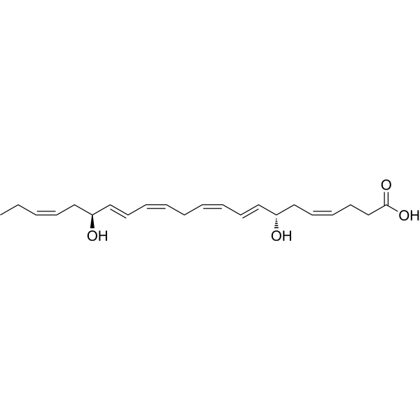
Resolvin D5

Based on 1 Customer Validation
Resolvin D5 is an anti-inflammatory and analgesic agent produced in M2 macrophages. Resolvin D5 alleviates Paclitaxel (HY-B0015)-induced mechanical allodynia and inflammatory pain by activating the GPR32 receptor, with gender specificity (effective only in male mice) and independence from TRPV1 or TRPA1 channels. Resolvin D5 attenuates LPS-induced ERK phosphorylation and NF-κB nuclear translocation, downregulates proinflammatory mediators such as IL-6 and CCL5, inhibits Th17 cell differentiation and osteoclastogenesis, promotes regulatory T cell differentiation, and shows no cytotoxicity to human monocytes. The level of Resolvin D5 is elevated in arthritic SKG mice, but Resolvin D5 has no effect on dendritic cell differentiation or M1 macrophage polarization, nor does it prevent ZyA-induced arthritis progression. Resolvin D5 is suitable for research related to chemotherapy-induced peripheral neuropathy, inflammatory pain and rheumatoid arthritis.
For research use only. We do not sell to patients.
- Purity : 98.3%
- CAS No.: 578008-43-2
- Formula: C22H32O4
- Molecular Weight:360.49
-
Storage:
-80°C

To place orders, for customer services and technical support, please contact: MedChemExpress USA
Tel: 609-228-6898 E-mail: [email protected] [email protected]


-
 Biological Activity
-
Chemical Information
- Protocols
- Purity & Documentation
- References
-
 Help & FAQs


-
HY-15898
Y-320Apoptosis Inducer -
HY-13219
TepoxalinCyclooxygenase/Lipoxygenase Inhibitor -
HY-N3460
IsorhoifolinFlavonoid Glycoside -
HY-120824
MofezolacCOX-1 Inhibitor -
HY-14519S
Methotrexate-d3Stable Isotope -

HY-130675A
15(S)-HEPEAsthma Marker -
HY-17506S
Azithromycin-d3Stable Isotope -
HY-125516
MCTR3Pro-resolving Mediator -
HY-117430
HymeglusinHMG-CoA Synthase Inhibitor -

HY-133976
Aceclofenac methyl esterAceclofenac Impurity -
HY-N0594
Deacetylasperulosidic AcidAntioxidant -

HY-13219R
Tepoxalin (Standard)COX Inhibitor


Biological Activity
Description
In Vitro
Resolvin D5 (100, 500 nM; 3-5 days) suppresses Th17 cell differentiation and facilitates Treg differentiation in CD4+ T cells isolated from SKG mouse spleens in a concentration-dependent manner[1].
Resolvin D5 (1-100 nM; 3 days) suppresses the proliferation of CD4+ T cells isolated from SKG mouse spleens in a concentration-dependent manner, with 100 nM inducing a significant reduction in proliferating cells[1].
Resolvin D5 (10-500 nM; 3 days) suppresses RANKL-induced osteoclast differentiation in bone marrow-derived macrophages from SKG mice in a concentration-dependent manner, with 10, 100, and 500 nM concentrations reducing TRAP+ MNC formation and downregulating key osteoclastogenesis-related genes[1].
MedChemExpress (MCE) has not independently confirmed the accuracy of these methods. They are for reference only. Further protocols information, click here.
-
Cell Line:Th17 cell
-
Concentration:1, 10, 100, 500 nM
-
Incubation Time:3-5 days
-
Result:Suppressed Th17 cell differentiation and facilitates Treg differentiation in CD4+ T cells isolated from SKG mouse spleens in a concentration-dependent manner (100-500 nM).
In Vivo
Resolvin D5 (100 ng; i.t.; single dose) produces a transient, male-specific reduction in chemotherapy-induced mechanical allodynia in wild-type, Trpv1 knockout, and Trpa1 knockout mice, with no analgesic effect in female mice[2].
Resolvin D5 (10 ng; i.t.; single dose) produces a male-specific reduction in formalin-induced Phase II inflammatory pain, with no analgesic effect in female mice or on Phase I pain responses[2].
MedChemExpress (MCE) has not independently confirmed the accuracy of these methods. They are for reference only.
-
Animal Model:CD1 mice with Chemotherapy-induced peripheral neuropathy (male and female, adult, 25-35 g); B6.129X1-Trpv1tm1Jul/J (male and female, Trpv1 knockout); B6;129P-Trpa1tm1Kykw/J (male and female, Trpa1 knockout)[2]
-
Dosage:100 ng
-
Administration:i.t.; single dose
-
Result:Significantly increased paw withdrawal threshold (reduced mechanical allodynia) in male wild-type, male Trpv1 knockout, and male Trpa1 knockout mice at 1 hour post-injection, with effects diminished by 3 hours post-injection.
Showed no significant change in paw withdrawal threshold in female wild-type, female Trpv1 knockout, or female Trpa1 knockout mice at any time point post-injection.
-
Animal Model:CD1 mice with Inflammatory pain (male and female, adult, 25-35 g)[2]
-
Dosage:10 ng
-
Administration:i.t.; single dose
-
Result:Significantly reduced the duration of licking and flinching during the Phase II inflammatory pain response (10-45 minutes post-formalin) in male mice, with no significant effect on Phase I pain behavior (0-10 minutes post-formalin).
Showed no significant change in Phase I or Phase II pain behavior in female mice.


Chemical Information
-
CAS No. 578008-43-2
-
Appearance Liquid
-
Molecular Weight 360.49
-
Formula C22H32O4
-
Color Colorless to light yellow
-
SMILES
CC/C=C\C[C@H](O)/C=C/C=C\C/C=C\C=C\[C@@H](O)C/C=C\CCC(O)=O
-
Structure Classification
-
Initial Source
-
Shipping
Shipping with dry ice.
-
Storage
-80°C

Protocols
-
Western Blot
Western blotting (WB) is a commonly used experimental method in molecular biology, biochemistry, and immunogenetics for identifying and quantifying target proteins. It combines gel electrophoresis with immunoassay, enabling researchers to analyze protein expression, post-translational modifications, and molecular weight.
-
Mammalian live/dead viability and cytotoxicity staining
Live/dead viability and cytotoxicity staining assays are based on the simultaneous detection of intracellular esterase activity in metabolically active (viable) cells and membrane integrity loss in non-viable cells. In commonly used dual-staining approaches, membrane-permeant fluorogenic substrates are converted by intracellular esterases into fluorescent products in live cells, while impermeant DNA-binding dyes selectively enter cells with compromised plasma membranes and label nucleic acids in dead or dying cells, enabling discrimination between viable and non-viable populations by fluorescence microscopy or flow cytometry.
-
Naïve CD4+ T-cell subset differentiation/polarization
Naïve CD4+ T-cell subset differentiation/polarization is an in vitro assay in which purified naïve CD4+ T cells are activated through TCR and CD28 costimulation and cultured with defined cytokines and neutralizing antibodies to generate Th0, Th1, Th2, Th17, or induced Treg-like populations. Differentiation is detected by subset-associated cytokines and transcription factors: IFN-γ/T-bet for Th1, IL-4/GATA3 for Th2, IL-17A/RORγt for Th17, and Foxp3 for induced Treg cells. The assay readout is usually generated by intracellular cytokine staining after restimulation, transcription-factor staining by flow cytometry, ELISA of secreted cytokines, or gene-expression analysis. The result reflects cytokine-directed lineage commitment or polarization rather than antigen-specific immune protection by itself.
-
Research Protocol for Inflammation-related Diseases
The NLRP3 inflammasome is a cytosolic innate immune signaling platform that integrates priming signals and danger-signal activation to promote caspase-1 activation, maturation of IL-1β and IL-18, and gasdermin D-mediated pyroptotic cell death. The core experimental logic is to determine whether inflammatory disease phenotypes are driven by increased NLRP3 expression, ASC-containing inflammasome assembly, caspase-1 cleavage, GSDMD cleavage, and extracellular release of IL-1β/IL-18 rather than by nonspecific cell injury alone. The pathway is strongly linked to inflammation-related disease phenotypes because monosodium urate crystals activate NALP3/NLRP3 inflammasome signaling in gout-like crystal inflammation, cholesterol crystals activate NLRP3 inflammasomes in atherogenesis models, and DSS-induced intestinal inflammation has been reported to involve NLRP3 inflammasome activity. However, experimental colitis studies also show context-dependent protective effects of NLRP3 inflammasome co
-
Research Protocol for Cancer Immunology
Cancer immunology studies how the immune system recognizes, suppresses, edits, or fails to eliminate malignant cells through tumor antigen release, antigen presentation, T-cell priming, immune trafficking, tumor-cell killing, and feedback inhibition in the tumor microenvironment. The cancer-immunity cycle links tumor antigenicity, dendritic-cell priming, CD8+ T-cell infiltration, cytotoxic function, and immune-checkpoint regulation to tumor rejection or immune escape. Immune-checkpoint pathways such as PD-1/PD-L1 and CTLA-4 suppress antitumor T-cell activity and can be therapeutically blocked, but many tumors remain resistant because of poor antigen presentation, weak T-cell infiltration, suppressive myeloid cells, regulatory T cells, and tumor-intrinsic immune-exclusion programs. Unresolved questions include which immune-cell states predict response, how tumor-intrinsic pathways exclude immune cells, how myeloid suppression limits checkpoint blockade, and which combination strategies
-
Cell Viability Determination by MTT Colorimetric Assay
The following protocol uses the MTT colorimetric assay as a classic literature-established method for assessing cell viability/metabolic activity in cultured mammalian cells. MTT[3-(4,5-dimethylthiazol-2-yl)-2,5-diphenyltetrazolium bromide] is reduced by metabolically active cells to a colored formazan product; the amount of formazan is quantified spectrophotometrically and provides an indirect measure of metabolically active viable cells. Importantly, MTT reduction reflects cellular oxidoreductase/metabolic activity rather than an absolute direct count of living cells, so changes in cellular metabolism can alter the signal independently of cell number.
-
Kinase activity and phosphorylation assays
Kinase activity assays measure the ability of kinases to transfer phosphate groups from ATP to specific substrates, while phosphorylation assays detect the presence and levels of phosphorylated proteins. Common methods include radiolabeled ATP incorporation (e. g. ,), ADP release detection via bioluminescence (e. g. ,[3]), enzyme-linked immunosorbent assays (ELISA) for phospho-specific epitopes (e. g. ,[6]), and microtiter-based formats for high-throughput screening (e. g. ,[8]). The ADP-Glo assay quantifies kinase activity by measuring ADP produced during phosphorylation using a luciferase-based system. Radiometric assays involve autoradiography or scintillation counting after incorporation of 32P-labeled ATP into substrate proteins. ELISA-based approaches rely on phospho-specific antibodies to detect activated kinases in cell lysates or purified samples.
-
Cytoplasmic-Nuclear Fractionated Protein Extraction
Cytoplasmic-nuclear fractionated protein extraction separates soluble cytoplasmic proteins from nuclear-enriched proteins by mild plasma-membrane permeabilization, differential centrifugation, washing of nuclei, and extraction of nuclear proteins for downstream immunoblotting or related molecular analysis. The readout is the relative abundance of a protein in cytoplasmic and nuclear fractions, commonly assessed by western blotting together with compartment markers such as tubulin or pyruvate kinase for cytoplasm and lamin, nucleoporin, hnRNP, H2AX, or Lamin B for nuclear fractions.
-
Collagen-Induced Arthritis
Collagen-induced arthritis (CIA) is an autoimmune murine model of rheumatoid arthritis in which immunization with type II collagen (CII) emulsified in an adjuvant induces a T cell- and autoantibody-driven inflammatory arthritis characterized by synovial hyperplasia, immune cell infiltration, and joint destruction. The model typically relies on genetically susceptible mouse strains (e. g. , DBA/1) and reproduces key features of human rheumatoid arthritis, including anti-collagen immune responses and progressive joint inflammation. Disease onset generally occurs within ~3-4 weeks after immunization, depending on antigen/adjuvant combinations and protocol variation. The immunopathology is driven by adaptive immune activation against CII, leading to systemic and local joint inflammation mediated by pro-inflammatory cytokines and effector immune cells, making CIA a standard preclinical platform for evaluating immunomodulatory and anti-arthritic interventions.
-
Cell Cytotoxicity Assay
Cytotoxicity assays are usually based on the assessment of cell membrane damage, which can also be indirectly detected by measuring cell viability. Detection methods include MTT assay, CKK-8 assay, LDH assay and ATP assay, etc.
-
Primary monocyte-to-macrophage differentiation
Primary human monocytes can be differentiated ex vivo into monocyte-derived macrophages by culturing purified blood monocytes for approximately 5-7 days in macrophage-supporting cytokine conditions; M-CSF commonly yields CD14^high/CD163^high macrophages, while GM-CSF yields a phenotypically distinct macrophage population, so the cytokine condition should be chosen according to the downstream model. The readout of successful differentiation is a combined change in morphology, adherence, surface phenotype, and function: differentiated macrophages become adherent, enlarge, acquire macrophage-associated markers such as CD14, CD68, CD163, CD206, or HLA-DR depending on culture condition, and show increased phagocytic capacity compared with starting monocytes.
-
Formalin-Induced Paw Inflammation/Nociceptive Inflammation
The formalin-induced paw inflammation/nociceptive test is a chemical persistent pain model in rodents in which subcutaneous injection of formalin into the hind paw produces spontaneous nocifensive behaviors such as flinching and licking. The response is classically biphasic, consisting of an early acute phase (Phase I) reflecting direct activation of peripheral nociceptors (particularly C-fiber afferents), followed by a later prolonged phase (Phase II) associated with central sensitization in the spinal dorsal horn driven by sustained afferent input and inflammatory signaling. This model is widely used to evaluate analgesic and anti-inflammatory interventions because it captures both peripheral nociception and central sensitization processes within a single assay system.
-
Protocol for Kinase activity and phosphorylation assays
Kinase activity assays measure transfer of phosphate from ATP to a protein or peptide substrate, generating phosphorylated substrate, ADP, or incorporated radiolabeled phosphate as the readout; phosphorylation assays measure site-specific phosphorylation in cells or tissues as a proxy for kinase-pathway activation, inhibition, or substrate regulation. Phosphorylation can be detected by phospho-specific Western blot, immunoprecipitation kinase assay, phospho-immunofluorescence, phospho-flow cytometry, luminescent ADP detection, radiolabeled ATP incorporation, or reporter-based pathway assays, and these readouts can be applied to cancer cells, primary neurons, mouse tumors, organoids, inflammatory macrophages, ferroptosis studies, and mitophagy studies when the kinase target is biologically relevant.
-
Monocyte-derived dendritic cell differentiation
Human monocyte-derived dendritic cells are generated by isolating PBMC-derived monocytes and culturing them with GM-CSF plus IL-4, which produces cells with dendritic-cell antigen-presenting properties, reduced monocyte phenotype, and increased dendritic-cell functional readouts such as antigen uptake, allogeneic T-cell stimulation, and expression of markers including HLA-DR, CD80, CD86, CD83, CD1a, or CD209 depending on protocol and maturation state. The main readout is phenotypic and functional differentiation: immature MoDCs are commonly evaluated by loss or reduction of CD14 with acquisition of dendritic-cell markers and antigen uptake capacity, whereas mature MoDCs are evaluated by increased CD83, CD80, CD86, HLA-DR, and T-cell stimulatory function after exposure to maturation stimuli such as TNF-α or a cytokine/PGE2 cocktail.
-
iPSC cell differentiation
Induced pluripotent stem cells (iPSCs) are a type of cell that has similar properties to embryonic stem cells through somatic cell reprogramming.
-
Osteoclast differentiation from monocyte/macrophage precursors
Osteoclast differentiation is an in vitro induction assay in which monocyte/macrophage-lineage precursors are exposed to macrophage colony-stimulating factor (M-CSF) and receptor activator of NF-κB ligand (RANKL), generating multinucleated osteoclasts that are commonly identified by tartrate-resistant acid phosphatase (TRAP) staining and functionally confirmed by resorption pits on dentin, bone, or mineralized substrates. M-CSF supports survival and expansion of osteoclast precursors, while RANKL binding to RANK drives osteoclast commitment, fusion, maturation, and resorptive function; osteoprotegerin inhibits this pathway by binding RANKL and preventing RANK activation. The main readouts are the number of TRAP-positive multinucleated cells, formation of F-actin rings, and resorbed surface area; TRAP-positive multinucleated cells indicate osteoclast differentiation, whereas pit formation on dentin, bone, or mineralized coating indicates functional bone-resorbing activity.


Purity & Documentation
-
Data Sheet (268 KB)
-
SDS (394 KB)
- English - EN (394 KB)
- Français - FR (394 KB)
- Deutsch - DE (394 KB)
- Norwegian - NO (394 KB)
- Español - ES (394 KB)
- Swedish - SV (394 KB)
- Italian - IT (394 KB)
- Korean - KR (394 KB)
- Portuguese - PT (394 KB)
-
Handling Instructions (2659 KB)
References
[1]. Yamada H, et al. Effect of resolvin D5 on T cell differentiation and osteoclastogenesis analyzed by lipid mediator profiling in the experimental arthritis. Sci Rep. 2021;11(1):17312. Published 2021 Aug 27. [Content Brief]
[2]. Luo X, et al. Resolvin D5 Inhibits Neuropathic and Inflammatory Pain in Male But Not Female Mice: Distinct Actions of D-Series Resolvins in Chemotherapy-Induced Peripheral Neuropathy. Front Pharmacol. 2019;10:745. Published 2019 Jul 5. [Content Brief]
[3]. Chun HW, et al. Resolvin D5, a Lipid Mediator, Inhibits Production of Interleukin-6 and CCL5 Via the ERK-NF-κB Signaling Pathway in Lipopolysaccharide-Stimulated THP-1 Cells. J Microbiol Biotechnol. 2020;30(1):85-92. [Content Brief]

Calculators
Concentration (start) × Volume (start) = Concentration (final) × Volume (final)


Resolvin D5 Related Classifications
HY-145491 Related Classifications
 Powered by Bioz