



Soyasaponin Ab
Based on 1 publication(s) in Google Scholar
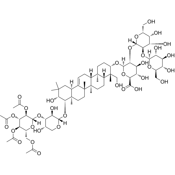
Soyasaponin Ab is an orally active soyasaponin. Soyasaponin Ab inhibits PPARγ transcriptional activity. Soyasaponin Ab induces apoptosis in high concentrations. Soyasaponin Ab exerts anti-obesity, anti-oxidation, anti-inflammation, anti-aging effects. Soyasaponin Ab prevents Scopolamine (HY-N0296)-induced memory impairment.
For research use only. We do not sell to patients.
- Purity : 98.23%
- CAS No.: 118194-13-1
- Formula: C67H104O33
- Molecular Weight:1437.52
-
Storage:
4°C, protect from light
* In solvent : -80°C, 6 months; -20°C, 1 month (protect from light)

To place orders, for customer services and technical support, please contact: MedChemExpress USA
Tel: 609-228-6898 E-mail: [email protected] [email protected]


-
 Biological Activity
-
Chemical Information
-
Solvent & Solubility
- Protocols
- Purity & Documentation
- References
-
 Help & FAQs


Publications Citing Use of MedChemExpress (MCE) Soyasaponin Ab
More

Biological Activity
Description
In Vitro
Soyasaponin Ab (25-100 μM, 8 days ) decreases triglyceride accumulation in a dose-dependent manner in 3T3-L1 adipocytes[1].
Soyasaponin Ab (25-100 μM, 24 h) suppresses the transcriptional activity of peroxisome proliferator-activated receptor γ (PPARγ) in HEK 293T cells[1].
Soyasaponin Ab (50-100 μM, 8 days ) markedly inhibits adipocyte differentiation and expression of various adipogenic marker genes (including adiponectin, ADD1/SREBP1c, aP2, Fas, and resistin) through the downregulation of the adipogenesis-related transcription factors PPARγ and C/EBPα in 3T3-L1 adipocytes[1].
Soyasaponin Ab (1-100 μM, 1 h) shows concentration dependent inhibition of lipid peroxidation in liposomes (IC50 = 14.5 μM)[2].
Soyasaponin Ab (10-50 μM, 48 h) regulates translocation of Nrf2 and protein expressions of the phase II antioxidant enzyme HO-1 and NQO1, through the ERK1/2 signaling pathway in HepG2 cells[2].
Soyasaponin Ab (1-400 μM, 48 h or 10 days) shows little toxicity below 50 μM and reduces the formation of colonies at the dose that above 50 μM and induces apoptosis in HepG2 cells[2].
Soyasaponin Ab (1-10 μM, 16 h or 4 h) significantly reduces Lipopolysaccharides (LPS) (HY-D1056)-stimulated IL-1β, TNF-α and TLR4 expression and PGE2 and NO production in peritoneal macrophages[3].
MedChemExpress (MCE) has not independently confirmed the accuracy of these methods. They are for reference only. Further protocols information, click here.
-
Cell Line:HepG2 cells
-
Concentration:50, 100, 200, 400 μM
-
Incubation Time:10 days
-
Result:Reduced the formation of colonies in dose-dependent manner, especially in 200 and 400 μM treatment groups.
-
Cell Line:HepG2 cells
-
Concentration:1, 5, 10, 20, 50, 100, 200, 400 μM
-
Incubation Time:48 h
-
Result:The cytotoxicity represented a slight increase after treated with higher concentrations after 48 h, especially for the cells treated over 200 μM. There were some morphological changes including loss of cellular geometry and reduction in proliferation of cells after a 100 μM or more treatment. No obvious change on the cell morphology of the groups treated less than 50 μM.
-
Cell Line:HepG2 cells
-
Concentration:50, 100, 200, 400 μM
-
Incubation Time:48 h
-
Result:Induced some extent of apoptosis in cells in a dose-dependent manner.
-
Cell Line:3T3-L1
-
Concentration:50, 100 μM
-
Incubation Time:8 days
-
Result:Effectively suppressed PPARγ and CEBPα mRNA expression. Markedly reduced expression of various adipogenic marker genes, including adiponectin, ADD1/SREBP1c, aP2, Fas, and resistin.
-
Cell Line:3T3-L1
-
Concentration:50, 100 μM
-
Incubation Time:8 days
-
Result:Significantly suppressed the expression of PPARγ and CEBPα proteins at 50 and 100 μM.
In Vivo
Soyasaponin Ab (5-40 mg/kg; p.o.; one hour before the trial) significantly prevents Scopolamine (HY-N0296)-induced memory impairment in male ICR mice[4].
MedChemExpress (MCE) has not independently confirmed the accuracy of these methods. They are for reference only.
-
Animal Model:Male ICR mice (18-22 g, 5 weeks)[4]
-
Dosage:5, 10, 20, 40 mg/kg
-
Administration:Oral gavage (p.o.); One hour before the trial, and memory impairment was induced by intraperitoneal injection of Scopolamine (0.9 mg/kg) 30 min after oral administrations of test agents.
-
Result:Restored memory impairment to 86% at a dose of 10 mg/kg of untreated normal control mice in the passive avoidance task. Restored spontaneous alteration, which was lowered by Scopolamine on the Y-maze task. Significantly shortened the escape latencies on the fifth day.
-
Animal Model:Male institute of cancer research (ICR) mice (20-22 g, 4 weeks)[3]
-
Dosage:10, 20 mg/kg
-
Administration:Oral gavage (p.o.); daily for 5 days
-
Result:Inhibited TNBS-induced body weight reduction, colon shortening, macroscopic score, and myeloperoxidase activity. Inhibited the expression of pro-inflammatory cytokines (TNF-α, IL-1β, IL-6). Ameliorated changes of TNBS-treated mouse colons which showed increased neutrophils, massive bowel edema, dense infiltration of the superficial layers of the mucosa, and epithelial cell disruption by large ulcerations. Inhibited the expression of TLR4, COX-2, and iNOS and the phosphorylation of IRAK1, IKK-β and p65, although it reversed IRAK-1 expression.


Chemical Information
-
CAS No. 118194-13-1
-
Appearance Solid
-
Molecular Weight 1437.52
-
Formula C67H104O33
-
Color White to off-white
-
SMILES
C[C@]12[C@]3(C([C@@]4([H])[C@](C)([C@@H]([C@H](O)C(C)(C)C4)O[C@@]5([H])[C@@H]([C@H]([C@@H](O)CO5)O[C@@]6([H])[C@@H]([C@H]([C@H](OC(C)=O)[C@@H](COC(C)=O)O6)OC(C)=O)OC(C)=O)O)CC3)=CC[C@]1([H])[C@@]7([C@@]([C@](C)([C@@H](O[C@@]8([H])[C@@H]([C@H]([C@H](O)[C@@H](C(O)=O)O8)O)O[C@@]9([H])[C@@H]([C@H]([C@@H](O)[C@@H](CO)O9)O)O[C@]%10([H])O[C@@H]([C@@H](O)[C@H](O)[C@H]%10O)CO)CC7)CO)([H])CC2)C)C
-
Structure Classification
-
Initial Source
-
Shipping
Room temperature in continental US; may vary elsewhere.
-
Storage
4°C, protect from light
* In solvent : -80°C, 6 months; -20°C, 1 month (protect from light)


Publications (1)
-
Journal Impact Factor
-
Most Recent
-
Foods
Development of Extraction Method for Determination of Saponins in Soybean-Based Yoghurt Alternatives: Effect of Sample pH. [Abstract]2023 May 27;12(11):2164. PMID: 37297409


Solvent & Solubility
In Vitro:
DMSO : 100 mg/mL (69.56 mM; Need ultrasonic; Hygroscopic DMSO has a significant impact on the solubility of product, please use newly opened DMSO)
Please refer to the solubility information to select the appropriate solvent. Once prepared, please aliquot and store the solution to prevent product inactivation from repeated freeze-thaw cycles.
Storage method and period of stock solution: -80°C, 6 months; -20°C, 1 month (protect from light). When stored at -80°C, please use it within 6 months. When stored at -20°C, please use it within 1 month.
Please refer to the solubility information to select the appropriate solvent. Once prepared, please aliquot and store the solution to prevent product inactivation from repeated freeze-thaw cycles.
Storage method and period of stock solution: -80°C, 6 months; -20°C, 1 month (protect from light). When stored at -80°C, please use it within 6 months. When stored at -20°C, please use it within 1 month.
Concentration (start) × Volume (start) = Concentration (final) × Volume (final)
In Vivo:
Select the appropriate dissolution method based on your experimental animal and administration route.
- For the following dissolution methods, please ensure to first prepare a clear stock solution using an In Vitro approach and then sequentially add co-solvents:
- To ensure reliable experimental results, the clarified stock solution can be appropriately stored based on storage conditions. As for the working solution for In Vivo experiments, it is recommended to prepare freshly and use it on the same day.
- The percentages shown for the solvents indicate their volumetric ratio in the final prepared solution. If precipitation or phase separation occurs during preparation, heat and/or sonication can be used to aid dissolution.
Add each solvent one by one: 10% DMSO 40% PEG300 5% Tween-80 45% Saline
Solubility: ≥ 2.5 mg/mL (1.74 mM); Clear solution
This protocol yields a clear solution of ≥ 2.5 mg/mL (saturation unknown).
Taking 1 mL working solution as an example, add 100 μL DMSO stock solution (25.0 mg/mL) to 400 μL PEG300, and mix evenly; then add 50 μL Tween-80 and mix evenly; then add 450 μL Saline to adjust the volume to 1 mL.
Preparation of Saline: Dissolve 0.9 g sodium chloride in ddH₂O and dilute to 100 mL to obtain a clear Saline solution.
Add each solvent one by one: 10% DMSO 90% (20% SBE-β-CD in Saline)
Solubility: ≥ 2.5 mg/mL (1.74 mM); Clear solution
This protocol yields a clear solution of ≥ 2.5 mg/mL (saturation unknown).
Taking 1 mL working solution as an example, add 100 μL DMSO stock solution (25.0 mg/mL) to 900 μL 20% SBE-β-CD in Saline, and mix evenly.
Preparation of 20% SBE-β-CD in Saline (4°C, storage for one week): 2 g SBE-β-CD powder is dissolved in 10 mL Saline, completely dissolve until clear.
In Vivo Dissolution Calculator
Please enter the basic information of animal experiments:
-
-
-
-
Recommended: Prepare an additional quantity of animals to account for potential losses during experiments.
Please enter your animal formula composition:
-
%DMSO +
Recommended: Keep the proportion of DMSO in working solution below 2% if your animal is weak.
-
%+
-
+%Tween-80 + +
-
%Saline +
The co-solvents required include: DMSO, . All of co-solvents are available by MedChemExpress (MCE). , Tween 80. All of co-solvents are available by MedChemExpress (MCE).
Working solution concentration: 0.22 mg/mL
Method for preparing stock solution: mg drug dissolved in μL DMSO. Stock solution concentration: mg/mL. * In solvent : -80°C, 6 months; -20°C, 1 month (protect from light)
1. Take μL DMSO stock solution;
2. Add μL .
μL , mix evenly;
3. Then add μL Tween 80, mix evenly;
4. Then add μL
Please ensure that the stock solution in the first step is dissolved to a clear state, and add co-solvents in sequence. You can use ultrasonic heating (ultrasonic cleaner, recommended frequency 20-40 kHz), vortexing, etc. to assist dissolution.

Protocols
-
Dual Luciferin reporter gene assay
Luciferin reporter gene assay is a reporting system to detect the activity of Firefly Luciferase using luciferin as a substrate, which is often used in the research of miRNA target gene verification and promoter transcriptive activity regulation. Dual luciferase usually refers to Firefly luciferase and Renilla luciferase.
-
Apoptosis
Apoptosis, also called programmed cell death, is generally characterized by distinct morphological characteristics.
-
TUNEL staining for apoptotic DNA fragmentation
TUNEL staining detects DNA strand breaks by using terminal deoxynucleotidyl transferase to add labeled nucleotides to exposed 3′-OH DNA termini, generating either microscopic staining in fixed cells or tissue sections, or fluorescence/cytometric signal in cell suspensions. TUNEL positivity reflects DNA fragmentation but should not be interpreted alone as definitive apoptosis, because TUNEL can also label necrotic, autolytic, mechanically damaged, or DNA-repair-associated DNA breaks.
-
Annexin V plus membrane-impermeant dye apoptosis staining
Annexin V-based apoptosis assays rely on the detection of phosphatidylserine (PS) externalization from the inner leaflet of the plasma membrane to the outer leaflet, an early biochemical hallmark of apoptosis. Fluorescently labeled Annexin V binds PS in a calcium-dependent manner, enabling identification of early apoptotic cells by flow cytometry or fluorescence microscopy. When combined with a membrane-impermeant DNA-binding dye (e. g. , propidium iodide), this approach allows discrimination between viable (Annexin V−/dye−), early apoptotic (Annexin V+/dye−), and late apoptotic or necrotic (Annexin V+/dye+) cell populations by assessing membrane integrity and PS exposure.
-
Apoptosis Solutions
Apoptosis is a regulated, generally non-lytic cell-death pathway that removes unwanted, damaged, infected, or abnormal cells through coordinated morphological changes, caspase activation, DNA fragmentation, and membrane remodeling. The intrinsic apoptosis pathway is controlled mainly by mitochondrial outer membrane permeabilization, BCL-2 family proteins, cytochrome c release, apoptosome formation, caspase-9 activation, and downstream executioner caspase-3/7 activation. The extrinsic apoptosis pathway is initiated by death receptors such as Fas, TNFR, and TRAIL receptors, which recruit adaptor proteins and activate caspase-8 before engaging executioner caspases or mitochondrial amplification through BID cleavage. Apoptosis is linked to many phenotypes, including cancer cell killing, tissue homeostasis, immune regulation, neurodegeneration, infection response, and treatment-induced cytotoxicity; unresolved questions include how apoptosis interacts with necroptosis, pyroptosis, ferroptos
-
Research Protocol for Inflammation-related Diseases
The NLRP3 inflammasome is a cytosolic innate immune signaling platform that integrates priming signals and danger-signal activation to promote caspase-1 activation, maturation of IL-1β and IL-18, and gasdermin D-mediated pyroptotic cell death. The core experimental logic is to determine whether inflammatory disease phenotypes are driven by increased NLRP3 expression, ASC-containing inflammasome assembly, caspase-1 cleavage, GSDMD cleavage, and extracellular release of IL-1β/IL-18 rather than by nonspecific cell injury alone. The pathway is strongly linked to inflammation-related disease phenotypes because monosodium urate crystals activate NALP3/NLRP3 inflammasome signaling in gout-like crystal inflammation, cholesterol crystals activate NLRP3 inflammasomes in atherogenesis models, and DSS-induced intestinal inflammation has been reported to involve NLRP3 inflammasome activity. However, experimental colitis studies also show context-dependent protective effects of NLRP3 inflammasome co
-
Research Protocol for Metabolic Diseases
AMP-activated protein kinase, AMPK, is a conserved cellular energy sensor that responds to reduced cellular energy status and coordinates metabolism by increasing ATP-generating catabolic pathways while suppressing ATP-consuming anabolic processes. In metabolic disease research, the AMPK pathway is experimentally relevant because it regulates hepatic lipid synthesis, fatty acid oxidation, glucose production, skeletal-muscle glucose disposal, mTORC1-linked biosynthesis, autophagy, mitochondrial homeostasis, and whole-body energy balance. The central pathway logic is that energy stress, metformin, exercise-like stimulation, or direct AMPK activators increase AMPKα Thr172 phosphorylation and downstream substrate phosphorylation, including ACC and RAPTOR. Phosphorylation of ACC suppresses lipogenesis and supports fatty acid oxidation, whereas phosphorylation of RAPTOR suppresses mTORC1 signaling and links cellular energy status to growth and protein synthesis control. The pathway is linked


Purity & Documentation
-
Data Sheet (291 KB)
-
SDS (252 KB)
- English - EN (252 KB)
- Français - FR (252 KB)
- Deutsch - DE (252 KB)
- Norwegian - NO (252 KB)
- Español - ES (252 KB)
- Swedish - SV (252 KB)
- Italian - IT (252 KB)
- Korean - KR (252 KB)
- Portuguese - PT (252 KB)
-
Handling Instructions (2659 KB)
References
[1]. Yang SH, et al. Soyasaponins Aa and Ab exert an anti-obesity effect in 3T3-L1 adipocytes through downregulation of PPARγ. Phytother Res. 2015 Feb;29(2):281-7. [Content Brief]
[3]. Lee IA, et al. Soyasaponin Ab ameliorates colitis by inhibiting the binding of lipopolysaccharide (LPS) to Toll-like receptor (TLR)4 on macrophages. J Agric Food Chem. 2011 Dec 28;59(24):13165-72. [Content Brief]
[4]. Hong SW, et al. Soyasaponins Ab and Bb prevent scopolamine-induced memory impairment in mice without the inhibition of acetylcholinesterase. J Agric Food Chem. 2014 Mar 5;62(9):2062-8. [Content Brief]


Complete Stock Solution Preparation Table
Please refer to the solubility information to select the appropriate solvent. Once prepared, please aliquot and store the solution to prevent product inactivation from repeated freeze-thaw cycles.
Storage method and period of stock solution: -80°C, 6 months; -20°C, 1 month (protect from light). When stored at -80°C, please use it within 6 months. When stored at -20°C, please use it within 1 month.
| Optional Solvent | Concentration Solvent Mass | 1 mg | 5 mg | 10 mg | 25 mg |
|---|---|---|---|---|---|
| DMSO | 1 mM | 0.6956 mL | 3.4782 mL | 6.9564 mL | 17.3911 mL |
| 5 mM | 0.1391 mL | 0.6956 mL | 1.3913 mL | 3.4782 mL | |
| 10 mM | 0.0696 mL | 0.3478 mL | 0.6956 mL | 1.7391 mL | |
| 15 mM | 0.0464 mL | 0.2319 mL | 0.4638 mL | 1.1594 mL | |
| 20 mM | 0.0348 mL | 0.1739 mL | 0.3478 mL | 0.8696 mL | |
| 25 mM | 0.0278 mL | 0.1391 mL | 0.2783 mL | 0.6956 mL | |
| 30 mM | 0.0232 mL | 0.1159 mL | 0.2319 mL | 0.5797 mL | |
| 40 mM | 0.0174 mL | 0.0870 mL | 0.1739 mL | 0.4348 mL | |
| 50 mM | 0.0139 mL | 0.0696 mL | 0.1391 mL | 0.3478 mL | |
| 60 mM | 0.0116 mL | 0.0580 mL | 0.1159 mL | 0.2899 mL |


Soyasaponin Ab Related Classifications
HY-N3026 Related Classifications
 Powered by Bioz