



Tomaralimab

Based on 1 Customer Validation
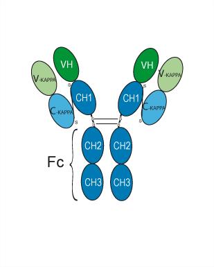
Tomaralimab (OPN-305) is a humanized anti-TLR2 IgG4 monoclonal antibody. Tomaralimab inhibits TLR2, MyD88, NLRP3, and reduces pro-inflammatory cytokine (IL-1β, IL-6, IL-8) production. Tomaralimab reduces tau pathology. Tomaralimab improves cognition, atopic dermatitis. Tomaralimab has anticancer effects on pancreatic ductal adenocarcinoma. Tomaralimab is being studied in myelodysplastic syndrome (MDS), atopic dermatitis, pancreatic ductal adenocarcinoma, Alzheimer's disease, and myocardial ischemia/reperfusion injury.
For research use only. We do not sell to patients.
- Purity : 99.42%
- CAS No.: 1449294-76-1
- Molecular Weight:145.32 kDa
-
Storage:
Please store the product under the recommended conditions in the Certificate of Analysis.


To place orders, for customer services and technical support, please contact: MedChemExpress USA
Tel: 609-228-6898 E-mail: [email protected] [email protected]


-
 Biological Activity
-
Chemical Information
- Protocols
- Purity & Documentation
- References
-
 Help & FAQs






Biological Activity
Description
Isotype
Human IgG4 kappa
Recommend Isotype Controls
Species Reactivity
Human
|
IL-1β |
IL-6 |
IL-8 |
NLRP3 |
In Vitro
Tomaralimab (100-500 nM; 20 h) dose-dependently inhibits oligomeric tau-induced TLR2 activation in HEK-Blue hTLR2 cells, reducing SEAP levels [1].
MedChemExpress (MCE) has not independently confirmed the accuracy of these methods. They are for reference only.
-
Cell Line:THP-1 cells
-
Concentration:100 nM, 500 nM
-
Incubation Time:24 h, 48 h
-
Result:Decreased the protein levels of TLR2, MyD88, NLRP3.
-
Cell Line:THP-1 cells
-
Concentration:100 nM, 500 nM
-
Incubation Time:24 h, 48 h
-
Result:Decreased the mRNA levels of proinflammatory cytokines (TNF-α, IL-1β, IL-6, IL-8) induced by oligomeric tau.
In Vivo
Tomaralimab (25 mg/kg; i.v.; every other day; until day 19) attenuates DNCB‑induced atopic dermatitis (AD)‑like skin lesions in BALB/c mice[2].
Tomaralimab (10 mg/kg; i.p.; twice weekly) suppresses tumor growth and reduces PCNA-positive proliferating cells in NSG mice with human pancreatic ductal adenocarcinoma (PDAC) xenografts[3].
Tomaralimab (1.56-12.5 mg/kg; i.v.; 15 min before reperfusion) dose-dependently reduces infarct size by up to 45% and preserves cardiac function in porcine myocardial ischemia/reperfusion models[5].
MedChemExpress (MCE) has not independently confirmed the accuracy of these methods. They are for reference only.
-
Animal Model:BALB/c mice (male, 18-23 g, ~7 weeks old) with DNCB-induced atopic dermatitis[2]
-
Dosage:25 mg/kg
-
Administration:Intravenous injection (i.v.); every other day; until day 19
-
Result:Reduced epidermal thickness, mast cell/macrophage infiltration, and pro-inflammatory cytokines (TSLP, IL-4, IL-13).
Increased filaggrin expression.
Clinical Trial
| NCT Number | Sponsor | Condition | Start Date |
Phase

|
|---|---|---|---|---|
| NCT01329991 | 2011-05 | PHASE1 |
Gene ID
Accession
Conjugated
Unconjugated
Reconsititution
The product can be reconstituted/diluted with sterile PBS or saline.
Format
-
Human IgG4 kappa
Application
ELISA, FACS, Functional assay
Verified Bioactivity
-
 Immobilized Human TLR2 Protein can bind Tomaralimab. The EC50 for this effect is 649.63 ng/mL.
 -
 Flow cytometric analysis of 1.5X10⁶ THP-1 cells with Tomaralimab (HY-P99555, red). Cells were fixed with 4% paraformaldehyde. Then stained with the primary antibody at 1/200 dilution for an hour at 4℃. Goat Anti-Human IgG H&L (AF488) (HY-P83776) was used as the secondary antibody at 1/1,000 dilution for 30 minutes at 4℃. Human IgG4 (S228P) kappa, Isotype Control (HY-P99003, blue) was used as the isotype control, cells without incubation with primary antibody were used as the unlabeled control (black).


Chemical Information
-
CAS No. 1449294-76-1
-
Appearance Liquid
-
Molecular Weight 145.32 kDa
-
Color Colorless to light yellow
-
SMILES
[Tomaralimab]
-
Synonyms
OPN-305
-
Shipping
Shipping with dry ice.
-
Formulation
Please refer to the lot-specific COA for specific buffer information.
-
Storage
Please store the product under the recommended conditions in the Certificate of Analysis.


Purity & Documentation
-
Data Sheet (263 KB)
-
SDS (251 KB)
- English - EN (251 KB)
- Français - FR (251 KB)
- Deutsch - DE (251 KB)
- Norwegian - NO (251 KB)
- Español - ES (251 KB)
- Swedish - SV (251 KB)
- Italian - IT (251 KB)
- Korean - KR (251 KB)
- Portuguese - PT (251 KB)
-
Inhibitory Antibodies User Guide (603 KB)
References
[1]. Kim Y, et al. TLR2 immunotherapy suppresses neuroinflammation, tau spread, and memory loss in rTg4510 mice. Brain Behav Immun. 2024 Oct;121:291-302. [Content Brief]
[2]. Yeo H, et al. Therapeutic potential of a systemically applied humanized monoclonal antibody targeting Toll‑like receptor 2 in atopic‑dermatitis‑like skin lesions in a mouse model. Biomed Rep. 2024 Dec 30;22(3):41. [Content Brief]
[3]. Lundy J, et al. TLR2 activation promotes tumour growth and associates with patient survival and chemotherapy response in pancreatic ductal adenocarcinoma. Oncogene. 2021 Oct;40(41):6007-6022. [Content Brief]
[5]. Arslan F, et al. Treatment with OPN-305, a humanized anti-Toll-Like receptor-2 antibody, reduces myocardial ischemia/reperfusion injury in pigs. Circ Cardiovasc Interv. 2012 Apr;5(2):279-87. [Content Brief]

Calculators
Concentration (start) × Volume (start) = Concentration (final) × Volume (final)


Tomaralimab Related Classifications
HY-P99555 Related Classifications
 Powered by Bioz