



Wasabi Receptor Toxin
Wasabi Receptor Toxin is a cell-penetrating scorpion toxin. Wasabi Receptor Toxin is the activator for TRPA1 ion channel with EC50 in nanomolar level, and prolongs the channel open time, but reduces Ca2+ permeability. Wasabi Receptor Toxin causes thermal hypersensitivity and mechanical allodynia in rats, without triggering neurogenic inflammation.
For research use only. We do not sell to patients.
- CAS No.: 2569291-86-5
- Formula: C164H245N45O53S5
- Molecular Weight:3855.29
-
Storage:
Please store the product under the recommended conditions in the Certificate of Analysis.

To place orders, for customer services and technical support, please contact: MedChemExpress USA
Tel: 609-228-6898 E-mail: [email protected] [email protected]


-
 Biological Activity
-
Chemical Information
- Protocols
- Purity & Documentation
- References
-
 Help & FAQs




Biological Activity
Description


Chemical Information
-
CAS No. 2569291-86-5
-
Molecular Weight 3855.29
-
Formula C164H245N45O53S5
-
Synonyms
WaTx
-
Sequence
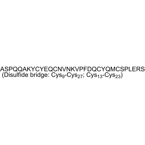
Ala-Ser-Pro-Gln-Gln-Ala-Lys-Tyr-Cys-Tyr-Glu-Gln-Cys-Asn-Val-Asn-Lys-Val-Pro-Phe-Asp-Gln-Cys-Tyr-Gln-Met-Cys-Ser-Pro-Leu-Glu-Arg-Ser (Disulfide bridge: Cys9-Cys27; Cys13-Cys23)
-
Sequence Shortening
ASPQQAKYCYEQCNVNKVPFDQCYQMCSPLERS (Disulfide bridge: Cys9-Cys27; Cys13-Cys23)
-
Shipping
Room temperature in continental US; may vary elsewhere.
-
Storage
Please store the product under the recommended conditions in the Certificate of Analysis.

Protocols
-
Two-electrode voltage clamp in Xenopus oocytes
Two-electrode voltage clamp measures whole-oocyte membrane current from Xenopus oocytes expressing exogenous ion channels, receptors, or transporters; one intracellular microelectrode senses membrane voltage, and the second injects current so the amplifier can hold the membrane at command voltages while recording the compensating current as the functional readout. The method is suited to Xenopus oocytes because their large size supports microinjection and intracellular electrode impalement, but the large membrane area can limit voltage-clamp speed and accuracy, especially for large or fast currents.
-
Transepithelial/transendothelial electrical resistance assay
TEER measures electrical resistance across epithelial or endothelial monolayers cultured on permeable supports, and the readout reflects ionic conductance through the cell barrier, especially the paracellular pathway regulated by junctional integrity. TEER can be measured without destroying the monolayer and is commonly used before or during transport, permeability, barrier-disruption, and barrier-maturation experiments. TEER values are influenced by biological maturation and technical conditions; reported factors include temperature, medium formulation, passage number, electrode geometry, membrane properties, and junctional length during early monolayer maturation. Therefore, TEER should be interpreted with blank-insert subtraction, area normalization, repeated readings, and, when possible, orthogonal barrier readouts such as FITC-dextran flux or tight-junction staining.
-
Ca2+ Staining Technique
Ca2+ staining is an experimental technique that utilizes specific fluorescent probes (such as Fluo-4 AM, Fura-2, etc.) to qualitatively or quantitatively detect dynamic changes in intracellular Ca2+ concentrations; this is achieved by monitoring the changes in fluorescent signals generated when these probes bind to free intracellular calcium ions. The underlying principle relies primarily on the presence of chelating groups within the probe's molecular structure that possess high affinity for calcium ions.
-
Research Protocol for Inflammation-related Diseases
The NLRP3 inflammasome is a cytosolic innate immune signaling platform that integrates priming signals and danger-signal activation to promote caspase-1 activation, maturation of IL-1β and IL-18, and gasdermin D-mediated pyroptotic cell death. The core experimental logic is to determine whether inflammatory disease phenotypes are driven by increased NLRP3 expression, ASC-containing inflammasome assembly, caspase-1 cleavage, GSDMD cleavage, and extracellular release of IL-1β/IL-18 rather than by nonspecific cell injury alone. The pathway is strongly linked to inflammation-related disease phenotypes because monosodium urate crystals activate NALP3/NLRP3 inflammasome signaling in gout-like crystal inflammation, cholesterol crystals activate NLRP3 inflammasomes in atherogenesis models, and DSS-induced intestinal inflammation has been reported to involve NLRP3 inflammasome activity. However, experimental colitis studies also show context-dependent protective effects of NLRP3 inflammasome co


Purity & Documentation
References

Calculators
Concentration (start) × Volume (start) = Concentration (final) × Volume (final)


Wasabi Receptor Toxin Related Classifications
HY-P5914 Related Classifications
 Powered by Bioz